A STUDY OF MR ANGIOGRAPHY CHARACTERISTICS IN ACUTE ISCHEMIC STROKE IN TERTIARY CARE HOSPITAL IN NORTHERN INDIA
- Neelam Kumari Sahu , Consultant Neurology Indraprastha Apollo Hospitals New Delhi-110076
- Pushpendra Nath Renjen , Sr. Consultant Neurologist & Academic C Institute of Neurosciences, Indraprastha Apollo Hospitals New Delhi-110076
- Harsh Rastogi , Sr. Consultant Department of Interventional Neuroradiology Indraprastha Apollo Hospitals New Delhi-110076
- Avinash Goswami , Consultant Neurology, Indraprastha Apollo Hospital, New Delhi-110076
- Dinesh Mohan Chaudhari , Consultant Neurology, Institute of Neurosciences Indraprastha Apollo Hospitals, New Delhi– 110076
Article Information:
Abstract:
Objective: Stroke is second-leading cause of death and steno-occlusive arterial disease is commonest cause of ischemic-stroke worldwide. Time of flight (TOF) MRA technique gives an idea of the overall cerebral-vascular-channel morphology and has important prognostic and prophylactic value in the management. The primary objective is to study occurrence of occlusion versus stenosis in MR-angiography study in acute-ischemic-stroke patients, and other characteristics including size and location of infarct, location of involved arterial-territory, their correlation and associated risk-factors. The frequency of size of infarct compared to degree of stenosis was also studied.Methods: 110 acute-ischemic-stroke subjects were enrolled during March 2021 to March 2022 on the basis of inclusion and exclusion criteria. 13 patients were excluded due to incomplete evaluation. Thus, 97 acute-ischemic-stroke subjects were finally and evaluated on the basis of history, physical examination, and investigations as per stroke protocol. The data was analyzed using SPSS 22.0 and R.3.2.0. Results: Mean age of acute-ischemic-stroke subjects was 60.927±14.182 years, large artery atherosclerosis (53%) was commonest subtype, MCA being most commonly involved (45.05%). In 45.36 % ischemic stroke patients there was no steno-occlusion, while 24.74 % and 29.90 % patients had stenosis and occlusion respectively. In patients with no steno-occlusive disease, 59 % and 41 % had small and medium sized infarcts respectively.Conclusion: MRA provides early diagnosis of occlusive/stenotic arterial disease and demonstrated vascular lesions show a high correlation with infarct distribution. Our study re-affirms that large artery atherosclerosis is most frequent stroke subtype in ischemic-stroke patients in India.
Keywords:
Article :
INTRODUCTION:
World Health Organization defines stroke as ―a clinical syndrome of rapidly developing clinical signs of focal disturbance of cerebral function, lasting more than 24 hours, or leading to death, with before a no apparent cause, other than that of vascular origin [1]. The American Heart Association/American Stroke Association published an updated definition of stroke in 2013 [2] and defined ischemic stroke as an episode of neurological dysfunction caused by focal cerebral, spinal, or retinal infarction, and hemorrhagic stroke as, rapidly developing clinical signs of neurological dysfunction attributable to a focal collection of blood within the brain parenchyma or ventricular system that is not caused by trauma. Globally, Stroke is the second-leading cause of death and the third-leading cause of death and disability combined according to latest Global Burden of Disease study in 2019 (GBD-2019) [3]. The study also showed that there were 101 million (93.2–111) prevalent cases and 6.55 million (6.00–7.02) deaths due to stroke reported worldwide in 2019. From 1990 to 2019, incident cases of stroke increased by 67–73 %, prevalent cases increased by 83–88 %, deaths increased by 31–55 %, and DALYs increased by 22–42 % [3]. In India [4], prevalence rate of stroke ranges from 44-150/100000, while 30 days case fatality rate ranges from 18-46.3%. Around 10-15% of all strokes occur in the young (<40 years of age). Several risk factors have been identified for stroke. Non-modifiable ones include age, sex, race, and ethnicity, while modifiable ones are hypertension, diabetes mellitus, current smoking, overweight, diet, physical inactivity, dyslipidemia, alcohol consumption and cardiac illness [5-7]. Ischemic stroke can be classified on the basis of various classification systems, like, TOAST (Trial of Org 10172 in Acute Stroke Treatment) [8], CCS (Causative classification system) [9], ASCO (Atherothrombosis, Small Vessel Disease, Cardiac pathology, Other) [10] , and CISS (Chinese Ischemic Stroke Classification) [11]. Among these, TOAST classification is the most widely used classification system for subtyping ischemic stroke [12-14]. Studies have shown racial differences in the location of large vessel atherosclerotic disease [15-25]. ICAD and ECAD can be evaluated by magnetic resonance angiography (MRA) and other invasive angiography techniques [26] . Currently, the acute management of Ischemic Stroke patients, who present within a time window, includes Intravenous thrombolysis and/or Mechanical Thrombectomy [27-38]. Similar to the differences in acute management of patients with intracranial large vessel occlusion compared to other ischemic stroke subtypes, secondary preventive treatment for these patients is also different. SAMMPRIS trial [23] demonstrated superiority of aggressive medical therapy comprising of dual antiplatelets for 3 months followed by single antiplatelet, over intracranial stenting. For extracranial atherosclerotic disease, carotid endarterectomy or carotid stenting is recommended [39, 40, 41] . Steno-occlusive arterial disease is the commonest cause of ischemic stroke all over the world where the thromboembolic causes account for about 85% of stroke cases [42] . A high incidence of the disease was reported in African and Asian populations [42] . Proper and accurate assessment of arterial diseases that underlie development of stroke is essential in patient management and follow-up. The protocols of MR imaging in stroke patients are not standard across institutions [43] . Adding magnetic resonance angiography (MRA) to the imaging protocol improves both diagnosis and clinical management [43]. MRA can track the changes in the vessel lumen with time. It assesses the risk of future events by identifying whether there is occlusive arterial disease, localizing the exact site of occlusion and by determining the pathology underlying the stroke such as atherosclerosis or dissection [44] . MRA can also identify other vascular lesions such as malformation, aneurysms and arterial compression [44] . Time of flight (TOF) MRA technique gives the managing physician an idea of the overall cerebral vascular channel morphology and thus has an important prognostic and prophylactic value in the patient’s management [43,44]. Moreover, MRA has a potentially important role in appropriate patient selection for intra-arterial fibrinolysis, in patient’s follow-up and monitoring of therapeutic efficacy [45]. A normal or near normal MRA of the intra cranial arteries can effectively exclude the possibility of a high-grade arterial stenosis [44] . In the current study we assessed the MR Angiography pattern (occlusion versus stenosis) in evaluation of patients with acute ischemic stroke and also determined various characteristics (size and location of infarct and the arterial territory involved) and studied the various risk factors and their association with different MR angiographic characteristics of infarct and associated blood vessels.
METHODS:
We studied ischemic stroke patients aged >18 years, admitted at our facility. A total of 110 ischemic stroke patients were enrolled, out of which 13 patients were excluded due to incomplete evaluation. Thus, a total of 97 ischemic stroke patients were studied for our primary objective (sample size was 92). We included patients with >18 years with acute ischemic stroke within 7 days of onset. We excluded patients with hemorrhagic stroke, stroke mimics- like brain tumors, toxic or metabolic disorders (hypoglycemia, hypercalcemia, hyponatremia, uremia, hepatic encephalopathy, and hyperthyroidism), infectious disorders (meningitis, encephalitis), psychological disorders, migraine, seizures and demyelinating disorders, patients with history of previous ischemic, hemorrhagic stroke or sinus thrombosis and patients unwilling to give consent for participation in the study. Patients were assessed on the basis of history, physical examination, and investigations as per proforma. Ischemic stroke patients were classified as per TOAST classification. ICAD or ECAD or Coexisting ICAD & ECAD was identified on the basis of MR Angiography. Stenosis that was ≥50% was classified as significant large vessel atherosclerosis [24, 44,46, 47, 48,49]. MRA findings which signify affection of intra cranial/ extra cranial arteries were recorded as stenosis (segmental flow gap or luminal stenosis >50%), occlusion (non-visualized vessel segment with absent distal flow), atherosclerotic changes of the vessels and presence of plaques [50].
The MR studies were carried out within 1 week of admission. Patients were scanned on a 3T MR imaging system and 3D TOF –MR Angiography (Time of flight –MR Angiography). The sequences used were multislab (5 slabs for IC and 8 slabs for EC regions) 3-dimensional study based on time of flight principle. MRAs were reviewed by a single Neuroradiologist, who was blinded to the clinical details. Brain MRI was acquired using 3 Tesla. The infarct size was categorized as small (2 cm), medium (2-4 cm), and large (4 cm) according to its largest diameter in one plane on Diffusion Weighted Imaging (DWI), T1 Weighted Imaging (T1WI), T2 Weighted Imaging (T2WI), Fluid Attenuated Inversion Recovery technique (FLAIR). Assessment was done for the intracranial vessels: middle cerebral arteries (MCAs), anterior cerebral arteries (ACAs), intracranial internal carotid arteries (ICAs), posterior cerebral arteries (PCAs), vertebral arteries (VAs), and the basilar artery (BA). MRA findings which signify affection of intra cranial/ extra cranial arteries were recorded as stenosis (segmental flow gap or luminal stenosis >50%), occlusion (nonvisualized vessel segment with absent distal flow), atherosclerotic changes of the vessels and presence of plaques. MRA findings were recorded and correlated with distribution of signal abnormalities seen in the MRI sequences. According to location, the infarcts were categorized into cortical, lacunar, territorial and watershed. The risk factors of stroke, age, sex, hypertension, diabetes mellitus, obesity (BMI) and pattern of infarcts were correlated with any MRA abnormality (IC MRA, EC MRA, or both) separately. Data was analysed using Statistical Package for Social Sciences (SPSS) version 21, IBM Inc. Level of statistical significance was set at p-value.
RESULTS:
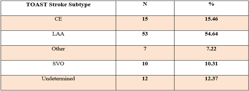
Mean age of acute ischemic stroke subjects was 60.927±14.182 years and prevalence of ischemic stroke was observed to increase with increasing age. Highest prevalence of ischemic stroke was observed in the age group of 71-80 years (31 %), followed by age group of 61-70 years (26.8 %), while lowest prevalence was observed in the age group of 20-30 years (3.3%). Gender wise distribution was observed to be insignificant. Large Artery Atherosclerosis (53%) was the commonest subtype of acute ischemic stroke in our study cohort, followed by strokes of cardio embolic etiology (15%).
Table:1 Distribution of TOAST Stroke Subtypes among Acute Ischemic Stroke Patients
56 patients (58%) of acute ischemic stroke had significant large vessel atherosclerotic disease, while 41 patients (42 %) did not have any. Isolated ICAD was seen in 38.14%, while isolated ECAD was less prevalent (9.3% patients). 10.31 % patients had coexisting ICAD and ECAD. No significant statistical difference in the gender composition of patients having ICAD, ECAD, Coexisting ICAD & ECAD or no significant Large Vessel Atherosclerotic Disease. DM and hypertension were the most prevalent risk factor in subjects with acute ischemic stroke (60.82%) and (60.82%) respectively, 56.7% patients had BMI>25, 52.5 % had dyslipidemia.
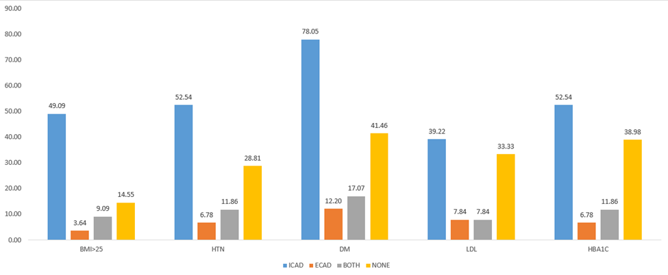
Figure:1 Distribution of Risk Factors among ICAD, ECAD, Coexisting ICAD & ECAD, and No Large Vessel Disease
Significantly a greater number of subjects with ICAD were diabetic (52.54%) and hypertensive (52.54%), as compared to subjects with ECAD or subjects with coexisting ICAD & ECAD or those without Large Vessel Atherosclerotic Disease. BMI>25 was observed in 49.09 % of subjects with isolated ICAD. Significant co-relation between BMI>25 and HbA1C > 6.5, in patients with ischemic stroke in which large vessel atherosclerotic disease was present.
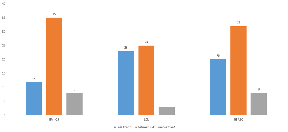
Figure:2 Correlation of Infarct Size with BMI, LDL, and HbA1C Levels
42.2 % ischemic stroke patients had infarct size 4 cm. P-value was 0.001 and result was significant for infarct size and BMI >25, while p-value was not significant for infarct size and patients with dyslipidemia and diabetes in ischemic stroke patients.
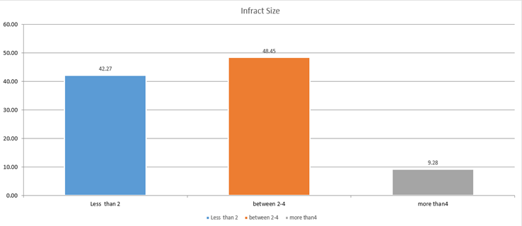
Figure:3 Distribution of Infarct Size among Acute Ischemic Stroke Patients
45.36 % ischemic stroke patients there was no steno-occlusion, while 24.74 % and 29.90 % patients had stenosis and occlusion in MR angiography respectively. 59 % and 41 % with no steno-occlusive disease ischemic stroke patient had small and medium sized infarcts respectively. 41.6 % and 58.2 % patients with stenotic lesion in their MR angiography study had small and medium sized infarct respectively. 65 % and 3 % patients with complete occlusion in MR angiography study had medium and small sized infarct respectively. while 31 % patients with complete occlusion in MR angiography study showed large sized infarct, none of the steno-occlusive or stenosis patients had large infarct in MR angiography study. Thus, this frequency table shows there is some relation between level of occlusion and size of infarct.
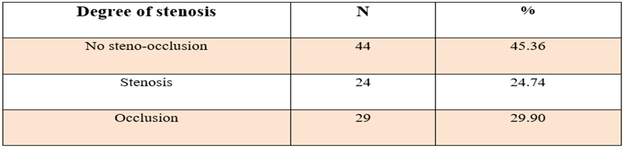
Table:2 Degree of Steno-Occlusive Disease on MR Angiography
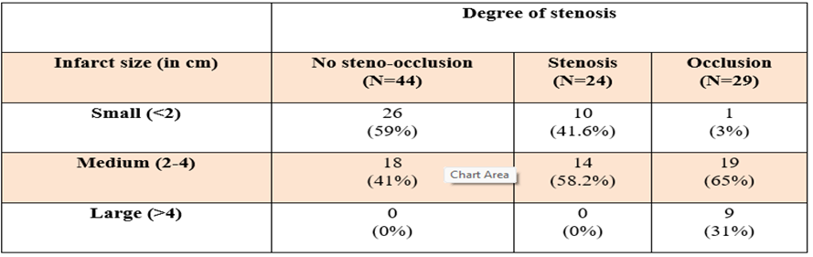
Table:3 Correlation between Infarct Size and Degree of Stenosis/Occlusion on MR Angiography
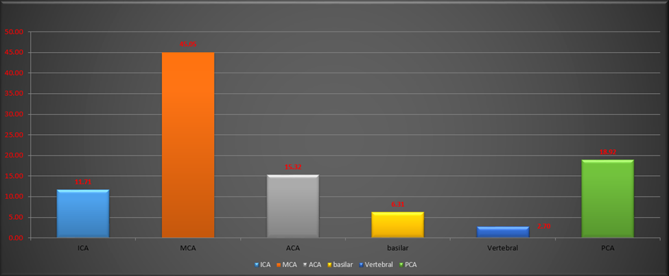
Figure:4 Distribution of Involved Arterial Territories in Acute Ischemic Stroke Patients
57.73 % ischemic stroke patients had plaque in the MR angiography study involving multiple arterial segments, while 23.7 % and 18.56 % patients had mild atheromatous changes and none respectively. 10. 94.5 % patients with ischemic stroke who did not have any atheromatous changes in their MR angiography study had no steno-occlusion and 5.5 % patients with no atheromatous changes had stenosis while none had occlusion. Similarly, 95.6 % patients with mild atheromatous changes had no steno-occlusion while 4.4 % patients had stenosis and none had occlusion. While 51.8 % patients with ischemic stroke with plaque in their MR angiography study revealed occlusion of artery, 39.3 % patients with plaque had stenosis and 8.9% patients had no steno-occlusion in their MR angiography study. Most commonly involved territory was that of MCA (45.05%), followed by PCA territory (18.9%). Least commonly affected territory was that of vertebral (3.0%).
DISCUSSION :
Stroke is the second-leading cause of death and the third-leading cause of death and disability combined according to the latest GBD-2019 study [3]. Steno-occlusive arterial disease is the commonest cause of ischemic stroke all over the world. Proper and accurate assessment of arterial diseases that underlie development of stroke is essential in patient management and follow-up. Large artery atherosclerosis (LAA) is one of the commonest pathophysiological mechanism for ischemic stroke and includes patients with significant stenosis (>50%). The protocols of MR imaging in stroke patients are not standard across institutions [8]. Adding magnetic resonance angiography (MRA) to the imaging protocol improves both diagnosis and clinical management [44,50] . MRA can track the changes in the vessel lumen with time. It also assesses the risk of future events by identifying whether there is occlusive arterial disease, localizing the exact site of occlusion and by determining the pathology underlying the stroke such as atherosclerosis or dissection [12]. MRA can also identify other vascular lesions such as malformation, aneurysms and arterial compression. Time of flight (TOF) MRA technique gives the managing physician an idea of the overall cerebral vascular channel morphology and thus has an important prognostic and prophylactic value in the patient’s management [51,52]. Moreover, MRA has a potentially important role in appropriate patient selection for intraarterial fibrinolysis, in patient’s follow-up and monitoring of therapeutic efficacy [44,45]. A normal or near normal MRA of the intra cranial arteries can effectively exclude the possibility of a high-grade arterial stenosis. Population-based data of stroke subtypes is needed to identify and implement appropriate resources for stroke management. This study was conducted with the primary objective to look for the occurrence of occlusion versus stenosis in MR angiography study in acute ischemic stroke patients, and various other characteristics including size and location of infarct, location of involved arterial territory, their correlation with each other and associated risk factors.
A total of 110 patients were enrolled, out of which 13 patients were excluded due to incomplete evaluation thus, 97 acute ischemic stroke subjects were finally analysed (estimated sample size was 92).
In this study, mean age of acute ischemic stroke subjects was 60.927±14.182 years (Table 1). Similar results have been reported by Wang Y et al [24] (61.91±11.24 years), Stephanie P Jones et al [53] (62.2 years), PN Sylaja et al [54] (58.3±14.7 years), C Venkata S Ram et al [55] (56.3±11.7 years), and Paramdeep Kaur et al [56] (59±15 years).
Most of the acute ischemic stroke subjects in this study were males (72%), while females were 28%, (Table 3). Similarly, higher percentage of male patients was reported by Kaul S et al [57] (78.3% males), Wang Y et al [24] (67.8 % males), C Venkata S Ram et al [55] (72.3% males), Raghuvanshi S [58] (74.28% males and 25.71% females).
This study re-affirmed the fact that LAA is the most prevalent stroke subtype in Asians and Indian population (Table 4). Prevalence of LAA in this study was 53% followed by cardio-embolic (15%). Undetermined etiology was seen in 12%, small vessel occlusion in 10%, while Other causes were identified in only 7% ischemic stroke subjects. Adamski et al [12] , and Cole JW et al [13] showed that LAA is responsible for approximately 15-20% of all ischemic strokes worldwide, while limited studies from India suggests that LAA is the most common underlying pathology in ischemic stroke patients- Wasay M et al [7] , Kaul S et al [57] , and Sylaja PN etal [54] reported LAA in 30-40%, 37.6%, and 29.9% of Indian acute ischemic stroke patients respectively.
TOAST categories did not have significant difference in gender composition across all the stroke subtypes (Table 5), although commonest ischemic stroke subtype in both males and females was LAA (60 % and 41% respectively). As per results of our study, 56 patients (58%) of acute ischemic stroke had significant large vessel atherosclerotic disease, while 41 patients (42 %) did not have any. Isolated ICAD was seen in 38.14%, while isolated ECAD was less prevalent (9.3% patients). 10.31 % patients had coexisting ICAD and ECAD (Table 6). Thus, overall ICAD (Isolated plus Coexisting with ECAD) was seen in 47 ischemic stroke subjects, while overall ECAD (Isolated plus Coexisting with ICAD) was seen in 19 ischemic stroke subjects (48.4% and 19.6% of total ischemic stroke subjects respectively). Wang Y et al [24] reported similar results as our study- 37.5% patients of acute ischemic stroke had isolated ICAD, 9.1% had coexisting ICAD & ECAD, while 4.9% had isolated ECAD. Indian data for differential prevalence of ICAD and ECAD is quite limited. Ghosh M et al [59] reported results similar to our study- 52.78% patients had ICAD, while ECAD was found in 13.89% patients, and in majority of patients with ECAD, concurrent ICAD was present (80%). Kaul S et al [57] reported that out of total patients with significant large vessel atherosclerosis, 78.3% had ICAD and 21.7% had ECAD. Kumar G et al [44] reported 56.3% patients with ECAD and 63.6% patients with ICAD, although they also included patients with <50 % stenosis.
This study shows that 42.2 % ischemic stroke patients had infarct size 4 cm (Table 9) and co-relation between infarct size and various risk factors in ischemic stroke patients. 63.5% ischemic stroke patients with BMI > 25 had medium sized infarct (2-4 cm) while 14.5% and 22 % had large (>4 cm) and small (25, while p-value was not significant for infarct size and patients with dyslipidemia and diabetes in ischemic stroke patients (Table 10). Co-relation between infarct size and hypertension in ischemic stroke patients. 54 % hypertensive patients had medium sized (2-4 cm) infarct, 38 % and 13 % ischemic stroke patients had small (4cm) sized infarct respectively, while 50 %, 47% and only 2 % non-hypertensive patients medium, small and large sized infarct respectively (Table 11). In this study 45.36 % ischemic stroke patients there was no steno-occlusion, while 24.74 % and 29.90 % patients had stenosis and occlusion in MR angiography respectively (Table 12). In similar study of acute intracranial exclusively steno-occlusive arterial disease by Moustafa et al [60] patients were assessed using MRA, 61 % patients had occlusion in MR angiography while 39 % patients had stenosis. This study shows that 59 % and 41 % with no steno-occlusive disease ischemic stroke patient had small (4 cm), none of the steno-occlusive or stenosis patients had large sized (>4 cm) infarct in MR angiography study. (Table 13). A study by Alagoz AN et al [61] of 47 acute stroke patients who studied significant correlation between the infarct volume of the patients as measured by diffusion-weighted magnetic resonance imaging (DW MRI), degree of carotid stenosis as identified by carotid computed tomography angiography (CTA), while in our study we used MR angiography. They had done grouping of infarct volume as group A: 1.5–5 cm2; group B: 5–10 cm2; and group C: over 10 cm2. The North American Symptomatic Carotid Endarterectomy Trial (NASCET) classification was used in the evaluation of carotid stenosis using carotid CTA. According to the grouping, Group 1 had 50–69% occlusion, Group 2 had 70–99% occlusion, and Group 3 had complete occlusion. A significant correlation was observed between the percentage of carotid artery stenosis and infarct volume (p50% and occlusion was non-visualized vessel segment with absent distal flow [60]. In this study 57.73 % ischemic stroke patients had plaque in the MR angiography study involving multiple arterial segments, while 23.7 % and 18.56 % patients had mild atheromatous changes and none respectively (Table 14). In a similar study by Moustafa etal [60], Mild atherosclerotic changes were detected in multiple arterial segments in 42 % patients. Definite atheromatous plaques were detected at the left ICA in 3 cases and in Right ICA in 2 cases. Kumar G etal [44] also showed similar results. Sylaja PN et al [54] and Moustafa etal [60] observed that MCA was the most common site of large vessel occlusion followed by IC ICA, while least common were ACA and basilar. In this study 94.5 % patients with ischemic stroke who did not have any atheromatous changes in their MR angiography study had no steno-occlusion and 5.5 % patients with no atheromatous changes had stenosis while none had occlusion. Similarly, 95.6 % patients with mild atheromatous changes had no steno-occlusion while 4.4 % patients had stenosis and none had occlusion. While 51.8 % patients with ischemic stroke with plaque in their 46 MR angiography study revealed occlusion of artery, 39.3 % patients with plaque had stenosis and 8.9% patients had no steno-occlusion in their MR angiography study (Table 15). In this study, most commonly involved territory was that of MCA (45.05%), followed by PCA territory (18.9%). Least commonly affected territory was that of vertebral (3.0%) (Table 16). Ghosh M et al [59] reported similar results with 55.56% MCA and 11.11% ACA, and 36.11% PCA territory strokes. While Sylaja PN et al [54] reported even higher MCA territory strokes (78.9% patients).
BIBLIOGRAPHY:
1. Investigators WMPP: The world health organization Monica project (monitoring trends and determinants in cardiovascular disease): a major international collaboration. J Clin Epidemiol. 1988; 41(2):105–114. doi: 10.1016/0895-4356(88)90084-4.
2. Sacco RL, Kasner SE, Broderick JP, Caplan LR, Connors JJ, Culebras A et al. An updated definition of stroke for the 21st century: a statement for healthcare professionals from the American Heart Association/American Stroke Association. Stroke. 2013 Jul; 44(7): 2064-89. doi: 10.1161/STR.0b013e318296aeca.
3. GBD 2019 Diseases and Injuries Collaborators. Global burden of 369 diseases and injuries in 204 countries and territories, 1990-2019: a systematic analysis for the Global Burden of Disease Study 2019. Lancet. 2020 Oct 17; 396(10258):1204-1222. doi: 10.1016/S0140-6736(20)30925-9.
4. Khurana S, Gourie-Devi M, Sharma S, Kushwaha S. Burden of Stroke in India During 1960 to 2018: A Systematic Review and Meta-Analysis of Community Based Surveys. Neurol India. 2021 May-Jun; 69(3):547-559. doi: 10.4103/0028-3886.317240.
5. O'Donnell MJ, Xavier D, Liu L, Zhang H, Chin SL, Rao-Melacini P et al; INTERSTROKE investigators. Risk factors for ischaemic and intracerebral haemorrhagic stroke in 22 countries (the INTERSTROKE study): a case-control study. Lancet. 2010 Jul 10;376(9735):112-23. doi: 10.1016/S0140-6736(10)60834-3.
6. Virani SS, Alonso A, Aparicio HJ, Benjamin EJ, Bittencourt MS, Callaway CW et al. American Heart Association Council on Epidemiology and Prevention Statistics Committee and Stroke Statistics Subcommittee. Heart Disease and Stroke Statistics-2021 Update: A Report From the American Heart Association. Circulation. 2021 Feb 23; 143(8):e254-e743. doi: 10.1161/CIR.0000000000000950.
7. Wasay M, Khatri IA, Kaul S. Stroke in South Asian countries. Nat Rev Neurol. 2014 Mar; 10(3):135-43. doi: 10.1038/nrneurol.2014.13.
8. Adams HP Jr, Bendixen BH, Kappelle LJ, Biller J, Love BB, Gordon DL et al. Classification of subtype of acute ischemic stroke. Definitions for use in a multicenter clinical trial. TOAST. Trial of Org 10172 in Acute Stroke Treatment. Stroke. 1993 Jan;24(1):35-41. doi: 10.1161/01.str.24.1.35.
9. Ay H, Benner T, Arsava EM, Furie KL, Singhal AB, Jensen MB et al. A computerized algorithm for etiologic classification of ischemic stroke: the Causative Classification of Stroke System. Stroke. 2007 Nov;38(11):2979-84. doi: 10.1161/STROKEAHA.107.490896.
10. Amarenco P, Bogousslavsky J, Caplan LR, Donnan GA, Hennerici MG. New approach to stroke subtyping: the A-S-C-O (phenotypic) classification of stroke. Cerebrovasc Dis. 2009;27(5):502-8. doi: 10.1159/000210433.
11. Gao S, Wang YJ, Xu AD, Li YS, Wang DZ. Chinese ischemic stroke subclassification. Front Neurol. 2011 Feb 15;2:6. doi: 10.3389/fneur.2011.00006.
12. Adamski, M. G., Baird, A. E. Genetics of Stroke. Emery and Rimoin’s Principles and Practice of Medical Genetics. 2013;1–20. doi:10.1016/b978-0-12-383834-6.00130-0.
13. Cole JW. Large Artery Atherosclerotic Occlusive Disease. Continuum (Minneap Minn). 2017; 23(1, Cerebrovascular Disease):133–157. doi:10.1212/CON.0000000000000436.
14. Sato S, Uehara T, Hayakawa M, Nagatsuka K, Minematsu K, Toyoda K. Intra- and extracranial atherosclerotic disease in acute spontaneous intracerebral hemorrhage. J Neurol Sci. 2013 Sep 15;332(1-2):116-20. doi: 10.1016/j.jns.2013.06.031.
15. Leng X, Hurford R, Feng X, Chan KL, Wolters FJ, Li L et al. Intracranial arterial stenosis in Caucasian versus Chinese patients with TIA and minor stroke: two contemporaneous cohorts and a systematic review. J Neurol Neurosurg Psychiatry. 2021 Mar 30;92(6):590–7. doi: 10.1136/jnnp-2020-325630.
16. Lee SJ, Cho SJ, Moon HS, Shon YM, Lee KH, Kim DI et al. Combined extracranial and intracranial atherosclerosis in Korean patients. Arch Neurol. 2003 Nov;60(11):1561-4. doi: 10.1001/archneur.60.11.1561.
17. White H, Boden-Albala B, Wang C, Elkind MS, Rundek T, Wright CB et al. Ischemic stroke subtype incidence among whites, blacks, and Hispanics: the Northern Manhattan Study. Circulation. 2005 Mar 15;111(10):1327-31. doi: 10.1161/01.CIR.0000157736.19739.D0.
18. Holmstedt CA, Turan TN, Chimowitz MI. Atherosclerotic intracranial arterial stenosis: risk factors, diagnosis, and treatment. Lancet Neurol. 2013 Nov;12(11):1106-14. doi: 10.1016/S1474-4422(13)70195-9.
19. Qureshi AI, Caplan LR. Intracranial atherosclerosis. Lancet. 2014 Mar 15;383(9921):984-98. doi: 10.1016/S0140-6736(13)61088-0.
20. Gorelick P, Wong KS, Liu L. Epidemiology. Front Neurol Neurosci. 2016;40:34-46. doi: 10.1159/000448272.
21. Bos D, Portegies ML, van der Lugt A, Bos MJ, Koudstaal PJ, Hofman A et al. Intracranial carotid artery atherosclerosis and the risk of stroke in whites: the Rotterdam Study. JAMA Neurol. 2014 Apr;71(4):405-11. doi: 10.1001/jamaneurol.2013.6223.
22. Wong KS, Li H. Long-term mortality and recurrent stroke risk among Chinese stroke patients with predominant intracranial atherosclerosis. Stroke. 2003 Oct;34(10):2361-6. doi: 10.1161/01.STR.0000089017.90037.7A.
23. Chimowitz MI, Lynn MJ, Derdeyn CP, Turan TN, Fiorella D, Lane BF et al; SAMMPRIS Trial Investigators. Stenting versus aggressive medical therapy for intracranial arterial stenosis. N Engl J Med. 2011 Sep 15;365(11):993-1003. doi: 10.1056/NEJMoa1105335.
24. Wang Y, Zhao X, Liu L, Soo YO, Pu Y, Pan Y et al. CICAS Study Group. Prevalence and outcomes of symptomatic intracranial large artery stenoses and occlusions in China: The Chinese Intracranial Atherosclerosis (CICAS) Study. Stroke 2014; 45:663-9. Doi: 10.1161/STROKEAHA.113.003508.
25. Romano JG, Prabhakaran S, Nizam A, Feldmann E, Sangha R, Cotsonis G et al; MyRIAD Investigators. Infarct Recurrence in Intracranial Atherosclerosis: Results from the MyRIAD Study. J Stroke Cerebrovasc Dis. 2021 Feb; 30(2):105504. doi: 10.1016/j.jstrokecerebrovasdis.2020.105504.
26. Bots ML, Hoes AW, Koudstaal PJ, Hofman A, Grobbee DE. Common carotid intima-media thickness and risk of stroke and myocardial infarction: the Rotterdam Study. Circulation. 1997 Sep 2;96(5):1432-7. doi: 10.1161/01.cir.96.5.1432.
27. National Institute of Neurological Disorders and Stroke rt-PA Stroke Study Group. Tissue plasminogen activator for acute ischemic stroke. N Engl J Med. 1995 Dec 14;333(24):1581-7. doi: 10.1056/NEJM199512143332401.
28. Hacke W, Kaste M, Bluhmki E, Brozman M, Dávalos A, Guidetti D et al. ECASS Investigators. Thrombolysis with alteplase 3 to 4.5 hours after acute ischemic stroke. N Engl J Med. 2008 Sep 25; 359(13):1317-29. doi: 10.1056/NEJMoa0804656.
29. Berkhemer OA, Fransen PS, Beumer D, van den Berg LA, Lingsma HF, Yoo AJ et al; MR CLEAN Investigators. A randomized trial of intraarterial treatment for acute ischemic stroke. N Engl J Med. 2015 Jan 1; 372(1):11-20. doi: 10.1056/NEJMoa1411587.
30. Campbell BC, Mitchell PJ, Kleinig TJ, Dewey HM, Churilov L, Yassi N et al; EXTEND-IA Investigators. Endovascular therapy for ischemic stroke with perfusion-imaging selection. N Engl J Med. 2015 Mar 12;372(11):1009-18. doi: 10.1056/NEJMoa1414792.
31. Goyal M, Demchuk AM, Menon BK, Eesa M, Rempel JL, Thornton J et al; ESCAPE Trial Investigators. Randomized assessment of rapid endovascular treatment of ischemic stroke. N Engl J Med. 2015 Mar 12;372(11):1019-30. doi: 10.1056/NEJMoa1414905.
32. Saver JL, Goyal M, Bonafe A, Diener HC, Levy EI, Pereira VM et al; SWIFT PRIME Investigators. Stent-retriever thrombectomy after intravenous t-PA vs. t-PA alone in stroke. N Engl J Med. 2015 Jun 11;372(24):2285-95. doi: 10.1056/NEJMoa1415061.
33. Jovin TG, Chamorro A, Cobo E, de Miquel MA, Molina CA, Rovira A et al; REVASCAT Trial Investigators. Thrombectomy within 8 hours after symptom onset in ischemic stroke. N Engl J Med. 2015 Jun 11;372(24):2296-306. doi: 10.1056/NEJMoa1503780.
34. Nogueira RG, Jadhav AP, Haussen DC, Bonafe A, Budzik RF, Bhuva P et al; DAWN Trial Investigators. Thrombectomy 6 to 24 Hours after Stroke with a Mismatch between Deficit and Infarct. N Engl J Med. 2018 Jan 4;378(1):11-21. doi: 10.1056/NEJMoa1706442.
35. Albers GW, Marks MP, Kemp S, Christensen S, Tsai JP, Ortega-Gutierrez S et al; DEFUSE 3 Investigators. Thrombectomy for Stroke at 6 to 16 Hours with Selection by Perfusion Imaging. N Engl J Med. 2018 Feb 22;378(8):708-718. doi: 10.1056/NEJMoa1713973.
36. Lee JS, Hong JM, Lee KS, Suh HI, Demchuk AM, Hwang YH et al. Endovascular therapy of cerebral arterial occlusions: intracranial atherosclerosis versus embolism. J Stroke Cerebrovasc Dis. 2015; 24:2074–80. doi: 10.1016/j.jstrokecerebrovasdis.2015.05.003.
37. Goyal M, Menon BK, Van Zwam WH, Dippel DWJ, Mitchell PJ, Demchuk AM et al, HERMES collaborators. Endovascular thrombectomy after large-vessel ischaemic stroke: a meta-analysis of individual patient data from five randomised trials. Lancet. 2016; 387:1723–31. doi: 10.1016/S0140-6736(16)00163-X.
38. Sundar K, Panwar A, Yagaval DR, Huded V, Sylaja PN. Mission thrombectomy2020 (MT2020)—India’s biggest healthcare challenge yet. J Stroke Med. (2020)3:62–71. doi: 10.1177/251660852098427.
39. Aguiar de Sousa D, von Martial R, Abilleira S, Gattringer T, Kobayashi A, Gallofré M et al. Access to and delivery of acute ischaemic stroke treatments: A survey of national scientific societies and stroke experts in 44 European countries. Eur Stroke J. 2019 Mar;4(1):13-28. doi: 10.1177/2396987318786023.
40. Rerkasem A, Orrapin S, Howard DP, Rerkasem K. Carotid endarterectomy for symptomatic carotid stenosis. Cochrane Database Syst Rev. 2020 Sep 12;9(9):CD001081. doi: 10.1002/14651858.CD001081.pub4.
41. Kleindorfer DO, Towfighi A, Chaturvedi S, Cockroft KM, Gutierrez J, Lombardi-Hill D et al. 2021 Guideline for the Prevention of Stroke in Patients With Stroke and Transient Ischemic Attack: A Guideline From the American Heart Association/American Stroke Association. Stroke. 2021 Jul;52(7):e364-e467. doi: 10.1161/STR.0000000000000375.
42. Cole W. A Physico-Medical Essay Concerning the Late Frequency of Apoplexies Together With a General Method of Their Prevention and Cure: In a Letter to a Physician. Oxford, United Kingdom; The Theater; 1869. Reprinted by: New York, NY: Classics of Neurology & Neurosurgery Library; 1995.
43. Kim JT, Yoo SH, Kwon JH, Kwon SU, Kim JS. Subtyping of ischemic stroke based on vascular imaging: analysis of 1,167 acute, consecutive patients. J Clin Neurol. 2006 Dec;2(4):225-30. doi: 10.3988/jcn.2006.2.4.225.
44. Kumar G, Kalita J, Kumar B, Bansal V, Jain SK, Misra UK. Magnetic resonance angiography findings in patients with ischemic stroke from North India. J Stroke CerebrovascDis. 2010; 19(2):146 152. Doi: 10.1016/j.jstrokecerebrovasdis.2009.03.012.
45. O'Donnell MJ, Chin SL, Rangarajan S, Xavier D, Liu L, Zhang Het al; INTERSTROKE investigators. Global and regional effects of potentially modifiable risk factors associated with acute stroke in 32 countries (INTERSTROKE): a case-control study. Lancet. 2016 Aug 20; 388(10046):761-75. doi: 10.1016/S0140-6736(16)30506-2.
46. Man BL, Fu YP, Chan YY, Lam W, Hui AC, Leung WH et al. Lesion patterns and stroke mechanisms in concurrent atherosclerosis of intracranial and extracranial vessels. Stroke. 2009; 40:3211–3215. Doi:10.1161/STROKEAHA.109.557041.
47. Chung PW, Won YS. Cerebral Arterial Stenosis in Patients with Spontaneous Intracerebral Hemorrhage. J Korean Neurosurg Soc. 2017; 60(5):511–517. Doi: 10.3340/jkns.2016.1011.003.
48. Kim JS, Caplan LR, Wong KS. Intracranial atherosclerosis: Pathophysiology, diagnosis and treatment. Frontiers of Neurology and Neuroscience. 2016;40:34-43. DOI: 10.1159/000448309.
49. Chimowitz MI, Lynn MJ, Howlett-Smith H, Stern BJ, Hertzberg VS, Frankel MR et al; Warfarin-Aspirin Symptomatic Intracranial Disease Trial Investigators.(WASID) Comparison of warfarin and aspirin for symptomatic intracranial arterial stenosis. N Engl J Med. 2005 Mar 31;352(13):1305-16. doi: 10.1056/NEJMoa043033.
50. Moustafa E.M. Radwan, Khaled O. Aboshaera, Magnetic resonance angiography in
51. evaluation of acute intracranial steno-occlusive arterial disease,The Egyptian Journal of
52. Radiology and Nuclear Medicine,Volume 47, Issue 3,2016,Pages 903-908,
53. Ng YS, Stein J, Ning M, Black-Schaffer RM. Comparison of clinical characteristics and functional outcomes of ischemic stroke in different vascular territories. Stroke. 2007 Aug;38(8):2309-14. doi: 10.1161/STROKEAHA.106.475483.
54. Turan TN, Makki AA, Tsappidi S, Cotsonis G, Lynn MJ, Cloft HJ et al; WASID Investigators. Risk factors associated with severity and location of intracranial arterial stenosis. Stroke. 2010 Aug;41(8):1636-40. doi: 10.1161/STROKEAHA.110.584672.
55. Stephanie P Jones et al, Shahar E, Iso H, Chambless LE, Rosamond WD, Sharrett AR et al. Carotid artery wall thickness and risk of stroke subtypes: the atherosclerosis risk in communities study. Stroke. 2011 Feb;42(2):397-403. doi: 10.1161/STROKEAHA.110.592261.
56. Sylaja PN, Pandian JD, Kaul S, Srivastava MVP, Khurana D, Schwamm LH et al. Ischemic Stroke Profile, Risk Factors, and Outcomes in India: The Indo-US Collaborative Stroke Project. Stroke. 2018 Jan;49(1):219-222. doi: 10.1161/STROKEAHA.117.018700.
57. Ram CVS, Kumar S, Renjen PN, Kumar GP, Swaminathan J, Reddy CR et al. Risk factors predisposing to acute stroke in India: a prospective study. J Hypertens. 2021 Nov 1;39(11):2183-2189. doi: 10.1097/HJH.0000000000002915.
58. Kaur P, Verma SJ, Singh G, Bansal R, Paul BS, Singla M et al. Stroke profile and outcome between urban and rural regions of Northwest India: Data from Ludhiana population-based stroke registry. Eur Stroke J. 2017 Dec;2(4):377-384. doi: 10.1177/2396987317724052.
59. Kaul S, Alladi S, Jabeen SA, Bandaru VS, Ankem U, Mekala S et al. Intracranial atherosclerosis is the most common stroke subtype: Ten-year data from hyderabad stroke registry (India). Ann Indian Acad Neurol 2018; 21:209-13. Doi: 10.4103/aian.AIAN_86_18.
60. Raghuvanshi S. A Study of Clinical Profile and Subtypes of Acute Ischemic Stroke in a Tertiary Care Center. Int J Sci Stud 2016;4(5):128-131. DOI: 10.17354/ijss/2016/444
61. Ghosh M, Ghosh K, Chatterjee A, Bhattacharya A, Acharya A, Chakraborty S et al. Correlation of intracranial atherosclerosis with carotid stenosis in ischemic stroke patients. Ann Indian Acad Neurol. 2015 Oct-Dec;18(4):412-4. doi: 10.4103/0972-2327.165473.
62. Moustafa E.M. Radwan, Khaled O. Aboshaera, Magnetic resonance angiography in
63. evaluation of acute intracranial steno-occlusive arterial disease,The Egyptian Journal of
64. Radiology and Nuclear Medicine,Volume 47, Issue 3,2016,Pages 903-908,
65. Alagoz AN, Acar BA, Acar T, Karacan A, Demiryürek BE. Relationship Between Carotid Stenosis and Infarct Volume in Ischemic Stroke Patients. Med Sci Monit. 2016 Dec 16;22:4954-4959. doi: 10.12659/msm.898112. PMID: 27984560; PMCID: PMC5189723.