Diagnostic Accuracy of Non-Contrast MRI for Detecting Endometrial Carcinoma: A Cross-Sectional Study Using Histopathology as Gold Standard
- Rabia Ibrahim , Department of Radiology, LUMHS Jamshoro, Pakistan.
- Adnan Ahmed Qureshi , Professor, Department of Radiology, LUMHS Jamshoro, Pakistan.
Article Information:
Abstract:
Endometrial cancer represents the most prevalent malignant gynecologic condition. Therefore, rapid and precise diagnosis are required to appropriately manage the disease. MRI can be used as an alternative tool in areas where resources are limited, as in the case of Pakistan, to complement histopathology. Objective: To evaluate the diagnostic ability of MRI for identifying endometrial carcinoma when histopathology is considered the gold standard. Methods: Cross-sectional study carried out over six months at the Radiology Department of Liaquat University Hospital in Hyderabad, from August 2025 to November 2025. The sample consisted of 227 women aged from 18 – 65 years presenting clinical suspicion of endometrial carcinoma. These women underwent pelvic MRI using T1 -, T2- and diffusion-weighted sequences without contrast. The images obtained were analyzed by a senior radiologist who had no access to histopathological data. Histopathology from endometrial biopsy served as the gold standard. Indices relating to diagnostics were computed including sensitivity, specificity, positive predictive value (PPV), negative predictive value (NPV) and overall diagnostic accuracy. The receiver-operating characteristic (ROC) curve analysis was also performed; consequently the area under the curve (AUC) was determined. Results: The average age of subjects was 49.6 ± 8.7 years, while 62.6% of them were post-menopausal. Carcinoma was diagnosed in 102 subjects (44.9%) on histopathology. The use of MRI resulted in identification of 110 positive results among which there were 94 true positives, 16 false positives, eight false negatives and 109 true negatives. Consequently, the sensitivity, specificity, PPV, NPV and overall accuracy of MRI were 92.1%, 87.2%, 85.5%, 93.2% and 89.9%. The AUC was 0.90 demonstrating an excellent diagnostic capability of MRI. Analysis of subgroups indicated slight improvement in accuracy within postmenopausal subjects versus premenopausal subjects (91.5% versus 87.1%). Conclusion: Multiparametric MRI without contrast exhibited high diagnostic capabilities for the detection of endometrial carcinoma showing great strength in its ability to rule out disease with a very good negative predictive value allowing confident exclusion of disease in cases in which clinical suspicion exists. Therefore, these studies indicate that MRI could serve as a useful and viable additional diagnostic tool along with histopathology and preoperative evaluation in tertiary care centers.performed in the first case, confirming a low-flow vascular lesion.
Keywords:
Article :
INTRODUCTION:
Endometrial carcinoma (EC) is the leading gynaecologic malignancy globally and has an increasing incidence. It is estimated that there will be over 417,000 new cases and approximately 97,000 related deaths attributed to EC in 2020. [1] Postmenopause women are at highest risk; however, risk factors include obesity, exposure to unopposed estrogen, nulliparity, and genetic disorders such as Lynch syndrome. [2]
Corpus uteri cancer represents a significant proportion of gynecologic malignancies diagnosed in Pakistan. Uterine cancers represented 33.8% of all gynecologic malignancies in data obtained from a tertiary hospital located in Karachi. [3] This emphasizes the need to enhance diagnostic approaches available within the local population. Accurate diagnosis prior to treatment remains essential since prognosis is largely dependent upon tumor grade, depth of myometrial invasion by the tumor, presence of cervical invasion and lymphovascular invasion. [4]
First-line diagnostic methods commonly utilized include transvaginal ultrasound (TVUS) and endometrial sampling (i.e., Pipelle biopsy or dilatation and curettage). Both methods possess shortcomings: TVUS may have low specificity in distinguishing malignant from benign masses; and endometrial sampling may miss focal or heterogeneous disease. An analysis of 18 studies including a total of 1,548 patients found that the combined sensitivity and specificity for the detection of deep myometrial invasion utilizing TVUS was 76.6% and 87.4%, respectively. Conversely, MRI offered pooled sensitivity and specificity of 81.1% and 83.8%, respectively. [5]
Due to its ability to provide better detail of soft tissues and allow for evaluation in multiple planes, MRI provides an ideal method for assessing the extent of tumor spread, depth of myometrial invasion by tumors, and cervical involvement. Pooled sensitivities ranging from 74-79% and specificities ranging from 81-82% have been reported in meta-analyses for MRI in identifying tumors with deep myometrial invasion and those classified as high-grade. [6] Results from a study conducted at Liaquat University of Medical & Health Sciences (LUMHS), Jamshoro, indicated that MRI achieved a sensitivity of 90%, specificity of 82% for diagnosing cervical stromal invasion with a positive predictive value (PPV) of 86% and negative predictive value (NPV) of 97%. [7]
However, diagnostic accuracy varies significantly among studies due to differences in MRI protocols used, level of experience possessed by radiologists performing evaluations and variation in patient populations assessed. To validate MRI's potential use in detecting EC relative to histopathology, which currently serves as the gold standard for this purpose, additional studies are required.
Therefore, the primary objective of the current study was to evaluate the diagnostic accuracy of MRI for detecting endometrial carcinoma among women evaluated based upon clinical suspicion at a Pakistani tertiary care facility using histopathology as the reference standard.
Objective
To determine the diagnostic accuracy of magnetic resonance imaging (MRI) in detecting endometrial carcinoma, using histopathology as the gold standard, among women presenting with clinical suspicion of the disease in a tertiary care setting in Pakistan.
METHODOLOGY:
This was a cross-sectional descriptive study conducted at the Department of Radiology, Liaquat University Hospital, Hyderabad, from August 2025 to November 2025. Ethical approval was obtained from the College of Physicians and Surgeons of Pakistan (Approval Ref. CPSP/REU/RAD-2018-164-2697). Written informed consent was taken from all participants. A total of 227 female patients were recruited using non-probability consecutive sampling. Sample size was calculated using expected sensitivity of 90.4% and specificity of 66.1%, with 95% confidence level and 10% precision, using standard diagnostic test sample size formulas.
Eligibility Criteria
The study recruited women aged 18 to 65 years that presenting with clinical suspicion of endometrial carcinoma (e.g., abnormal uterine bleeding, postmenopausal bleeding) and reported an irregular menstrual cycle (bleeding, heavy menstrual bleeding, etc.), or post menopausal bleeding. Participants were excluded based on the criteria of being in the middle of their menstrual cycle at the time of recruitment, the use of hormones for medical purposes, pregnancy.
Procedure
Patients eligible for inclusion had a clinical examination; then an MRI scan of their pelvis which used conventional sequences (T1-weighted, T2-weighted and diffusion weighted) was undertaken. A senior radiologist with at least three years’ experience since completing training, reviewed all images taken from the MRI. All findings were either categorized as “positive” for endometrial cancer or “negative”. Positive MRI findings were those that indicated abnormal thickening of the endometrium in terms of signal characteristics and/or diffusion restriction indicative of malignancy. Endometrial biopsies were also performed from each patient enrolled and examined under light microscopy by a Consultant Histopathologist to confirm the diagnosis (the gold standard). The radiologist did not know what the histopathological results were prior to reviewing his own interpretations.
Data Analysis
Statistical analyses were conducted using SPSS version 21.0. For qualitative variables such as the frequency distribution for menopausal status, marital status, and MRI and histopathological results; frequencies and percentages are reported. For quantitative variables such as age and symptom duration in years; the means with standard deviations will be used to describe these data. Sensitivity, specificity, PPV, NPV, and total accuracy of the MRI examination versus histopathology (the gold standard) were calculated to assess the diagnostic performance of MRI. Stratified analysis was planned to investigate potential confounders that may affect the diagnostic performance of MRI, namely age, parity, and menopause status. Diagnostic accuracy measures will be recalculate after stratifying on those confounding factors.
RESULTS:
A total of 227 female subjects that met the entry requirements for this trial entered into the clinical trials; the average age of all subjects at time of enrollment to the clinical trial was 49.6 ± 8.7 years old. These included a total of 142 (62.6%) postmenopausal females and a total of 85 (37.4%) premenopausal females. The majority of females presented with abnormal uterine bleeding as their primary symptom (a total of 158 patients or 69.6%); secondary symptoms included postmenopausal bleeding (total of 51 patients, 22.5%) and excessive menstrual bleeding (total of 18 patients or 7.9%).
Table 01: Basic Demographics
|
Variable |
Value |
|
Mean Age (years) |
49.6 ± 8.7 |
|
Premenopausal |
85 (37.4%) |
|
Postmenopausal |
142 (62.6%) |
|
Abnormal uterine bleeding |
158 (69.6%) |
|
Postmenopausal bleeding |
51 (22.5%) |
|
Heavy menstrual bleeding |
18 (7.9%) |
MRI vs. Histopathology Findings
Histopathology confirmed endometrial carcinoma in 102 patients (44.9%), while 125 patients (55.1%) had benign findings (including hyperplasia without atypia, polyps, or normal endometrium). MRI identified endometrial carcinoma in 110 patients. Compared with histopathology, MRI correctly diagnosed 94 true positives and 109 true negatives. There were 16 false positives and 8 false negatives.
Table 02: 2 x 2 Contngency Table
|
Histopathology Positive |
Histopathology Negative |
|
|
MRI Positive |
94 |
16 |
|
MRI Negative |
8 |
109 |
Table 03: Diagnostic Performance of MRI vs Histopathology
|
Measure |
Value |
|
Sensitivity |
92.10% |
|
Specificity |
87.20% |
|
Positive Predictive Value (PPV) |
85.50% |
|
Negative Predictive Value (NPV) |
93.20% |
|
Overall Accuracy |
89.90% |
Effect Modifiers
Subgroup analysis demonstrated that diagnostic accuracy was slightly higher in postmenopausal women (91.5%) compared to premenopausal women (87.1%). No significant difference was observed with respect to parity or age stratification.
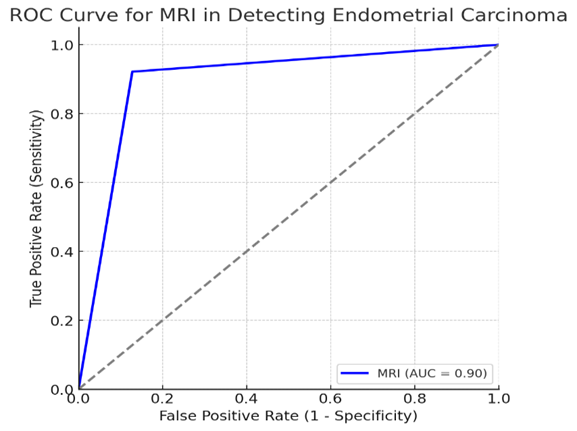
Figure 01: ROC Curve
Figure 1 presents the receiver operating characteristic (ROC) curve for MRI in detecting endometrial carcinoma. The area under the curve (AUC) was 0.90, indicating excellent discriminatory ability of MRI in distinguishing malignant from non-malignant cases. ROC curve analysis was performed to evaluate diagnostic performance, and area under the curve (AUC) was calculated
DISCUSSION :
MRI demonstrated high diagnostic performance sensitivity of 92.1%, specificity of 87.2%, PPV of 85.5%, NPV of 93.2%, and an overall accuracy of 89.9% —for diagnosing endometrial carcinoma compared to histopathology in the present cohort of 227 women assessed for suspected endometrial carcinoma. The values obtained demonstrate that in our tertiary care center, MRI is a noninvasive diagnostic tool for identifying endometrial carcinoma and aids significantly in downstream decision making regarding patient treatment. Sensitivity is well above many other studies currently available. Specificity is consistent with pooled specificities found in multiple studies assessing the use of MRI for preoperative diagnosis.
Sensitivity is consistent with or exceeds sensitivity found in several regional studies. One Karachi-based tertiary-center study found MRI sensitivity to be 95.8% and specificity to be 77.9% for deep myometrial invasion and 83.3% / 80% for endocervical invasion; although the primary focus was staging, the study shows MRI's ability to strongly "rule-in" disease locally. [8]
Another Pakistani based single-center study found MRI sensitivity to be 92.9%, specificity to be 88.1%, and accuracy to be 89.3% for identifying cervical stromal invasion, similar to the current study's overall accuracy. [9] Both these local studies along with the current study's results support the conclusion that MRI functions similarly in our context, regardless of the different types of imaging equipment and protocols utilized.
Internationally, two recent meta-analysis comparing Trans-Vaginal Ultrasound (TVUS) and MRI have determined pooled sensitivities for MRI for Deep Myometrial Invasion (DMI) at approximately 0.81, and pooled specificities at 0.84. TVUS were often similar in terms of sensitivity, but less so in terms of specificity. While both studies had some limitations, our point estimates for sensitivity were slightly better than those for MRI, and slightly worse than those for TVUS in terms of specificity. However, a 2022 Clinical Radiology study examining Low Grade Endometrial Carcinoma suggested MRI was able to obtain greater than 20% better specificity than TVUS for similar sensitivity, indicating MRI is advantageous in terms of ruling out extensive invasion. [10, 11] This pattern mirrors our high Negative Predictive Value (NPV = 93.2%) which would be valuable in clinical scenarios where negative MRI could obviate invasive procedures in select cases.
Beyond traditional sequences, Diffusion Weighted Imaging (DWI) has continued to expand MRI's capabilities. A 2024 prospective study published in BMC demonstrated excellent inter-observer agreement of ADC measurements and demonstrated quantitative DWI can help improve preoperative risk stratification. [12] Additionally, there is evidence through meta-analytic means that DWI can perform at or near levels of Dynamic Contrast Enhanced MRI (DCE-MRI) for DMI assessment; however, technique level factors (e.g., reduced field-of-view vs. high resolution methods) appear to affect accuracy. [13, 14] Given that we used a non-contrast protocol intentionally for practical purposes, our results demonstrate that the combination of T1/T2/DWI can yield accuracy within the range of what has been reported using multi-parametric approaches, providing additional support for the practical application of T1/T2/DWI across variable resources.
Regarding Cervical Stromal Invasion (CSI), pooled estimates show high specificity (>0.95) but only moderate sensitivity (∼0.55) for MRI. This finding reflects the challenges associated with detecting small breaches into the stroma. [15] We believe that the increased sensitivity seen in this study may reflect selection bias due to symptomatology; the inclusion of DWI consistently throughout all scans and the level of expertise among readers; as well as differences in disease spectrum and prevalence affecting test statistics. A very large 2025 multicenter analysis (n ≈ 2,020) further demonstrated that MRI significantly outperforms CT and serves as a complementary modality to biopsy for CSI assessment, highlighting the impact that image quality, tumor morphology, and reader expertise have on performance. [16]
Radiomics technology is rapidly evolving how MRI provides information prior to surgery. A 2025 systematic review reported pooled sensitivity of 81% and pooled specificity of 91% for radiomics-augmented multiparametric MRI assessing DMI. [11] Also, a 2025 BMC Cancer Study created MRI-radiomics predictive models capable of non-invasively predicting LVSI—a critical prognostic indicator that is generally only known post-operatively—using MRI. [17] Together these studies suggest a developing paradigm in which standard sequences (including DWI) will serve as robust base-line tools to detect disease—similar to what was seen in our study—and radiomics will aid in risk stratification and possibly diminish the need for contrast or invasive staging modalities in future workflows.
Key strengths of this study include (i) uniform reference standard provided by histopathology; (ii) larger-than-most single-site sample size from Pakistan; and (iii) systematic control over confounding variables. An important strength was utilization of a non-contrast sequence demonstrating its feasibility in a real world setting where contrast utilization may be limited by access costs or contraindication – yet still obtaining high accuracy comparable to literature mean for DMI and CSI. [10], [12-14]
Limitations include no analysis for inter-reader variability; potential spectrum bias due to evaluation of symptomatic women; and no formal evaluation of lymph nodes: DWI has shown significant promise for evaluating nodal metastases with sensitivity ∼81% and specificity ∼89%; however, DWI is imperfect and may benefit from hybrid techniques.
Clinical implications and future directions
Our results indicate a very high NPV (NPV = 93.2%), indicating that MRI can reliably rule out disease in a subset of patients who have equivocal ultrasound findings; this could decrease the number of immediate invasive samples. The excellent sensitivity and overall accuracy of MRI further support its use as an adjunctive imaging tool for MRI-guided surgical planning. Going forward, developing standardized non-contrast multi-parametric MRI protocols (e.g. T2/ DWI), and integrating validated radiomics-based features into these MRI protocols may provide additional improvements in accuracy while maintaining accessibility and cost-effectiveness. Future prospective multicenter studies conducted in Pakistan would help determine how best to sequence diagnostic modalities when utilizing both harmonized non-contrast MRI and transvaginal ultrasound (TVUS), including three-dimensional TVUS (which various meta-analyses suggest can perform as well as or better than MRI for DMI and CSI in skilled hands); in addition, such studies will assist in determining the cost utility of each modality within our healthcare system..
CONCLUSION :
The use of MRI (DWI with T1/T2) showed high diagnostic performance compared with histopathological examination. The study found sensitivity was 92.1%, specificity was 87.2%, positive predictive value (PPV) was 85.5% and negative predictive value (NPV) was 93.2%, which means MRI had an overall accuracy rate of 89.9%. This supports MRI as a good alternative option to detect endometrial carcinoma. MRI is also easy to perform and does not require the use of intravenous dye routinely, therefore it may be useful to augment assessment when there are equivocal results or when further information would aid in the evaluation of the extent of disease. The results suggest that MRI could potentially decrease the number of invasive studies performed on some patients.
Future research will need to confirm these results using larger numbers of participants from multiple sites. It will also be important to evaluate inter-rater reliability and develop multi-parametric MRI protocols without contrast and possibly incorporate AI-based radiomics algorithms to improve both diagnostic capabilities and the cost effectiveness in our specific context.
Funding
This research received no specific grant from any funding agency in the public, commercial, or not-for-profit sectors. Departmental resources at the Department of Radiology and the Histopathology Laboratory, Liaquat University Hospital, supported routine clinical imaging and tissue processing.
Competing Interests / Rights
The authors declare no competing interests (financial or non-financial). No sponsor or third party had any role in study design, data collection, analysis, interpretation, manuscript preparation, or the decision to submit for publication. The authors retain full rights to the work and its publication and had unrestricted access to the data.
BIBLIOGRAPHY:
1. Sung H, Ferlay J, Siegel RL, Laversanne M, Soerjomataram I, Jemal A, et al. Global Cancer Statistics 2020: GLOBOCAN estimates of incidence and mortality worldwide for 36 cancers in 185 countries. CA Cancer J Clin. 2021;71(3):209-249.
2. Feng T, Li W, Wang Q, Yang J, Shen F. Global, regional, and national burden of uterine cancer among women aged 50 years and older from 1990 to 2021: A systematic analysis for the Global Burden of Disease Study 2021. J Health Popul Nutr. 2025;44:208.
3. Afridi HK, Zahid NA. Frequency of gynecological malignancies in a tertiary care centre in Karachi, Pakistan. Liaquat Natl J Cancer Care. 2021;3(1):3-6.
4. Madár I, Szabó A, Vleskó G, Fülöp J, Bódizs B, Lánczi LI, et al. Diagnostic accuracy of transvaginal ultrasound and magnetic resonance imaging for the detection of myometrial infiltration in endometrial cancer: a systematic review and meta-analysis. Cancers (Basel). 2024;16(5):907.
5. Alcázar JL, Domínguez-Pellicer J, Juez L, Caparrós M, Iturra A, Ajossa S, et al. MRI and transvaginal ultrasound for assessment of myometrial invasion in endometrial cancer: a systematic review and meta-analysis. Ultrasound Obstet Gynecol. 2022;59(5):659-669.
6. Dappa E, Elger T, Hasenburg A, Battista MJ, Harter P, Herrmann K, et al. The value of advanced MRI techniques in the assessment of endometrial carcinoma: A systematic review. Cancers (Basel). 2020;12(2):297.
7. Sheeba A, Sahito AA, Surahyo P, Baloch M, Gul N, Ashraf H. Diagnostic accuracy of MRI pelvis in detection of cervical invasion in patients of endometrial carcinoma: A study at LUMHS Jamshoro. Pak J Med Health Sci. 2021;15(2):923-926.
8. Raza M, Saba K, Khan AH, et al. Accuracy of MRI in detection of depth of myometrial invasion and cervical invasion in endometrial carcinoma taking histopathology as gold standard. Liaquat Natl J Cancer Care. 2022;4(1):15-20.
9. Jaleel F, Zehra T, Tariq MU, et al. Diagnostic accuracy of pelvic MRI for determination of the cervical stromal invasion in endometrial carcinoma. J Coll Physicians Surg Pak. 2021;31(10):1175-1180. (AKU repository).
10. Madár I, Szabó A, Vleskó G, et al. Diagnostic accuracy of transvaginal ultrasound and magnetic resonance imaging for deep myometrial invasion in endometrial cancer: systematic review and meta-analysis. Cancers (Basel). 2024;16(5):907.
11. Zhang X, Li Y, Wang J, et al. Development and external validation of a multiparametric MRI radiomics nomogram for deep myometrial invasion in endometrial cancer. Acad Radiol. 2025;32(8):e271–e280.
12. Kamel Y, Nageeb A, Abdelrahman A, et al. Diagnostic utility of apparent diffusion coefficient in preoperative endometrial cancer assessment: a prospective study. BMC Med Imaging. 2024;24:60.
13. Ota T, Tsuboyama T, Onishi H, et al. Diagnostic accuracy of MUSE-DWI and rFOV-DWI vs DCE-MRI for myometrial invasion in endometrial cancer. Radiol Med. 2023;128:629-643.
14. Zhou C, Wang G, Che C. Comparison of DWI and DCE-MRI for myometrial and cervical invasion and FIGO staging in endometrial cancer. Eur J Gynaecol Oncol. 2024;45(1):124-131.
15. Deng L, Wang Y, Zhang Q, et al. MRI for assessment of cervical invasion in endometrial carcinoma: meta-analysis. J Cancer. 2021;12(3):754-761.
16. Yang Y, Ye Z, Zhao Y, Li Z. MRI, CT and biopsy for preoperative cervical stromal invasion in endometrial carcinoma: a 2,020-patient retrospective study. Front Oncol. 2025;15:1548436.
17. Ma W, Meng W, Yin J, et al. MRI radiomics and clinical models to predict lymphovascular space invasion in endometrial cancer. BMC Cancer. 2025;25:796.
18. Nougaret S, Addley HC, Colombo PE, et al. Diffusion-weighted MRI for nodal metastases in gynecologic malignancies: diagnostic performance and pitfalls. Clin Radiol. 2017;72(10):888.e1-888.e13.
19. Alcázar JL, Domínguez-Pellicer J, Juez L, et al. Three-dimensional TVUS vs MRI for deep myometrial and cervical invasion: systematic review and meta-analysis. Ultrasound Obstet Gynecol. 2022;60(2):178-190.
20. Ghaith MM, El-Agwany AS, El Sayed AM. TVUS versus MRI in preoperative assessment of endometrial cancer: diagnostic accuracy and clinical implications. Clin Radiol. 2023;78(5):e323-e331.
21. The ASCO Post Staff. Advances in endometrial cancer detection and treatment: highlights from SGO 2025. The ASCO Post. 2025 Mar 17 (updated 2025 Jun 27).