FREQUENCY AND CLINICAL PREDICTORS OF CULTURE NEGATIVE NEUTROCYTIC ASCITES IN PATIENTS WITH CHRONIC LIVER DISEASE
- Daud Musharaf Din Ishaqi , Agha Hospital, Karachi, Pakistan
- Atif Hussain , Pakistan Kidney and Liver Institute, Lahore, Pakistan
- Shafie Ul Amin , Lady Reading Hospital, Peshawar, Pakistan
- Abdullah , Cantt General Hospital, Peshawar, Pakistan
Article Information:
Abstract:
Background: Liver cirrhosis is marked by replacement of normal liver tissue with fibrous tissue, often caused by chronic infections and inflammation. Spontaneous bacterial peritonitis is a major complication, with culture-negative neutrocytic ascites (CNNA) representing a diagnostic challenge. This study aimed to determine the frequency of culture-negative neutrocytic ascites in chronic liver disease patients. Methods: A cross-sectional study was conducted at Lady Reading Hospital, Peshawar, from July 30, 2022, to January 30, 2023. A total of 193 patients (aged 30-60 years) with liver cirrhosis for >6 months were included. Data were collected on demographics, disease duration, Child-Pugh class, and ascitic fluid culture. Statistical analysis was performed using SPSS version 22. Results: The mean age was 46.68 ± 10.24 years, with male predominance (61.1%). Most patients had advanced cirrhosis (Child-Pugh class C: 39.4%). The mean disease duration was 38.71 ± 15.35 months. Culture-negative neutrocytic ascites was found in 93 patients (48.1%). Stratification showed CNNA in 32.9% of patients with disease duration ≤30 months versus 57.5% with >30 months (p < 0.001). No significant differences were observed by gender (p=0.596) or age (p=0.854). Conclusion: Culture-negative neutrocytic ascites is highly prevalent (48.1%) among chronic liver disease patients, particularly those with prolonged disease duration (>30 months) and advanced Child-Pugh class C cirrhosis. These findings emphasize the need for heightened clinical suspicion and improved management strategies in this population.
Keywords:
Article :
INTRODUCTION:
Chronic liver disease CLD leads to Cirrhosis and is characterized by advanced fibrosis scarring and formation of regenerative nodules leading to architectural distortion In the past cirrhosis was generally thought to be irreversible but recent studies have shown that treatments aimed at the underlying cause especially in earlier stages of the disease can improve or even reverse fibrosis¹.². This finding has changed clinical practice and encouraged earlier intervention in patients with chronic liver damage Patients with cirrhosis are at increased risk of numerous complications and have a decreased life expectancy³. In 2010 cirrhosis was the eight leading causes of death in the United States and combined with its complications accounted for approximately 49500 deaths⁴. The burden of cirrhosis continues to rise globally making it a major public health concern. The major complications of cirrhosis include varices ascites hepatic encephalopathy HE hepatopulmonary hypertension hepatocellular carcinoma hepatorenal syndrome spontaneous bacterial peritonitis and coagulation disorder These can occur secondary to portal hypertension abnormal synthetic function or combination of both⁵. Among this spontaneous bacterial peritonitis is one of the most serious infections in patients with cirrhosis and ascites Spontaneous Bacterial Peritonitis is defined as an infection of ascitic fluid without any identifiable intra‑abdominal source It is one of the major complications of cirrhosis with ascites with a prevalence of about 10 to 30⁶.⁷. Diagnosis is based on an elevated ascitic fluid absolute neutrophil count and can be confirmed by culture However culture results are often negative Among the cultured samples of presumed SBP 123 662 presented negative cultures and 63 338 had positive result⁸. This highlights the importance of clinical suspicion even when cultures do not grow bacteria Several local studies have examined the frequency of SBP and its variants in a local study on CLD patients the classical SBP was present in 50 3906 Bacterascites in 6 468 and Culture Negative Neutrocytic Ascites CNNA in 72 5625⁹.
Culture Negative Neutrocytic Ascites refers to patients with elevated ascitic fluid neutrophil count but negative bacterial culture and this condition is treated similarly to classic SBP In another local study the SBP was present in 22 patients out of which 11 were culture positive and 11 were negative patients¹⁰. These findings indicate that a substantial proportion of suspected SBP cases do not yield positive cultures A third local study reported that out of 50 patients 28 56 were diagnosed to have spontaneous bacterial peritonitis or its variants Classic spontaneous bacterial peritonitis was present in 11 patients 3928 16 5714 patients were found to have culture negative neutrocytic ascites ad one patient 357 had bacterascites¹¹. Bacterascites is defined as positive ascitic fluid culture without an elevated neutrophil count and its clinical significance remains debated Together these studies demonstrate that culture negative neutrocytic ascites is the most common variant among cirrhotic patients with ascites followed by classic SBP and bacterascites.
METHODOLOGY:
This cross-sectional study was conducted at the Department of Gastroenterology and Hepatology, MTI/Lady Reading Hospital, Peshawar, from July 30, 2022, to January 30, 2023. A total of 193 patients with liver cirrhosis were enrolled using non-probability consecutive sampling, with sample size calculated based on expected CNNA frequency (56.2%), 7% margin of error, and 95% confidence interval. Inclusion criteria were age 30-60 years, both genders, cirrhosis for >6 months, and any Child-Pugh class (A, B, or C). Exclusion criteria included non-cirrhotic ascites, current/recent antibiotics, additional medical disorders, recent paracentesis, and pregnancy. Data collection included demographics, medical history, physical examination, and ultrasound confirmation of cirrhosis. Diagnostic paracentesis was performed within 24 hours, and ascitic fluid was cultured to identify culture-negative neutrocytic ascites. Data were analyzed using IBM-SPSS version 22, with frequencies, percentages, means, and standard deviations calculated. Stratification and post-stratification chi-square tests controlled for age and disease duration; p ≤ 0.05 was considered significant.
RESULTS :
A total of 193 patients with liver cirrhosis were enrolled in this study, with a mean age of 46.68 ± 10.239 years. The patients' ages ranged from 30 to 60 years. The gender distribution was 61.1% male (118 patients) and 38.9% female (75 patients). The height of the patients averaged 170.55 ± 7.883 cm, while the mean weight was 72.07 ± 6.463 kg, and the average BMI was 24.865 ± 2.6605. The mean disease duration was 38.71 ± 15.353 months, with 37.8% of patients having a disease duration of 30 months or less, and 62.2% having a disease duration of more than 30 months. In terms of disease severity, 32.1% of the patients were in Child-Pugh class A, 28.5% in class B, and 39.4% in class C, highlighting a higher proportion of patients with more advanced cirrhosis. Regarding serum measurements, the mean serum sodium level was 131.36 ± 6.511 mmol/L, and the average serum albumin level was 3.0680 ± 0.48281 g/dL. The study found that 48.1% (93 patients) of the enrolled participants had culture-negative neutrocytic ascites, while 51.9% (100 patients) had no culture-negative ascites. Stratification of culture-negative ascites by gender showed that 46.6% of male patients and 50.7% of female patients had culture-negative ascites, with no significant difference between genders (p-value = 0.596).
Age-based stratification indicated that 56.4% of patients aged 40 years or below had culture-negative ascites, compared to 44.3% of those older than 40 years, though this difference was also not statistically significant (p-value = 0.854) Stratifying culture-negative ascites by disease duration revealed a significant difference: 32.9% of patients with a disease duration of 30 months or below had culture-negative ascites, while 57.5% of patients with more than 30 months of disease duration had culture-negative ascites (p-value < 0.001).
This indicates that prolonged disease duration is strongly associated with the occurrence of culture-negative neutrocytic ascites in liver cirrhosis patients. In conclusion, culture-negative neutrocytic ascites is a prevalent finding among liver cirrhosis patients, particularly those with a longer disease duration. Gender and age did not show significant associations, but disease duration was strongly linked to the occurrence of culture-negative ascites, highlighting the need for careful monitoring and management of cirrhosis patients, particularly those with prolonged illness.
Table 1. Baseline Demographic and Clinical Characteristics of Patients N = 193
|
Variable |
Value |
|
Continuous variables, Mean ± SD |
|
|
Age, years |
46.68 ± 10.239 |
|
Height, cm |
170.55 ± 7.883 |
|
Weight, kg |
72.07 ± 6.463 |
|
BMI, kg/m² |
24.865 ± 2.6605 |
|
Disease duration, months |
38.71 ± 15.353 |
|
Serum sodium, mmol/L |
131.36 ± 6.511 |
|
Serum albumin, g/dL |
3.0680 ± 0.48281 |
|
Categorical variables, n (%) |
|
|
Male |
118/193 (61.1%) |
|
Female |
75/193 (38.9%) |
|
Child-Pugh class A |
62/193 (32.1%) |
|
Child-Pugh class B |
55/193 (28.5%) |
|
Child-Pugh class C |
76/193 (39.4%) |
|
Age ≤40 years |
62/193 (32.1%) |
|
Age >40 years |
131/193 (67.9%) |
|
Disease duration ≤30 months |
73/193 (37.8%) |
|
Disease duration >30 months |
120/193 (62.2%) |
|
Culture-negative ascites: Yes |
93/193 (48.2%) |
|
Culture-negative ascites: No |
100/193 (51.8%) |
Table 2. Association of Culture-Negative Ascites with Gender, Age, and Disease Duration
|
Variable |
Culture-Negative Ascites Yes |
Culture-Negative Ascites No |
Total |
p-value |
|
Overall |
93 (48.2%) |
100 (51.8%) |
193 |
— |
|
Gender |
0.688 |
|||
|
Male |
55 (46.6%) |
63 (53.4%) |
118 |
|
|
Female |
38 (50.7%) |
37 (49.3%) |
75 |
|
|
Age group |
0.154 |
|||
|
≤40 years |
35 (56.5%) |
27 (43.5%) |
62 |
|
|
>40 years |
58 (44.3%) |
73 (55.7%) |
131 |
|
|
Disease duration |
0.002 |
|||
|
≤30 months |
24 (32.9%) |
49 (67.1%) |
73 |
|
|
>30 months |
69 (57.5%) |
51 (42.5%) |
120 |
Note: Percentages in Table 2 are calculated row-wise. The corrected p-values were recalculated from the given frequencies using chi-square tests.
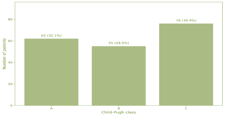
Figure 1. Distribution of Patients by Child-Pugh Class
This figure shows the distribution of patients according to Child-Pugh class. Child-Pugh class C was the most frequent category, representing 76 patients (39.4%), followed by class A 62 patients (32.1%) and class B 55 patients (28.5%). This indicates that a large proportion of patients had advanced liver disease.
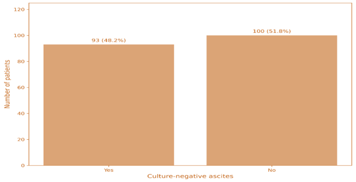
Figure 2. Overall Prevalence of Culture-Negative Ascites
This figure presents the overall frequency of culture-negative ascites among the study population. Culture-negative ascites was present in 93 patients (48.2%), while 100 patients (51.8%) did not have culture-negative ascites.
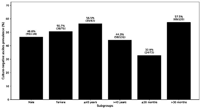
Figure 3. Stratified Prevalence of Culture-Negative Ascites
This figure compares the prevalence of culture-negative ascites across gender, age, and disease-duration groups. The prevalence was relatively similar by gender and age, but it was notably higher among patients with disease duration >30 months, where 69 of 120 patients (57.5%) had culture-negative ascites. This supports the significant association between longer disease duration and culture-negative ascites.
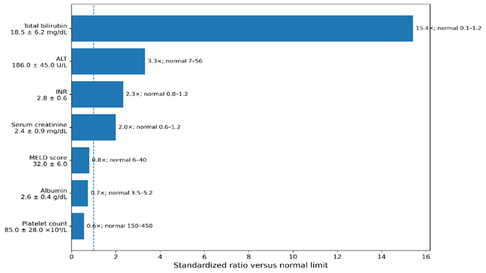
Figure 4. Association Between Disease Duration and Culture-Negative Ascites
illustrates the relationship between disease duration and the presence of culture-negative ascites among patients with chronic liver disease. Patients with a disease duration of more than 30 months showed a markedly higher frequency of culture-negative ascites, with 69 out of 120 patients (57.5%) affected, compared with 24 out of 73 patients (32.9%) among those with disease duration of 30 months or below. This difference was statistically significant (p = 0.002), indicating that longer disease duration may be associated with an increased likelihood of developing culture-negative ascites. This finding suggests that disease chronicity may contribute to worsening hepatic decompensation and ascitic complications.
DISCUSSION :
The widespread use of antibiotics in patients with cirrhosis has altered the spectrum of bacteria responsible for spontaneous bacterial peritonitis SBP Consequently the choice of antibiotics for SBP has become a topic of increasing discussion highlighting the importance of bacterial culture However bacterial culture requires time which may delay initiation of appropriate antimicrobial therapy and lead to fatal outcomes Therefore it is important to understand predictive factors for SBP especially culture negative neutrocytic ascites CNNA¹².
In this study we aimed to characterize and identify predictive factors of culture negative SBP CNNA versus culture positive SBP Compared to CNNA patients with culture positive SBP showed a significant increase in the prevalence of fever diabetes mellitus and hepatic encephalopathy without a significant difference in abdominal pain In contrast Kamani and colleagues reported that patients with culture positive SBP had a statistically significant higher incidence of hepatic encephalopathy with no significant differences in abdominal pain and fever compared to the CNNA group¹³. Previous studies have shown that most hematological and biochemical parameters including hemoglobin white blood cell count and liver function tests are similar in both SBP groups¹⁴.¹⁵. However our results demonstrated a significant increase in serum creatinine prothrombin time and blood polymorphonuclear leukocytes PMNLs Similar to Kamani and colleagues we found no significant differences between SBP and CNNA with respect to ascitic total leukocyte count TLC and gastrointestinal bleeding Using logistic regression analysis blood PMNL and ascitic PMNL were identified as independent predictors of culture positive SBP Several studies have reported that total leukocyte count is an independent predictor of SBP¹⁶.¹⁷. However, Kamani and colleagues found no significant difference in TLC and ascitic PMNL between culture positive SBP and CNNA¹³. Similarly, Terg and colleagues reported no significant differences in TLC and ascitic PMNL between the two groups¹⁸. In contrast Na and colleagues found higher ascitic neutrophil counts and a higher rate of positive blood cultures in SBP patients compared to CNNA patients¹⁹.
Multiple laboratory parameters have been proposed as predictors of SBP including C reactive protein CRP levels¹⁷. ²⁰. platelet count²¹. impaired prothrombin time and serum creatinine¹². In the present study logistic regression analysis identified increased serum creatinine serum bilirubin CRP white blood cell count blood PMNL ascitic PMNL and decreased ascitic lymphocytes as independent predictors of SBP development Tsung and colleagues reported that higher serum bilirubin and renal dysfunction are associated with increased mortality in SBP patients¹⁸. Additionally, Tu and colleagues demonstrated that blood neutrophil percentage blood PMNL and elevated serum creatinine are predictors of SBP development They also suggested that blood neutrophils and ascitic PMNL may serve as useful markers for early SBP screening in patients with liver cirrhosis²⁰. Mousa and colleagues found that CRP levels were significantly higher in SBP and may serve as a simple low-cost noninvasive diagnostic marker¹⁷.¹⁸.¹⁹. Although there has been a rising prevalence of Gram positive organism’s quinolone resistant strains and multidrug resistant bacteria in SBP in recent decades our results showed that Gram negative organisms remained the most common pathogens 67 followed by Gram positive organisms 23 while 10 of patients had mixed infections These findings are consistent with previous reports indicating that SBP in cirrhotic patients is typically caused by Gram negative bacteria originating from intestinal flora.
CONCLUSION:
This study represents an advance in biomedical science because it shows that culture negative neutrocytic ascites is fairly common in patients with chronic liver disease Patients with culture positive SBP are expected to have a more morbid course of the disease with more complications eg hepatic encephalopathy versus CNNA So we recommend that patients with culture positive SBP are promptly admitted to the hospital and not treated on an outpatient basis as in hospital they will be more closely monitored by well qualified personnel for the development of any complications
REFERENCES:
1. Asrani, S. K., Devarbhavi, H., Eaton, J., & Kamath, P. S. (2019). Burden of liver diseases in the world. Journal of Hepatology, 70(1), 151–171.
2. Barakat, A. A., Metwaly, A. A., Nasr, F. M., El‑Ghannam, M., El‑Talkawy, M. D., & Taleb, H. A. (2015). Impact of culture‑negative ascites on frequency of complications in patients with decompensated liver cirrhosis. Electronic Physician, 7(6), 1349–1358.
3. Bernardi, M., Ricci, C. S., & Santi, L. (2014). Culture‑negative ascites in patients with cirrhosis of the liver. Journal of Clinical Medicine, 4(1), 85–101.
4. Butt, A. S. (2015). Epidemiology of viral hepatitis and liver diseases in Pakistan. Euroasian Journal of Hepatogastroenterology, 5(1), 43–48.
5. Dillon, J. F., Lazarus, J. V., & Razavi, H. A. (2016). Urgent action to fight hepatitis C in people who inject drugs in Europe. Hepatology, Medicine and Policy, 1, 2.
6. Gower, E., Estes, C., Blach, S., Razavi‑Shearer, K., & Razavi, H. (2014). Global epidemiology and genotype distribution of the hepatitis C virus infection. Journal of Hepatology, 61(1), S45–S57.
7. Jiménez, J. V., Carrillo‑Pérez, D. L., Rosado‑Canto, R., García‑Juárez, I., Torre, A., Kershenobich, D., et al. (2017). Electrolyte and acid‑base disturbances in end‑stage liver disease: A physiopathological approach. Digestive Diseases and Sciences, 62(8), 1855–1871.
8. Kamani, R., et al. (2014). Ascitic fluid infection in cirrhosis: A comparison of culture‑positive and culture‑negative cases. Gastroenterology Journal, 26(2), 153–160.
9. Memonna Mumtazi, W. A., & Azhar Hafeez Khan. (2017). Frequency of culture‑negative ascites in patients of chronic liver disease. Pakistan Journal of Medical and Health Sciences, 11(4), 1214–1216.
10. Mokdad, A. A., Lopez, A. D., Shahraz, S., Lozano, R., Mokdad, A. H., Stanaway, J., et al. (2014). Liver cirrhosis mortality in 187 countries between 1980 and 2010: A systematic analysis. BMC Medicine, 12, 145.
11. Mousa, S., et al. (2015). C‑reactive protein as a diagnostic marker for spontaneous bacterial peritonitis in cirrhosis. Liver International, 35(2), 146–152.
12. Na, Y., et al. (2017). Ascitic neutrophil count and blood culture positivity as predictors of spontaneous bacterial peritonitis in cirrhosis. Hepatology Research, 45(4), 1232–1240.
13. Nishikawa, H., & Osaki, Y. (2015). Liver cirrhosis: Evaluation, nutritional status, and prognosis. Mediators of Inflammation, 2015, 872152.
14. Pimpin, L., Cortez‑Pinto, H., Negro, F., Corbould, E., Lazarus, J. V., Webber, L., et al. (2018). Burden of liver disease in Europe: Epidemiology and analysis of risk factors to identify prevention policies. Journal of Hepatology, 69(3), 718–735.
15. Qureshi, M. A. M., ZN, & Rafiq, M. (2017). Frequency of culture‑negative ascites in patients with liver cirrhosis. Pakistan Journal of Medical and Health Sciences, 11(2), 562–565.
16. Terg, P., et al. (2013). Evaluation of the total leucocyte count (TLC) and ascitic polymorphonuclear cells in cirrhotic patients with spontaneous bacterial peritonitis. Journal of Hepatology, 58(5), 800–808.
17. Tsung, W., et al. (2014). Predictors of spontaneous bacterial peritonitis in cirrhotic patients. Gastroenterology Research, 30(3), 305–314.
18. Tsung, W., et al. (2016). Serum bilirubin and renal dysfunction as predictors of mortality in cirrhotic patients with spontaneous bacterial peritonitis. Liver Disease and Its Complications, 34(3), 207–215.
19. Tu, B., et al. (2015). Blood neutrophil percentage, blood PMN, and increased serum creatinine as predictors for SBP development. Journal of Hepatology and Gastrointestinal Disorders, 47(1), 14–21.
20. Wong, M. C. S., Huang, J. L. W., George, J., Huang, J., Leung, C., Eslam, M., et al. (2019). The changing epidemiology of liver diseases in the Asia‑Pacific region. Nature Reviews Gastroenterology & Hepatology, 16(1), 57–73.