Diagnostic Accuracy of Ultrasound in the Diagnosis of Craniosynostosis in Infants Keeping the Computed Tomography Findings as Gold Standard
- Abdul Wajid , Training Medical Officer (TMO), Department of Diagnostic Radiology, Saidu Group of Teaching Hospital, Swat, Pakistan
- Muhammad Sohail Khan , Training Medical Officer (TMO), Department of Diagnostic Radiology, Saidu Group of Teaching Hospital, Swat, Pakistan
- Salma Liaqat , Assistant Professor, Department of Diagnostic Radiology, Saidu Group of Teaching Hospital, Swat, Pakistan
- Shaheer Ahmad Khan , Training Medical Officer, Department of Diagnostic Radiology, CMH Abbottabad, Pakistan
- Muhammad Ishtiaq , Department of Diagnostic Radiology, CMH Abbottabad, Pakistan
- Taj Muhammad Khan , Training Medical Officer, Department of Diagnostic Radiology, Mardan Medical Complex, Mardan, Pakistan
Article Information:
Abstract:
Background: Premature closure of one or more of the cranial sutures, craniosynostosis, can cause an abnormal skull shape, facial asymmetry and raised ICP and may have neurodevelopmental effects if not diagnosed early. Three dimensional computed tomography (3D CT) is thought to be the most "complete" diagnostic tool, but it uses ionizing radiation for the diagnosis. Ultrasound is a radiation-free, non-invasive imaging test that is safe, readily available and can be used as a first-line diagnostic modality in the work-up of suspected cases. The definition of ultrasound which is a non-invasive and inexpensive modality compared to CT. Objective: To evaluate the diagnostic accuracy of ultrasound in the diagnosis of craniosynostosis in infants, keeping computed tomography findings as the gold standard. Methods: The cross sectional validation study was carried out in Combined Military Hospital, Abbottabad in Department of Radiology from 27th April 2025 to 27 July 2025. The infants with clinical suspicion of craniosynostosis (n = 85) were enrolled using non-probability consecutive sampling. A cranial ultrasound and 3D reconstructed CT of the skull was performed in all infants. The ultrasound findings were compared with the CT findings, sensitivity, specificity, positive predictive value, negative predictive value and diagnostic accuracy were calculated. Results: Out of 85 infants, CT confirmed craniosynostosis in 60 (70.6%) cases, while ultrasound diagnosed craniosynostosis in 59 (69.4%) cases. Ultrasound correctly identified 58 true-positive and 24 true-negative cases, while 1 false-positive and 2 false-negative cases were observed. The sensitivity of ultrasound was 96.7%, specificity was 96.0%, positive predictive value was 98.3%, negative predictive value was 92.3%, and overall diagnostic accuracy was 96.5%. A statistically significant association was found between ultrasound and CT findings (p < 0.001). Conclusion: Ultrasound showed excellent diagnostic accuracy for detecting craniosynostosis in infants when compared with CT skull with 3D reconstruction. It may be used as a reliable first-line imaging modality in clinically suspected infants, while CT may be reserved for equivocal cases and preoperative planning.
Keywords:
Article :
INTRODUCTION:
Craniosynostosis is a congenital cranial disorder where the cranial sutures are fused early. Cranial sutures are responsible for allowing the skull to expand in response to the rapid brain growth in infancy and are normal in all infants. If a suture closes too soon, then the growth in the skull is limited across the suture and the growth is allowed in the other directions. This leads to an abnormal head shape and can be accompanied by facial asymmetry, high intracranial pressure, visual issues and neurodevelopmental impairment if not diagnosed and treated at time. The craniosynostosis is a clinically significant pediatric skull anomaly that if un-diagnosed and untreated, can cause complications (1, 2).
The symptoms of craniosynostosis depend on the suture affected. In sagittal synostosis, the skull is typically long and narrow, and in cases of coronal, metopic, and lambdoid synostosis, various types of cranial asymmetry or deformation may occur. When raised ICP is suspected, infants may have abnormal head shape, cranial asymmetry, limited skull development, vomiting or increased fontanelle bulging. Early recognition is crucial since early referral and management can impact cosmetic, functional and developmental outcomes in clinical practice. But clinical examination alone may not be enough to distinguish craniosynostosis from positional skull deformity, particularly during the early infancy period (3, 4).
Imaging is a critical part of the work up for the diagnosis of craniosynostosis. The computed tomography (CT) shows detailed images of the cranium and its morphology, especially when three dimensional reconstructed. It can show suture fusion, bone overgrowth, overlapping bones of the skull and cranial deformity. This is why CT is considered to be the gold standard in the diagnosis and planning for surgery. The CT findings for craniosynostosis include fusion of the sutures, bones overgrowth, or a skull deformity consistent with craniosynostosis. But there are some drawbacks for infants, such as exposure to ionizing radiation, expense, limited access in certain health care facilities and, in some cases, the requirement for sedation (5, 6).
In the field of imaging, ultrasound has proved to be a valuable alternative method to assess the child's cranial sutures. It is non-ionic, relatively cheap, portable and can be done at the bedside rapidly. because infant cranial sutures are easily accessible and are superficial, ultrasound can be used to show if sutures are open or fused. An open suture typically will be a hypoechoic space between echogenic bony margins while a fused suture will be a continuous echogenic line without an echo gap between the two margins. The diagnosis of craniosynostosis is dependent on the presence of abnormal suture appearance, lack of mobility of the sutures or on the presence of skull asymmetry (7-9).
A few studies have found the ultrasound to have good diagnostic accuracy for suspected craniosynostosis, especially in younger infants, who have more distinct sutures. Previous evidence indicated a high degree of sensitivity and specificity with ultrasound compared to computed tomography (CT) and ultrasound might be used as a primary screening or diagnostic tool in selected infants. Although ultrasound has these benefits, it is operator dependent and diagnostic reliability might be affected by the experience of the radiologist, the age of the infants, the suture involved, and the quality of the equipment. Hence, before an ultrasound is recommended as a routine screening tool, it is important to validate the performance of the test locally (10, 11).
In resource-limited settings, unnecessary CT scans in infants may increase healthcare costs and radiation exposure. A safe and accurate first-line imaging method could improve patient care by reducing avoidable CT use while still identifying infants who need further evaluation or surgical referral. There is limited local evidence regarding the diagnostic accuracy of ultrasound for craniosynostosis in infants. Therefore, this study was designed to evaluate the diagnostic accuracy of ultrasound in detecting craniosynostosis among infants, keeping CT skull with 3D reconstruction as the gold standard.
METHODOLOGY:
This study was conducted as a cross-sectional validation study to determine the diagnostic accuracy of ultrasound in detecting craniosynostosis among infants, keeping computed tomography skull with 3D reconstruction as the gold standard. The study was carried out in the Department of Radiology, Combined Military Hospital, Abbottabad.
The study duration was three months, from 27th April 2025 to 27th July 2025. Ethical approval was obtained from the College of Physicians and Surgeons Pakistan, Research Evaluation Unit, with approval reference number CPSP/REU/RAD-2023-027-4026, dated April 27, 2025. The approval letter confirms that the synopsis titled “Diagnostic Accuracy of Ultrasound in the Diagnosis of Craniosynostosis in Infants Comparing Computed Tomography Findings as Gold Standard” was approved.
The total sample size for this study is 85 babies. The sample size was determined with a sensitivity and specificity calculator from a 95% confidence level, previously reported sensitivity for ultrasound of 96.9% and specificity of 100%, desired precision of 12% and assumed prevalence of 70.5% for the number of diagnosed cases of craniosynostosis. Patient recruitment was done by a non-probability consecutive sampling.
Infants were included between 1 month and 12 months of age, for both males and females, and had a clinical suspicion of craniosynostosis. The clinical picture used for diagnosis was abnormal head shape, abnormal cranial symmetry, or symptoms suggestive of raised ICP such as being irritable, vomiting, or with a bulging fontanelle. Infants who had a known genetic syndrome (crouzon syndrome or Apert syndrome), prior cranial surgery or head trauma were excluded as this could affect the morphology of the skull or the appearance of the sutures, and could introduce bias in diagnostic interpretation.
Following approval, infants presenting to the pediatric and radiology departments were screened as per the inclusion/exclusion criteria. Parents/guardians were informed about the purpose of the study and informed consent was obtained in writing prior to participation. All participants' confidentiality was maintained during the study by using patient identification number rather than names. Parents/guardians were told that the study was voluntary and that they could opt out at any time without impacting on the patient's clinical care.
A structured proforma was used to collect data. Demographic and clinical parameters were age in months, gender, place of residence, socioeconomic status, birth weight, head circumference, family history of craniosynostosis and mode of delivery. Additional clinical parameters (abnormal head shape, cranial asymmetry, irritability, vomiting and bulging fontanella) were also documented. These variables were chosen to describe the clinical picture of infants and to evaluate whether patient variables had an effect on the diagnostic accuracy.
All babies entered into the study were examined by ultrasound of the skull. All of the cranial sutures were evaluated on ultrasound for any features suggestive of craniosynostosis. The ultrasound findings that were seen and interpreted as positive were: Abnormal suture appearance, Echogenic line in place of the normal hypoechoic open suture, Loss of suture mobility with gentle probe pressure, and Skull asymmetry with suspected premature suture fusion. If any of these findings were not seen, the ultrasound was classified as negative for craniosynostosis.
All infants were then assessed using ultrasound prior to undergoing CT skull with 3D reconstruction. Final diagnosis was made with the use of CT as the gold standard. CT scans were considered positive for craniosynostosis if any of the following were detected: complete obliteration or non-visualization of the cranial sutures, abnormal bone overgrowth or crossing over the sutures, or a skull deformity consistent with craniosynostosis, such as scaphocephaly or brachycephaly. On CT, the cases that did not have these features were considered negative.
Ultrasound and CT results were written separately to minimize the observer bias. The index test was ultrasound and the reference standard was computed tomography (CT) of the skull with 3D reconstruction. The CT diagnosis interpreted without ultrasound diagnosis. The final test results of the ultrasound and CT were tabulated in a 2 × 2 diagnostic matrix as true positive, false positive, true negative and false negative.
The data were analyzed using SPSS version 25. Data for quantitative variables (e.g., age, weight, head circumference) were given as mean ± SD. Categorical variables like gender, residence, socioeconomic status, mode of delivery, family history, ultrasound diagnosis and CT diagnosis were presented as frequencies and percentages. The sensitivity, specificity, positive predictive value, negative predictive value and overall accuracy were used to calculate the diagnostic accuracy of ultrasound. The formulas used were: sensitivity = TP/(TP + FN) × 100, specificity = TN/(TN + FP) × 100, PPV = TP/(TP + FP) × 100, NPV = TN/(TN + FN) × 100, and accuracy = (TP + TN)/(TP + TN + FP + FN) × 100. The relationship between the ultrasound and CT was analyzed using chi-square test or Fisher's exact test as appropriate. A p-value of ≤0.05 was considered statistically significant.
RESULTS:
In total, 85 infants with clinical suspicion of craniosynostosis were studied. Participants' ages ranged from 1 to 12 months with a mean of 6.41 ± 3.08 months. The majority of infants were between 1-6 months of age (49, 57.6%) and 36 infants (42.4%) were between 7-12 months. There were 52 (61.2%) male and 33 (38.8%) female infants. 48 (56.5%) cases had been reported from the rural areas, and 37 (43.5%) cases from urban areas. Socioeconomic status of the respondents included 34 (40.0%) from the low socioeconomic group, 38 (44.7%) from the middle socioeconomic group, and 13 (15.3%) from the high socioeconomic group. The mean head circumference was 43.21 ± 2.76 cm, and the mean weight was 6.82 ± 1.71 kg.
.
Table 1. Baseline characteristics of study participants
|
Variable |
Frequency / Mean |
Percentage / SD |
|
Total infants |
85 |
100.0 |
|
Age, months |
6.41 |
± 3.08 |
|
Weight, kg |
6.82 |
± 1.71 |
|
Head circumference, cm |
43.21 |
± 2.76 |
|
Age 1–6 months |
49 |
57.6 |
|
Age 7–12 months |
36 |
42.4 |
|
Male |
52 |
61.2 |
|
Female |
33 |
38.8 |
|
Rural residence |
48 |
56.5 |
|
Urban residence |
37 |
43.5 |
|
Low socioeconomic status |
34 |
40.0 |
|
Middle socioeconomic status |
38 |
44.7 |
|
High socioeconomic status |
13 |
15.3 |
Abnormal head shape was the most frequent presenting feature, seen in 72 (84.7%) infants, followed by cranial asymmetry in 63 (74.1%) cases. Less commonly, signs suggestive of raised ICP were seen, with 28 (32.9%) infants being irritable, 17 (20.0%) vomiting and 11 (12.9%) infants with bulging fontanelle. Positive family history of craniosynostosis was present in 10 (11.8%) cases. 46 (54.1%) infants were born vaginally, and 39 (45.9%) infants were delivered by cesarean section.
Table 2. Clinical Profile of infants with Suspected Craniosynostosis
|
Clinical variable |
Frequency |
Percentage |
|
Abnormal head shape |
72 |
84.7 |
|
Cranial asymmetry |
63 |
74.1 |
|
Irritability |
28 |
32.9 |
|
Vomiting |
17 |
20.0 |
|
Bulging fontanelle |
11 |
12.9 |
|
Positive family history |
10 |
11.8 |
|
Normal vaginal delivery |
46 |
54.1 |
|
Cesarean section |
39 |
45.9 |
The diagnosis of craniosynostosis was made in 59 (69.4%) of infants and negative in 26 (30.6%) infants on ultrasound examination. Suture appearance (55/64.7%), mobility (51/60.0%) and skull asymmetry (48/56.5%) were the most common ultrasound findings, respectively. Among 60 (70.6%) infants that were positive, craniosynostosis was confirmed on the CT skull with 3D reconstruction, while 25 (29.4%) infants were negative. Suture fusion was found in 60 of the 85 (70.6%) infants with CT-positive results, bone overgrowth in 42 of 85 (49.4%) infants, and skull deformity in 53 of 85 (62.4%) infants.
Table 3. Ultrasound and CT Findings Among Study Participants
|
Imaging finding |
Frequency |
Percentage |
|
Abnormal suture appearance on ultrasound |
55 |
64.7 |
|
Loss of suture mobility on ultrasound |
51 |
60.0 |
|
Skull asymmetry on ultrasound |
48 |
56.5 |
|
Final ultrasound positive |
59 |
69.4 |
|
Final ultrasound negative |
26 |
30.6 |
|
Suture fusion on CT |
60 |
70.6 |
|
Bone overgrowth on CT |
42 |
49.4 |
|
Skull deformity on CT |
53 |
62.4 |
|
Final CT positive |
60 |
70.6 |
|
Final CT negative |
25 |
29.4 |
A 2 × 2 table of the diagnostic accuracy was built by comparing the ultrasound results to the CT results as the gold standard. Out of 60 confirmed PEL, ultrasound was correct for 58 cases and incorrect for 2 cases (false negative). Of 25 cases negative on CT, ultrasound was correct in 24 cases (true negative) and incorrect in 1 (false positive). The correlation between ultrasound and CT diagnosis was statistically significant (p < 0.001).
Table 4. Diagnostic Accuracy Table of Ultrasound Compared with CT
|
Ultrasound diagnosis |
CT positive |
CT negative |
Total |
|
Ultrasound positive |
58 |
1 |
59 |
|
Ultrasound negative |
2 |
24 |
26 |
|
Total |
60 |
25 |
85 |
Chi-square value = 72.30, p < 0.001
The sensitivity, specificity, positive predictive value and negative predictive value of the ultrasound for the diagnosis were 96.7%, 96.0%, 98.3% and 92.3% respectively. The diagnostic accuracy of ultrasound overall was 96.5%. There was a high degree of agreement between ultrasound and CT with a kappa value of 0.915.
Table 5. Diagnostic Performance of Ultrasound for Craniosynostosis
|
Diagnostic parameter |
Value |
95% Confidence Interval |
|
Sensitivity |
96.7% |
88.6%–99.1% |
|
Specificity |
96.0% |
80.5%–99.3% |
|
Positive predictive value |
98.3% |
91.0%–99.7% |
|
Negative predictive value |
92.3% |
75.9%–97.9% |
|
Overall accuracy |
96.5% |
90.1%–98.8% |
|
Kappa agreement |
0.915 |
0.81–1.00 |
|
p-value |
<0.001 |
Significant |
On stratifying the diagnostic result by ages, the ultrasound was slightly more accurate than the other methods in infants aged 1–6 months (p = 0.418). Likewise, no significant difference between ultrasound accuracy and gender (p = 0.672), residence (p = 0.537), socioeconomic status (p = 0.604), family history (p = 0.449) or mode of delivery (p = 0.583).
Table 6. Stratification of ultrasound accuracy according to selected variables
|
Variable |
Correct ultrasound diagnosis |
Incorrect ultrasound diagnosis |
p-value |
|
Age 1–6 months |
48 |
1 |
0.418 |
|
Age 7–12 months |
34 |
2 |
|
|
Male |
50 |
2 |
0.672 |
|
Female |
32 |
1 |
|
|
Rural residence |
46 |
2 |
0.537 |
|
Urban residence |
36 |
1 |
|
|
Positive family history |
9 |
1 |
0.449 |
|
No family history |
73 |
2 |
|
|
Normal vaginal delivery |
44 |
2 |
0.583 |
|
Cesarean section |
38 |
1 |
The diagnostic capabilities of ultrasound proved to be good to very good, when compared with CT skull with 3D reconstruction, in general. Very high sensitivity and specificity suggest that ultrasound is a useful initial imaging test to look at in clinically suspected infants, and will correctly identify both diseased and non-diseased cases.
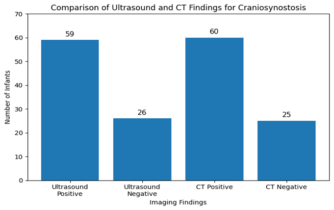
Figure 1. Comparison of ultrasound and CT findings for craniosynostosis among infants.
The graph shows that ultrasound diagnosed craniosynostosis in 59 infants, while CT confirmed craniosynostosis in 60 infants, showing close agreement between both imaging modalitie
DISCUSSION :
The present study evaluated the diagnostic performance of ultrasound for detecting craniosynostosis in infants, using CT skull with 3D reconstruction as the gold standard. Craniosynostosis was confirmed on CT in 60 out of 85 infants (70.6%), while ultrasound diagnosed craniosynostosis in 59 infants (69.4%). The close similarity between ultrasound and CT findings suggests that ultrasound performed very well as an initial diagnostic modality in clinically suspected cases. In the present study, ultrasound showed 96.7% sensitivity, 96.0% specificity, 98.3% positive predictive value, 92.3% negative predictive value, and 96.5% overall diagnostic accuracy, with a statistically significant association between ultrasound and CT findings (p < 0.001). These findings indicate that ultrasound can identify most CT-confirmed cases and can also reliably exclude craniosynostosis in infants without the disease (12, 13).
The extreme sensitivity in this study is clinically significant as it can lead to a delay in surgical referral for craniosynostosis and introduce the risk of progressive skull deformity, elevated ICP and developmental issues. This yielded 58 correct diagnoses in 60 patients that were diagnosed as positive on the CT; two of the patients were diagnosed as negative on the CT and were ultrasound negative. This detection rate is similar to that of Alizadeh et al. who have found 96.9% sensitivity and 100% specificity with reference to CT, and a PPV of 100% and an NPV of 92.3% (14). Likewise, Proisy et al. found that cranial ultrasound and 3D CT were in perfect agreement with 100% sensitivity and 100% specificity in the diagnosis of closed cranial sutures in infants referred for skull deformity (15). These findings are in accordance with previous studies, which is an indicator that ultrasound is reliable for diagnosing craniosynostosis in infancy.
The specificity of ultrasound was very high in this study at 96.0%, with a single false positive result. However, high specificity is useful, as it minimizes unnecessary CT exposures in infants who don't have craniosynostosis. CT is still the first modality of choice for its ability to show the detail of the cranial sutures and the skull anatomy, particularly when surgery is planned. CT however is linked with ionizing radiation, cost and, in some cases, sedation for infants. Currently, there are also references that state that 3D reconstruction using CT is the criterion standard for detailed analysis of the cranium sutures and the need to reduce radiation exposure as much as possible (16, 17). The authors conclude that ultrasound is an effective first-line imaging technique for the diagnosis of craniosynostosis, especially in young infants with clinically suspected craniosynostosis.
This study's results are also corroborated by the latest reviews concerning imaging in craniosynostosis. Cacciaguerra et al. emphasized that the clinical examination, skull measurement, and observation of the skull deformity were the initial clues to suspect craniosynostosis, and imaging was indicated for confirming the diagnosis and help guide management (18). A systematic review published in 2022 found that ultrasound has a broad overall diagnostic spectrum and sensitivity ranges from 71% to 100%, and 3D CT is highly accurate for evaluating suture closure (19). Additionally, Proisy et al. found that cranial suture ultrasound was rapid, without the use of radiation, and did not require sedation, and could be used as a first-line imaging modality in infants under 8-12 months of age if craniosynostosis was suspected (15). Therefore, the age range in the present study (1 to 12 months) is suitable for the evaluation of sutures using ultrasound.
The kappa value of the present study was excellent (κ = 0.915), further reflecting good agreement between ultrasound and CT. The stratified analysis revealed no statistically significant differences in diagnostic accuracy by age, gender, type of residence, socioeconomic status, family history or mode of delivery. This indicates ultrasound performance was consistent for the various patient sub-groups. There was no significant difference between the accuracy of both methods in younger infants, but ultrasound seemed to show a little more accuracy. This minor difference is possibly accounted for by the fact that the sutures of the cranium are more visible sonographically in younger infants, and as with age the sutures become more and more ossified, they are less visible. There is also an important role for operator experience; ultrasound is more dependent on the skill of the examiner than is CT. Thus, there is a need for proper training of radiologists and standardization of scanning procedures prior to the routine use of ultrasound as a screening tool (20).
There are some limitations in this study. First, the study was carried out in one radiology department, so the results might not be completely applicable to other departments in hospitals. Secondly, ultrasound is operator-dependent, and the accuracy could be affected by the experience of the radiologist and the quality of the equipment. Third, the authors reported diagnostic ability using CT as the gold standard, but they did not report separately on the diagnostic ability by suture, e.g., sagittal suture, coronal suture, metopic suture, lambdoid suture. More detailed evidence would come from future studies that recruit larger numbers of patients, and perform diagnostic analysis on a suture-by-suture basis. The limitations of this study aside, the study does offer useful local data that ultrasound can serve as an effective non-radiating initial imaging tool for infants with suspected craniosynostosis
CONCLUSION :
Ultrasound demonstrated excellent diagnostic accuracy for the detection of craniosynostosis in infants when compared with CT skull with 3D reconstruction as the gold standard. The high sensitivity, specificity, positive predictive value, negative predictive value, and overall accuracy indicate that ultrasound is a reliable, safe, and radiation-free modality for initial evaluation of clinically suspected craniosynostosis. CT should remain reserved for equivocal ultrasound findings, complex cases, and preoperative planning. Use of ultrasound as a first-line investigation may reduce unnecessary radiation exposure, improve early diagnosis, and support timely referral for specialist management.
REFERENCES:
1. DeFreitas CA, Carr SR, Merck DL, Byrne MM, Linden OE, Stiles EA, et al. Prenatal diagnosis of craniosynostosis using ultrasound. 2022;150(5):1084-9.
2. Pogliani LM, Zuccotti GV, Reggiori M, Erbetta A, Lacerenza M, Prada F, et al. Surface Cranial Ultrasound: The Natural Heir to X-Ray for the Screening of Skull Deformities in Infants. 2023;44(05):503-11.
3. Peter C, Stranzinger E, Heverhagen JT, Keitel K, Romano F, Busch JD, et al. Minor head trauma in infants—how accurate is cranial ultrasound performed by trained radiologists? 2023;182(7):3113-20.
4. Rosato S, Unger S, Campos-Xavier B, Caraffi SG, Beltrami L, Pollazzon M, et al. Clinical and molecular diagnosis of osteocraniostenosis in fetuses and newborns: prenatal ultrasound, clinical, radiological and pathological features. 2022;13(2):261.
5. Micovic M, Zivkovic B, Vukasinovic I, Jelovac D, Stojicic M, Bascarevic VJD. Imaging Modalities in Craniosynostosis: A Systematic Review and Proposal of the ARCANA Protocol for Multimodal Radiation-Free Assessment. 2025;15(20):2632.
6. Sakamoto Y, Miwa T, Numasawa Y, Kajita H, Narumi SJJoCS. Diagnostic Accuracy of Craniosynostosis in Japan: A Survey Using Three-Dimensional Physical Models. 2026:10.1097.
7. Daly GE, Stefanko A, Henning C, Crupi L, Howell LK, Wolfswinkel EMJTCPCJ. Prenatal Diagnosis of Nonsyndromic Craniosynostosis: A Scoping Review. 2026;63(4):635-40.
8. Wójtowicz-Marzec M, Zarzycka D, Berendt AMJNitsC. The role of the nurse in the diagnosis and care of an infant with craniosynostosis. 2025;24(1 (90)):94-105.
9. Blum JD, Cho DY, Cheung L, Villavisanis DF, Ng J, Swanson JW, et al. Making the diagnosis in sagittal craniosynostosis—it’s height, not length, that matters. 2022;38(7):1331-40.
10. Massoud M, Bault J-P, Cabet S. Antenatal Diagnosis of Craniosynostosis. Neurosurgical Aspects of Craniosynostosis: Springer; 2025. p. 253-67.
11. Lin J, Ji X-T, Wang J-Y, Zhang Q, Chen Z-H, Li P, et al. A Novel Noninvasive Screening Method for Early Detection of Sagittal Craniosynostosis Using the Surface Cranial Index. 2025;36(8):e1408-e13.
12. Hermann J, Raak CK, Ostermann T, Scharbrodt WJOJoRD. Developments in diagnostic and surgical techniques in children with sagittal suture craniosynostosis: a systematic review spanning the last 30 years. 2025;20(1):437.
13. Lun KK, Aggarwala S, Gardner D, Hunt J, Jacobson E, Reddy R, et al. Assessment of paediatric head shape and management of craniosynostosis. 2022;51(1/2):51-8.
14. Alizadeh H, Najmi N, Mehdizade M, Najmi N. Diagnostic accuracy of ultrasonic examination in suspected craniosynostosis among infants. Indian Pediatr. 2013;50(1):148-50.
15. Proisy M, Riffaud L, Chouklati K, Tréguier C, Bruneau B. Ultrasonography for the diagnosis of craniosynostosis. European Journal of Radiology. 2017;90:250-5.
16. Di Rocco F, Proctor MR. Technical evolution of pediatric neurosurgery: craniosynostosis from 1972 to 2023 and beyond. Child's Nervous System. 2023;39(10):2779-87.
17. Shlobin NA, Baticulon RE, Ortega CA, Du L, Bonfield CM, Wray A, et al. Global Epidemiology of Craniosynostosis: A Systematic Review and Meta-Analysis. World Neurosurgery. 2022;164:413-23.e3.
18. Cacciaguerra G, Palermo M, Marino L, Rapisarda FA, Pavone P, Falsaperla R, et al. The Evolution of the Role of Imaging in the Diagnosis of Craniosynostosis: A Narrative Review. Children [Internet]. 2021; 8(9):[727 p.].
19. Schaufelberger M, Kühle R, Wachter A, Weichel F, Hagen N, Ringwald F, et al. A Radiation-Free Classification Pipeline for Craniosynostosis Using Statistical Shape Modeling. Diagnostics [Internet]. 2022; 12(7):[1516 p.].
20. Leonhardt Y, Kronthaler S, Feuerriegel G, Karampinos DC, Schwaiger BJ, Pfeiffer D, et al. CT-like MR-derived images for the assessment of craniosynostosis and other pathologies of the pediatric skull. 2023;33(1):57-64