CALCIUM AND PHOSPHORUS SUPPLEMENTATION FOR THE PREVENTION OF METABOLIC BONE DISEASE IN PRETERM INFANTS
- Mian Ibrar Aleem , Department of Pediatrics, Saidu Teaching Hospital Swat, Pakistan
- Zahoor Ahmad , Department of Pediatrics, Saidu Teaching Hospital Swat, Pakistan
Article Information:
Abstract:
Preterm infants are at risk for metabolic bone disease (MBD) due to insufficient calcium and phosphorus accretion. Objective: To evaluate whether calcium and phosphorus supplementation prevents MBD and improves biochemical and growth outcomes in preterm infants. Methods: Fifty preterm infants (<37 weeks) were randomized into two equal groups. The supplemented group received calcium and phosphorus plus standard feeding; the control group received standard feeding only. Outcomes at 6 weeks included serum calcium, phosphate, alkaline phosphatase (ALP), growth, suspected MBD, radiological osteopenia, and fractures. Results: At 6 weeks, the supplemented group had significantly higher serum phosphate (5.4 vs. 4.9 mg/dL; p=0.04) and lower ALP (418 vs. 492 IU/L; p=0.02) compared to controls. Serum calcium was higher but not significant (9.3 vs. 9.0 mg/dL; p=0.11). Suspected MBD (16% vs. 32%), radiological osteopenia, fractures, and poor growth were less frequent in the supplemented group, though not statistically significant. Conclusion: Calcium and phosphorus supplementation improved biochemical markers of bone mineralization in preterm infants. Larger trials are needed to confirm clinical benefits.
Keywords:
Article :
INTRODUCTION:
Metabolic bone disease of prematurity is an important neonatal problem that occurs when bone mineralization is lower than expected for gestational and postnatal age. It is commonly associated with reduced bone mineral content, biochemical abnormalities, osteopenia, growth impairment, and, in severe cases, fractures.¹ The condition is especially relevant in preterm infants because fetal skeletal mineralization is most active during the third trimester. ² When birth occurs early, the infant misses a major part of transplacental calcium and phosphorus transfer, resulting in reduced mineral stores at birth.³
Calcium and phosphorus are the principal minerals required for skeletal growth and mineral deposition. Calcium contributes to bone structure and neuromuscular function, while phosphorus is essential for hydroxyapatite formation and energy metabolism.⁴ Inadequate intake or poor retention of either mineral may disturb bone formation and increase the risk of metabolic bone disease.⁵ Preterm infants are particularly vulnerable because their mineral requirements remain high after birth, while their nutritional intake may be limited by feeding intolerance, illness, delayed enteral feeding, or prolonged parenteral nutrition.⁶
Biochemical screening is important because metabolic bone disease may develop before obvious clinical or radiological signs appear. Low serum phosphate and elevated alkaline phosphatase are commonly used as early markers of impaired bone mineralization in preterm infants.⁷ Serum calcium may remain normal because it is tightly regulated, so phosphate and alkaline phosphatase are often more informative for early detection. ⁸ Radiographs may show osteopenia, metaphyseal changes, or fractures, but radiological changes usually appear later and may underestimate early disease. ⁹
Breast milk is the preferred nutrition for newborn infants because it provides immunological and developmental benefits. However, unfortified breast milk may not provide enough calcium and phosphorus for rapidly growing preterm infants.¹⁰ Therefore, mineral supplementation and human milk fortification have been recommended to improve mineral intake and support skeletal growth.¹¹ Despite these recommendations, the clinical benefit of routine calcium and phosphorus supplementation in different neonatal settings remains uncertain, especially in resource-limited hospitals where regular biochemical monitoring may not be consistently available.
This study was designed to evaluate the effect of calcium and phosphorus supplementation on metabolic bone disease, biochemical markers, and growth outcomes in preterm infants admitted to a neonatal unit in Swat, Pakistan..
METHODOLOGY:
The study was a prospective randomized clinical trial conducted over three months (March–June 2025) in the Pediatrics/Neonatology Department of Saidu Teaching Hospital, Swat, Pakistan. Fifty preterm infants (<37 weeks gestation) who tolerated enteral feeding and had parental consent were enrolled and randomized into two equal groups: 25 received calcium and phosphorus supplementation plus standard feeding, and 25 received standard feeding only, followed for six weeks. Infants with major congenital anomalies, known bone disorders, illness preventing feeding, prior supplementation, or high risk of loss to follow-up were excluded. Baseline and six‑week data included gestational age, birth weight, sex, feeding type, sepsis status, anthropometric measures (weight, length, occipitofrontal circumference), serum calcium, phosphate, and alkaline phosphatase. Outcomes included suspected metabolic bone disease, biochemical levels, radiological osteopenia, fractures, and growth gains. Statistical analysis used SPSS version 20, with Student’s t‑test for continuous variables and chi‑square test for categorical variables; p<0.05 was considered significant
RESULTS:
A total of 50 preterm infants were included in the study. Twenty-five infants were allocated to the supplemented group, and 25 were allocated to the control group. Both groups were comparable at baseline in demographic, clinical, anthropometric, and biochemical characteristics
Table 1. Baseline characteristics of study participants
|
Variable |
Supplemented Group (n = 25) |
Control Group (n = 25) |
p-value |
|
Gestational age (weeks), mean ± SD |
32.1 ± 1.8 |
31.9 ± 1.9 |
0.68 |
|
Birth weight (g), mean ± SD |
1510 ± 220 |
1488 ± 235 |
0.72 |
|
Male sex, n (%) |
14 (56.0) |
13 (52.0) |
0.78 |
|
Breast milk feeding, n (%) |
16 (64.0) |
15 (60.0) |
0.77 |
|
Formula/mixed feeding, n (%) |
9 (36.0) |
10 (40.0) |
0.77 |
|
Sepsis, n (%) |
6 (24.0) |
7 (28.0) |
0.75 |
|
Weight at enrollment (g), mean ± SD |
1495 ± 215 |
1478 ± 228 |
0.78 |
|
Length at enrollment (cm), mean ± SD |
40.8 ± 2.4 |
40.4 ± 2.6 |
0.57 |
|
OFC at enrollment (cm), mean ± SD |
29.3 ± 1.6 |
29.0 ± 1.7 |
0.53 |
|
Serum calcium (mg/dL), mean ± SD |
8.6 ± 0.5 |
8.5 ± 0.6 |
0.48 |
|
Serum phosphate (mg/dL), mean ± SD |
4.6 ± 0.7 |
4.5 ± 0.8 |
0.64 |
|
Alkaline phosphatase (IU/L), mean ± SD |
356 ± 82 |
362 ± 88 |
0.79 |
The absence of statistically significant baseline differences indicates that the two groups were comparable before supplementation
Table 2. Biochemical outcomes at 6 weeks
|
Variable |
Supplemented Group (n = 25) |
Control Group (n = 25) |
p-value |
|
Serum calcium (mg/dL), mean ± SD |
9.3 ± 0.6 |
9.0 ± 0.7 |
0.11 |
|
Serum phosphate (mg/dL), mean ± SD |
5.4 ± 0.8 |
4.9 ± 0.9 |
0.04 |
|
Alkaline phosphatase (IU/L), mean ± SD |
418 ± 96 |
492 ± 118 |
0.02 |
|
Low serum phosphate, n (%) |
4 (16.0) |
9 (36.0) |
0.10 |
|
Elevated alkaline phosphatase, n (%) |
5 (20.0) |
11 (44.0) |
0.07 |
At 6 weeks, serum phosphate was significantly higher and alkaline phosphatase was significantly lower in the supplemented group. Low serum phosphate and elevated alkaline phosphatase were less frequent in the supplemented group, although these differences were not statistically significant.
Table 3. Growth outcomes at 6 weeks
|
Variable |
Supplemented Group (n = 25) |
Control Group (n = 25) |
p-value |
|
Weight at 6 weeks (g), mean ± SD |
2525 ± 330 |
2388 ± 360 |
0.17 |
|
Weight gain (g), mean ± SD |
1030 ± 215 |
910 ± 230 |
0.07 |
|
Length at 6 weeks (cm), mean ± SD |
45.0 ± 2.7 |
44.2 ± 2.8 |
0.31 |
|
Length gain (cm), mean ± SD |
4.2 ± 1.0 |
3.8 ± 1.1 |
0.18 |
|
OFC at 6 weeks (cm), mean ± SD |
32.4 ± 1.7 |
31.9 ± 1.8 |
0.32 |
|
OFC gain (cm), mean ± SD |
3.1 ± 0.7 |
2.8 ± 0.8 |
0.16 |
The supplemented group showed greater gains in weight, length, and occipitofrontal circumference. However, these differences did not reach statistical significance.
Table 4. Metabolic bone disease and radiological findings at 6 weeks
|
Outcome |
Supplemented Group (n = 25) |
Control Group (n = 25) |
p-value |
|
Suspected metabolic bone disease, n (%) |
4 (16.0) |
8 (32.0) |
0.18 |
|
Radiological osteopenia, n (%) |
2 (8.0) |
5 (20.0) |
0.22 |
|
Fractures, n (%) |
0 (0.0) |
1 (4.0) |
0.31 |
Suspected metabolic bone disease, radiological osteopenia, and fractures were less frequent in the supplemented group. Although these differences were not statistically significant, all findings favored supplementation.
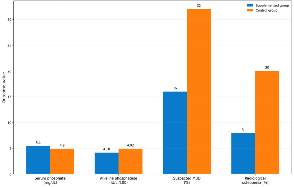
Figure: Six-week biochemical and clinical outcomes by study group serum phosphate, alkaline phosphatase, suspected metabolic bone disease, and radiological osteopenia
DISCUSSION :
This randomized clinical trial showed that calcium and phosphorus supplementation improved biochemical indicators of bone mineralization in preterm infants. Serum phosphate was significantly higher and alkaline phosphatase was significantly lower in the supplemented group after 6 weeks. These findings are clinically meaningful because hypophosphatemia and elevated alkaline phosphatase are commonly used markers of metabolic bone disease in premature infants.¹¹ The improvement in serum phosphate supports the biological role of phosphorus in hydroxyapatite formation and skeletal mineral deposition.¹² Phosphorus deficiency can limit bone mineralization even when calcium intake is adequate.¹³ Therefore, combined calcium and phosphorus supplementation is more physiologically appropriate than calcium supplementation alone, especially in rapidly growing preterm infants.¹⁴ The reduction in alkaline phosphatase in the supplemented group suggests reduced bone turnover and improved mineralization.¹⁵ Elevated alkaline phosphatase has been associated with poor skeletal mineralization and increased risk of osteopenia in preterm infants.¹⁶ In the present study, fewer supplemented infants had elevated alkaline phosphatase compared with controls, although the categorical difference did not reach statistical significance.
Serum calcium was higher in the supplemented group, but the difference was not statistically significant. This finding is expected because serum calcium is tightly regulated by hormonal mechanisms and may remain within the normal range despite inadequate bone mineralization.¹⁷ Therefore, serum calcium alone is not a reliable screening marker for metabolic bone disease of prematurity.
The supplemented group had a lower frequency of suspected metabolic bone disease, radiological osteopenia, and fractures compared with the control group. These differences were not statistically significant, likely because the study had a small sample size and a short follow-up duration.¹⁸ Bone mineral changes may require longer observation before they become radiologically apparent. In addition, radiographs were performed only when clinically indicated, which may have underestimated radiological osteopenia.
Growth outcomes also favored supplementation. Infants in the supplemented group gained more weight, length, and occipitofrontal circumference than those in the control group. Although these differences were not statistically significant, the direction of effect was consistent. Previous studies have shown that adequate mineral intake may support both skeletal development and overall growth in very low birth weight and preterm infants.¹⁹
The findings of this study are consistent with the concept that metabolic bone disease of prematurity is strongly influenced by substrate deficiency.²⁰ Preterm infants have high postnatal mineral requirements, and standard feeding may not always provide sufficient calcium and phosphorus. Early mineral supplementation may therefore help correct biochemical abnormalities and reduce later skeletal complications.²¹
This study has several strengths. It used a randomized design, included both biochemical and clinical outcomes, and addressed a locally relevant neonatal problem. It also assessed growth parameters and radiological findings, giving a broader view of supplementation effects. However, the study has limitations. The sample size was small, with only 25 infants in each group. The follow-up period was limited to 6 weeks. Radiographs were not performed routinely for all infants, and detailed intake of calcium, phosphorus, vitamin D, and fortified feeds was not quantified. Future studies should include larger samples, longer follow-up, standardized radiological assessment, and detailed nutritional intake monitoring..
CONCLUSION :
Calcium and phosphorus supplementation improved biochemical indicators of bone mineralization in preterm infants, particularly serum phosphate and alkaline phosphatase. The supplemented group also showed lower rates of suspected metabolic bone disease, radiological osteopenia, and fractures, along with better growth trends. However, several clinical and growth outcomes were not statistically significant, most likely due to the small sample size and short follow-up period.
These findings support the potential role of calcium and phosphorus supplementation in reducing metabolic bone disease risk among preterm infants. Larger randomized trials with longer follow-up are recommended to confirm these findings and guide routine supplementation practices in neonatal units..
Recommendations
1. Preterm infants should be screened for metabolic bone disease using serum phosphate and alkaline phosphatase.
2. Calcium and phosphorus supplementation should be considered in preterm infants at risk of poor bone mineralization.
3. Serum calcium should not be used alone to exclude metabolic bone disease.
4. Larger randomized trials should be conducted with longer follow-up and standardized radiological assessment.
5. Future studies should record exact mineral intake from feeding, fortification, and supplements.
Acknowledgment
The authors would like to thank the staff of the Pediatrics and Neonatology Department at Saidu Teaching Hospital Swat for their support and cooperation during data collection and follow-up of study participants. The authors are also grateful to the parents and families of the enrolled infants for their participation and trust.
Conflict of Interest
The authors declare that there is no conflict of interest regarding the publication of this study.
Funding Statement
This research received no external funding and was conducted without financial support from any public, commercial, or nonprofit organization.
Author Contributions
Mian Ibrar Aleem¹, Conceptualization, study supervision, methodology review, manuscript review, and final approval of the manuscript. Zahoor Ahmad¹* Data collection, data curation, statistical analysis, manuscript drafting, correspondence, and manuscript editing.
Ethical Approval
Ethical approval for this study was obtained from the Ethical Review Committee (ERC) of Saidu Teaching Hospital, Pakistan No: ERC/STH/2025/017), dated 15 January 2025
Informed Consent
Informed consent was obtained from the parents or legal guardians of all enrolled infants before participation in the study.
REFERENCES:
1. Faienza MF, D’Amato E, Natale MP, Grano M, Chiarito M, Brunetti G, D’Amato G. Metabolic bone disease of prematurity: diagnosis and management. Frontiers in Pediatrics. 2019;7:143.
2. Rustico SE, Calabria AC, Garber SJ. Metabolic bone disease of prematurity. Journal of Clinical & Translational Endocrinology. 2014;1(3):85–91.
3. Wang J, Chen W, Liu Y, et al. Risk factors for metabolic bone disease of prematurity: a meta-analysis. PLOS ONE. 2022;17(6):e0269180.
4. Mihatsch W, Thome U, Sáenz de Pipaon M. Update on calcium and phosphorus requirements of preterm infants and recommendations for enteral mineral intake. Nutrients. 2021;13(5):1470.
5. Embleton ND, Moltu SJ, Lapillonne A, van den Akker CHP, Carnielli VP, Fusch C, et al. Enteral nutrition in preterm infants: a position paper from the ESPGHAN Committee on Nutrition and invited experts. Journal of Pediatric Gastroenterology and Nutrition. 2023;76(2):248–268.
6. Perrone M, et al. Incidence and risk factors of metabolic bone disease in preterm infants: an observational study from a neonatal intensive care unit. Nutrients. 2022;14:3660.
7. Grover M, Kovacs CS, Krishnamurthy S. Invited mini review: metabolic bone disease of prematurity. Frontiers in Pediatrics. 2025;12:1455094.
8. Nationwide Children’s Hospital. Metabolic bone disease: NICU clinical pathway. 2023.
9. Graça MI, et al. Metabolic bone disease of prematurity: a report of five cases. Journal of Pediatric and Neonatal Individualized Medicine. 2019;8(1):e080120.
10. Brown JVE, Embleton ND, Harding JE, McGuire W. Multi-nutrient fortification of human milk for preterm infants. Cochrane Database of Systematic Reviews. 2016;(5):CD000343.
11. Picaud JC, Vincent M, Buffin R. Human milk fortification for preterm infants: a review. World Review of Nutrition and Dietetics. 2021;122:225–247.
12. Xu X, Ma H, Cheng S, Xue J. Effect of early preventive supplementation with calcium and phosphorus on metabolic bone disease in premature infants. BMC Pediatrics. 2024;24:171.
13. Kajohntridach P, et al. Calcium and phosphorus intake in very low birth weight infants and the association with growth and bone health outcomes. Pediatric Gastroenterology, Hepatology & Nutrition. 2025;28(5):320–329.
14. Ashford and St Peter’s Hospitals NHS Foundation Trust. Prevention and management of metabolic bone disease of prematurity guideline. 2023.
15. Liu X, et al. Analysis of clinical risk factors for metabolic bone disease of prematurity. Frontiers in Pediatrics. 2024;12:1345878.
16. Krishna SBM, et al. Incidence and risk factors for metabolic bone disease of prematurity. Indian Pediatrics. 2025;62:289–294.
17. Mutlu GY, et al. Metabolic bone disease of prematurity: report of four cases. Journal of Clinical Research in Pediatric Endocrinology. 2014;6(2):111–115.
18. Alalwani YJ, et al. Interventions for metabolic bone disease of prematurity. Seminars in Perinatology. 2024.
19. Fewtrell MS. Does early nutrition program later bone health in preterm infants? American Journal of Clinical Nutrition. 2011;94(6 Suppl):1870S–1873S.
20. Chitale R, Ferguson K, Talej M, et al. Early enteral feeding for preterm or low birth weight infants: a systematic review and meta-analysis. Pediatrics. 2022;150(Suppl 1):e2022057092E.
21. Western Scotland Neonatal Guideline Group. Metabolic bone disease in neonates guideline. 2025.