A comparative evaluation to determine the effect of Sodium Ascorbate and Aloe Vera on the push out bond strength of resin cement used to lute fiber post to radicular dentin: an in-vitro study
- Sandhya Kapoor Punia , Professor and Head of Department, Department of Conservative Dentistry and Endodontics, Darshan Dental College and Hospital, Rajasthan
- Payal Gokhe , PG Student, Department of Conservative Dentistry and Endodontics, Darshan Dental College and Hospital, Rajasthan
- Shilpi Kushwaha , Senior lecturer, Department of Conservative Dentistry and Endodontics, Darshan Dental College and Hospital, Rajasthan
- Yash Wadhwani , Senior lecturer, Department of Conservative Dentistry and Endodontics, Darshan Dental College and Hospital, Rajasthan
- Renu Kajla , Senior lecturer, Department of Conservative Dentistry and Endodontics, Darshan Dental College and Hospital, Rajasthan
- Swarnendra Nag , Senior lecturer, Department of Conservative Dentistry and Endodontics, Darshan Dental College and Hospital, Rajasthan
Article Information:
Abstract:
To evaluate and compare the effect of sodium ascorbate and Aloe vera on the push-out bond strength of fiber posts luted to radicular dentin. Materials And Method: Thirty intact permanent anterior teeth with single root and single canal were collected and examined under a stereomicroscope to eliminate the teeth with cracks or defects. Standardized root length of 13 mm was obtained by sectioning near the cemento-enamel junction using a slow-speed diamond disc. Working length was determined using stainless steel #15 K-file and canals were prepared using Universal hand Pro-taper instruments up to F4 under copious irrigation followed by obturation with F4 Pro-taper gutta-percha point and AH Plus sealer. Post space was drilled, 5 mm gutta-percha retained, and samples were randomly divided into three groups (n=10): Group A (Normal saline), Group B (Sodium Ascorbate), Group C (Aloe vera). Prepared post spaces were irrigated with 9 ml of the respective solutions for 3 minutes, followed by normal saline irrigation. Glass fiber Post (Reforpost #2) was luted with Calibra Universal Self-Adhesive Resin Cement for each sample followed by incubation for 24 hours. Samples sectioned into discs (2mm thick) from the middle third of the root. Discs were embedded and tested for push-out strength using a Universal testing machine. Data was analyzed using one-way and two-way ANOVA followed by post hoc Tukey’s test. Results: A statistically significant difference was observed among groups (p<0.05). Aloe vera demonstrated the highest bond strength, followed by sodium ascorbate, while saline showed the lowest values. Conclusion: Natural collagen crosslinking agents significantly enhance push-out bond strength of fiber posts, with Aloe vera showing superior performance.
Keywords:
Article :
INTRODUCTION:
Extensively damaged teeth treated with endodontic therapy are frequently restored using post-and-core systems. Adhesive resin cements are extensively utilized for the placement of posts within root canals.1 Despite their advantages, the most common cause of failure in resin-bonded fiber posts is bond degradation. The organic matrix of dentin is susceptible to deterioration, particularly when sodium hypochlorite (NaOCl), the most widely used irrigant in root canal treatments, is employed.2
Research indicates that bond failure may result from the activation of matrix metalloproteinases (MMPs) in dentin, triggered by the application of acids during adhesive procedures. These enzymes degrade collagen fibrils within the hybrid layer, compromising the bond integrity over time.3 One approach to enhance bond strength is the use of natural crosslinkers, which have been shown to improve dentin stiffness by increasing the number of intramolecular collagen crosslinks.4
Studies suggest that the reduction in bond strength caused by sodium hypochlorite can be counteracted by using Sodium Ascorbate (SA) or Vitamin C, both of which help restore bond strength. SA is a powerful antioxidant that aids in collagen synthesis by promoting the formation of hydroxyproline and hydroxylysine, which are crucial for collagen stability. Hydroxyproline stabilizes the collagen triple helix, while hydroxylysine facilitates intermolecular crosslinks, enhancing collagen's mechanical strength5
Aloe Barbadensis Miller (commonly known as Aloe vera), a perennial succulent resembling a cactus with green, fleshy, spiny, dagger-shaped leaves containing a clear, viscous gel, belongs to the Aloeaceae family (a subfamily of Asphodelaceae). It exhibits anti-MMP activity, particularly against MMP-2 and MMP-9, attributed to its anthraquinone content. Aloe vera has demonstrated a sustained beneficial effect by increasing collagen content which warranties its use as collagen cross linking agent.6
Hence, this study aims to evaluate and compare the push-out bond strength of fiber posts luted to radicular dentin using 10% Sodium ascorbate,10% Aloe vera as collagen cross-linking agents.
The null hypothesis states that the application of these collagen cross-linking agents does not improve the push-out bond strength of fiber posts to radicular dentin.
MATERIAL AND METHOD:
This in-vitro study was conducted in the Department of Conservative Dentistry and Endodontics, Darshan Dental College and Hospital, Udaipur, Rajasthan, India.
Teeth Selection:
A total of 30 extracted permanent anterior single rooted teeth with single canal were collected from the Department of Oral and Maxillofacial Surgery, Darshan Dental College and Hospital, Loyara, and were prepared for the study at the Department of Conservative Dentistry and Endodontics, Darshan Dental College and Hospital, Loyara, Udaipur.
Inclusion criteria:
The study involved the inclusion of teeth with single root and single canal and non carious permanent teeth with mature root apex, free from cracks.
Exclusion criteria:
The study involved the exclusion of teeth with any developmental defect and caries, restored teeth, endodontically treated teeth, resorption defects, teeth with immature apices, root fracture.
Sample Preparation:
All teeth were thoroughly cleaned using an ultrasonic scaler to remove debris, calculus, and stains and then stored in normal saline. Following the cleaning process, the teeth were examined under a stereomicroscope to identify any crack lines or defects in the roots.
The selected teeth were sectioned near the cemento-enamel junction using a slow-speed diamond disc under water coolant to obtain a standardized root length of 13 mm. The canal for each sample was accessed using a round bur attached to an air rotor handpiece and a #15 stainless steel K-file was passively introduced in the canal to check the apical patency and the working length (WL) was established 1mm short of the anatomical apex. This was followed by canal preparation with Universal Hand ProTaper NiTi (Dentsply Maillefer) instruments up to F4 under copious irrigation with normal saline. The prepared canals were dried using paper points and obturated with F4 gutta-percha and AH Plus sealer to ensure proper sealing of the root canal.
Following the obturation, the gutta-percha was carefully removed using a heated hand plugger, leaving only 5 mm of obturation material at the root end to prepare the space to recieve fiber post. The post space was prepared using the corresponding drill to ensure the proper fit and adaptation of the Angelus Size 2 Reforpost within the prepared canal space.
Grouping of Samples:
The teeth were then randomly divided into three groups, with each group containing n = 10 samples.
Group A: (Control group) Normal Saline
Group B: 10% Sodium Ascorbate
Group C: 10% Aloe Vera
Preparation of solution:
Preparation of 10% Sodium Ascorbate: To prepare a 10% Sodium Ascorbate solution, 10 grams of sodium ascorbate powder was weighed accurately and then dissolved in 100 ml of distilled water in a clean beaker. The mixture was stirred thoroughly until the sodium ascorbate powder was completely dissolved, resulting in a 10% sodium ascorbate solution.
Preparation of 10% Aloe Vera: To prepare 10% Aloe Vera solution, 10 grams of freshly obtained aloe vera gel was weight accurately and transferred it to a sterile beaker, followed by the gradual addition of distilled water to a final volume of 100 ml with continuous stirring to obtain a uniform mixture. The final extract was aliquoted into amber sterile bottles and stored at 4°C, with all preparations used within 24–72 hours to maintain optimal bioactivity and minimize microbial contamination.
The prepared post spaces of all the samples were irrigated with 9 ml of the respective solution for 3 minutes (3 ml/min). A final rinse with normal saline was performed, and canals were dried with paper points.
Fiber posts were cemented using a Calibra cement delivered through an automix syringe with an endodontic tip. Fiber post (Reforpost #2) was immediately cemented for each sample using Calibra Universal Self- Adhesive Resin Cement. The cement was allowed to self-cure, followed by light curing according to manufacturer instructions. Specimens were stored in an incubator at 37°C and 100% humidity for 24 hours to ensure complete polymerization.
Each root was horizontally sectioned using a diamond disc under water coolant to obtain 2-mm thick disc from the middle third. Specimens were embedded in self-cure acrylic resin, ensuring exposure of the post–dentin interface.
Push-out bond strength was measured using a Universal Testing Machine at a crosshead speed of 1.25 mm/min until bond failure occurred.
STATISTICAL ANALYSIS
The values obtained for push out bond strength were analyzed using one-way analysis of variance (one-way ANOVA) for inter-group comparisions followed by Post-Hoc Tukey test to evaluate the differences in the mean push out bond strength between samples with different collagen cross link. The level of significance was adopted as p < 0.05 and was considered significant.
RESULT:
This in-vitro study was conducted to evaluate and compare the effect of 10% Sodium Ascorbate and 10% Aloe vera on push out bond strength of resin cement used to lute fiber post to radicular dentin. The data obtained was tabulated and statistically analyzed using SPSS software (Version 21.0). The push-out bond strength was evaluated using one-way ANOVA for inter-group comparisons followed by Post hoc Tukey’s test for two different collagen cross-linking agents. A p-value of less than 0.05 was considered statistically significant, while values exceeding this threshold were deemed non-significant.
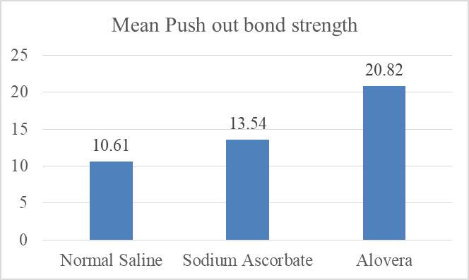
Group C (Aloe Vera) (20.820 ± 1.807) exhibited the highest push out bond strength followed by Group B (Sodium Ascorbate) (13.535 ± 1.670 MPa) and Group A (Normal Saline) (10.605 ± 1.543 MPa) exhibited the lowest push out bond strength. Order of the push out bond strength values of the 3 groups: Group C > Group B > Group A. (Table 1)
|
Table 1: Descriptive statistics of mean push out bond strength of all the experimental groups. |
|||
|
Groups |
Sample Size (N) |
Mean (MPa) |
SD |
|
Group A (Normal Saline) |
10 |
10.605 |
1.543 |
|
Group B (Sodium Ascorbate) |
10 |
13.535 |
1.670 |
|
Group C (Aloe Vera) |
10 |
20.820 |
1.807 |
|
Table 3: ONE WAY ANOVA for intergroup comparison of the influence of collagen cross linking agents on the push out bond strength of resin cement used to lute fiber post to radicular dentin for all experimental groups. |
|||||
|
|
Sum of Squares |
Df |
Mean Square |
F |
Sig. |
|
Between Groups |
1165.093 |
3 |
388.364 |
116.924* |
0.000 |
|
Within Groups |
252.435 |
76 |
3.322 |
||
|
Total |
1417.528 |
79 |
|
||
|
*p<0.05 is considered as statistically significant |
|||||
|
(I) Group |
Mean Difference (I-J) |
Std. Error |
Sig. |
95% Confidence Interval |
||
|
Lower Bound |
Upper Bound |
|||||
|
Normal Saline (Group A) |
Sodium Ascorbate |
-2.93000* |
0.576 |
0.000 |
-4.444 |
-1.416 |
|
Aloe vera |
-10.21500* |
0.576 |
0.000 |
-11.729 |
-8.701 |
|
|
Sodium Ascorbate (Group B) |
Normal Saline |
2.93000* |
0.576 |
0.000 |
1.416 |
4.444 |
|
Aloe vera |
-7.28500* |
0.576 |
0.000 |
-8.799 |
-5.771 |
|
|
Aloe vera (Group C) |
Normal Saline |
10.21500* |
0.576 |
0.000 |
8.701 |
11.729 |
|
Sodium Ascorbate |
7.28500* |
0.576 |
0.000 |
5.771 |
8.799 |
|
Graph 1: Bar graph showing the Mean push out bond strength of Group A (Normal Saline), Group B (Sodium Ascorbate) and Group C (Aloe Vera)
DISCUSSION:
Teeth that have undergone endodontic treatment are generally more prone to fractures than vital teeth. Restoration of such teeth can be performed using either direct or indirect techniques. When there is extensive loss of tooth structure, the use of an intracanal post becomes essential to provide adequate retention for the final restoration.7 The restoration of endodontically treated teeth may be achieved through multiple restorative strategies, such as coronal build-up with full-coverage crowns, post-and-core systems, adhesive partial-coverage restorations, with the choice of technique primarily guided by the quantity and quality of the remaining tooth structure and functional requirements.8
The post-and-core approach continues to be a fundamental technique for rebuilding extensively damaged tooth structure. But, custom cast metal posts are considerably more rigid, leading to adverse stress accumulation within the root and a higher likelihood of non-repairable root fractures. Their use also necessitates increased dentin removal, presents esthetic limitations, requires additional laboratory procedures, and complicates post removal during endodontic retreatment.9
The restoration of endodontically treated teeth using fiber posts has become a widely accepted approach due to their favorable mechanical properties and modulus of elasticity similar to dentin.10 However, the long-term success of such restorations is highly dependent on the quality and durability of the bond between resin cement and radicular dentin. The stability of the dentinal collagen matrix plays a crucial role in hybrid layer formation and adhesive longevity. Degradation of exposed collagen fibrils, primarily due to hydrolytic and enzymatic processes, may compromise bond strength over time. Therefore, strategies aimed at reinforcing collagen fibrils and enhancing dentin mechanical properties have gained considerable attention in adhesive dentistry.7
Naturally occurring collagen cross-linkers have demonstrated several advantages over conventional synthetic agents, making them a more promising choice for dentin biomodification. For instance, naturally occurring cross linkers provide dual benefits by not only inducing collagen cross-linking through their interaction with proline-rich proteins, but also by inhibiting proteolytic enzymes, thereby enhancing the mechanical stability and longevity of the collagen matrix. In contrast, although chemical agents such as chlorhexidine (CHX) can inhibit collagen degradation, they do not contribute to the structural reinforcement of demineralized collagen fibrils. Natural agents have shown superior performance even under acidic conditions and have been reported to maintain stable resin–dentin bonds over time due to their ability to interact with collagen, resist enzymatic breakdown, and inactivate proteases. On the other hand, synthetic cross-linkers such as glutaraldehyde can effectively stiffen collagen and improve dentin wettability, but their high cytotoxicity limits clinical applicability. Consequently, naturally derived cross-linkers, which offer biological compatibility, protease inhibition, and long-term stabilization of the collagen network, appear to be more favorable than traditional synthetic or chemical alternatives for enhancing resin–dentin bonding.11
The collagen cross linking agents used in the study were Sodium ascorbate and Aloe vera. Sodium ascorbate, a stable salt of ascorbic acid, acts as a strong antioxidant and is essential in the formation of hydroxyproline and hydroxylysine—two amino acids fundamental to collagen structure. Hydroxyproline is crucial for maintaining the stability of the collagen triple helix, while hydroxylysine contributes to the development of intermolecular crosslinks, thereby improving the tensile strength and durability of collagen fibers.11 A 10% concentration of Sodium Ascorbate was selected as Peterkofsky B et al. studied that this concentration provides effective collagen cross-linking and improves resin–dentin bond strength without adversely affecting dentin substrate properties. These biochemical functions highlight the significance of sodium ascorbate in preserving collagen stability and overall structural integrity.12
Aloe Barbadensis Miller (Aloe Vera), a perennial succulent of the Aloeaceae family, has thick, fleshy, lance-shaped leaves containing a clear, viscous gel. Both Aloe vera gel and aloins have been shown to effectively inhibit stimulated granulocyte MMP-2 and MMP-9.13 When applied to acid-etched dentin, Aloe barbadensis Miller has been shown to limit collagen breakdown and enhance the long-term performance of composite restorations. A 10% concentration of Aloe Vera was selected as Bhandari et al. studied that this concentration provides effective collagen cross-linking and improves resin–dentin bond strength without adversely affecting dentin substrate properties. 14
Hence, the aim of this study was to compare and evaluate the effects of Sodium Ascorbate and Aloe Vera on the push out bond strength of resin cement used to lute fiber post to radicular dentin.
Permanent anterior teeth were chosen for this study to ensure methodological consistency that could influence the experimental outcomes. These teeth typically present a straight canal morphology, characterized by a single canal path with uniform taper, minimal curvature, and the absence of complex anatomical features.
The extracted teeth were decoronated and standardized to a specific root length of 13 mm from the apex to ensure uniformity of samples and working length measured with K file. The ProTaper system (Dentsply Maillefer) of a convex triangular cross-section was utilized for root canal preparation because of its high shaping efficiency, flexibility. This geometry provides sharp cutting edges with minimal contact area against canal walls, improving cutting efficiency and debris removal during manual instrumentation. This system allows effective canal enlargement while maintaining the integrity of radicular dentin.15 Schäfer et al. demonstrated that the ProTaper system provides efficient shaping with minimal procedural errors, such as canal transportation or ledge formation.
Resin-based sealers are commonly used in endodontic practice because of their ability to bond to root dentin through the formation of resin tags, resulting in a unified “monoblock” structure. These materials are generally divided into epoxy resin–based and methacrylate-based groups. Epoxy resin–based sealers, first introduced by P. Castan in 1938, are known for their favorable handling and physical properties, including a long setting time, excellent flow, low solubility, minimal polymerization shrinkage, and superior adaptability to canal walls—making them a dependable option for root canal obturation.16
The bonding mechanism of epoxy resin sealers involves the formation of covalent bonds between the epoxide rings of the sealer and the exposed amino groups in the dentinal collagen matrix. Among the earliest materials of this type was AH 26, which, despite its effectiveness, showed drawbacks such as tooth discoloration (due to silver content) and formaldehyde release during setting. To address these limitations, AH Plus was developed, retaining the epoxy resin chemistry of AH 26 but replacing silver with amines to improve esthetics and biocompatibility. AH Plus has demonstrated excellent long-term performance as a root canal sealer because of its superior sealing ability, minimal solubility, and enhanced biological compatibility, attributed to advancements in adhesive technology.17
According to Bohrer et al., AH Plus is particularly recommended when a fiber post is to be placed, owing to its high bond strength. Its epoxy resin composition closely matches that of resin cements typically used for post luting, which promotes strong adhesion between the sealer and the post surface. This durable adhesive interface reduces the risk of post debonding or displacement compared with other sealer types. Furthermore, AH Plus exhibits minimal interference with the bonding properties of resin cements, allowing for optimal post retention.18 Hence, an epoxy resin–based sealer was selected for use in the present study.
In this study, single cone obturation technique was used that involves the use of single gutta percha point matching the prepared root canal together with the root canal sealer. According to a study done by Chen Z et al. showed that this technique shows many advantages over the cold lateral and warm vertical compaction technique such as limited root canal enlargement, simplified procedure, no catastrophic root fracture and thermal damage to the periodontal membrane.19
Post space was created using a Largo drill no. 4 for size 2 reforpost, maintaining a 5-mm apical seal from the root apex. The same instrument was also used to eliminate any remaining sealer residues adherent to the canal walls.20
Each sample was irrigated with collagen cross linking agent for 9 ml for 3 minutes (3ml/min). This duration allows sufficient chemical interaction with the dentin collagen matrix to form stable cross-links, enhancing mechanical properties and bond strength. Shorter times may not permit adequate cross-link formation, reducing the strengthening effect, while longer exposure does not significantly improve cross-linking and may risk over-stiffening the collagen, forming a surface barrier, or increasing cytotoxic effects. Therefore, 3–4 minutes is considered sufficient to maximize the agent’s benefit while remaining clinically feasible.21
Glass fiber posts are commonly used in the restoration of endodontically treated teeth because of their favorable biomechanical behavior and esthetic properties. A clinical study conducted by Ebert J. et al. reported a high success rate of 95%–97% with the use of glass fiber-reinforced resin posts. Their modulus of elasticity is similar to that of dentin, which enables more uniform stress distribution along the root and reduces the likelihood of catastrophic root fracture. Ferrari et al. reported that teeth restored with glass fiber posts exhibit improved stress dissipation and predominantly favorable, restorable failure patterns when compared with metallic posts, supporting their clinical reliability and long-term performance.22
In this study, the Angelus Reforpost size 2 (Angelus, Londrina, PR, Brazil), was selected due to its uniform diameter, which facilitates consistent engagement and accurate bond strength assessment. Composed of glass fiber–reinforced composite, it exhibits superior mechanical properties, including high flexural strength and dentin-like elasticity, which promote effective stress distribution and reduce the risk of root fracture. Additionally, its parallel-sided design with a tapered apical end enhances adaptation within the root canal, thereby improving retention and overall clinical performance.20
This study used Calibra Universal Self-Adhesive Resin Cement for fiber post cementation due to its superior bonding performance, simplified application, and lower technique sensitivity compared to conventional resin cements. Unlike traditional systems that require separate etching and bonding procedures, Calibra eliminates these additional steps, thereby minimizing the risks associated with moisture contamination and operator error (Sahafi et al., 2010).23 This cement achieves strong micromechanical retention through chemical interaction with dentin, providing reliable bond strength and long-term post retention. Furthermore, unlike conventional luting agents that are prone to hydrolytic breakdown, Calibra’s hydrophobic resin matrix enhances moisture resistance, thereby improving the durability and stability of the bonded interface (Perdigão et al., 2018).24 Considering these advantages, Calibra Universal Self-Adhesive Resin Cement was deemed the most appropriate material for this study, as it supports the objective of achieving consistent and reliable push-out bond strength in fiber post cementation.
In the present study, fiber posts were cemented immediately after post space preparation rather than following a delayed protocol, in order to optimize adhesion and bonding efficiency. Perdigão et al.,25 reported that immediate post cementation has been shown to maintain the integrity of the adhesive interface by minimizing the risk of contamination and preventing dentin surface deterioration, which may occur over time due to moisture variations and exposure to the oral environment.
All samples were incubated at 37°C and 100% humidity for 24 hours to ensure complete polymerization and optimal bonding of the fiber post to the radicular dentin, as adhesive materials continue to undergo post-curing reactions even after light activation.
2mm sectioned disc were mounted in cold-cure acrylic resin button of 12 mm diameter and 4 mm height to ensure proper stabilization and reproducibility during push out bond strength testing.
In this study, Push-out bond test was used to evaluate the adhesion between intraradicular materials and root dentin because it closely replicates the clinical forces responsible for post dislodgement along the long axis of the tooth.
The present study evaluated the effect of 10% Sodium ascorbate and 10% Aloe vera on the push-out bond strength of fiber posts luted with resin cement. The results demonstrated a statistically significant difference among the groups. The mean push-out bond strength values were Normal Saline (10.605 MPa), Sodium Ascorbate (13.535 MPa), Aloe Vera (20.820 MPa).
Among all groups, Aloe vera exhibited the highest bond strength, followed by Sodium ascorbate, and Normal saline.
Aloe vera demonstrated superior performance due to its rich composition of polyphenols, anthraquinones, vitamins, and Acemannan. These bioactive constituents induce intermolecular and intramolecular collagen cross-linking, enhance collagen stiffness, and inhibit MMP-2 and MMP-9 activity. Its dual mechanism collagen stabilization and enzymatic inhibition reduces nanoleakage, enhances resin infiltration, and promotes formation of a dense, stable hybrid layer. The presence of Acemannan further provides a scaffold that facilitates deeper monomer penetration. These synergistic effects explain the significantly higher bond strength observed.6
Sodium ascorbate showed improved bond strength compared to control but was inferior to other biomodifiers. Its primary action is antioxidant neutralization of free radicals generated during irrigation, thereby improving resin polymerization. Although it supports collagen stability via hydroxyproline and hydroxylysine synthesis, its collagen cross-linking capacity is comparatively weaker, explaining the moderate bond strength values.26
Normal saline, used as control, exhibited the lowest bond strength. Being chemically inert, it neither reinforces collagen nor inhibits enzymatic degradation. It serves only as a flushing agent without modifying the dentin substrate.27
Since collagen cross-linking agents significantly improved push-out bond strength compared to normal saline, the null hypothesis was rejected.
CONCLUSION:
Within the limitations of this in-vitro study, naturally derived collagen cross-linking agents significantly enhanced fiber post adhesion to radicular dentin. Aloe vera demonstrated the most favorable performance due to its combined collagen cross-linking, antioxidant, and MMP-inhibitory actions, whereas sodium ascorbate provided moderate enhancement primarily through antioxidant effects.
These findings suggest that biomodification of radicular dentin using natural cross-linkers can improve hybrid layer stability, reduce enzymatic degradation, and enhance long-term durability of fiber post restorations.
REFERENCES:
1. Raj M, Kumar S, Alagarsamy V, Charkravathy D. Evaluation of effect of various endodontic irrigants and intra canal medicaments on push out bond strength to root canal dentin – An in vitro study. International Journal of Contemporary Dentistry 2011;2(5):128–139
2. Mohammadi, Zahed. Sodium hypochlorite in endodontics: an update review. International Dental Journal 2008;58(6):329-341.
3. Breschi, L., Mazzoni, A., Ruggeri, A., Cadenaro, M., Di Lenarda, R., Dorigo, E.D. S. Dental adhesion review: aging and stability of the bonded interface. Dental Materials 2008;24(1):90-101.
4. Joseph, Varsha M., Manuel S. Thomas, Kishore Ginjupalli, M. Kundabala. Effect Of 3% riboflavin on the adhesion of dental composite resin to etched dentin. Research Journal of Pharmaceutical, Biological and Chemical Sciences 2016;7(3):1829-1834.
5. Prasansuttiporn, Taweesak, Masatoshi Nakajima, Sitthikorn Kunawarote, Richard
M. Foxton, Junji Tagami. Effect of reducing agents on bond strength to NaOCl- treated dentin. Dental Materials 2011;27(3):229-234.
6. Goel S, Sinha DJ, Singh UP, Ahuja U, Haider N, Sharma N. Comparative evaluation of effect of chlorhexidine, Azadirachta indica (neem), and Aloe barbadensis miller (Aloe vera) on resin-dentin bond stabilization using shear bond testing: An: in vitro: study. Journal of Conservative Dentistry 2019 May 1;22(3):300-304.
7. Ebert, Johannes, Andrea Leyer, Oliver Günther, Ulrich Lohbauer, Anselm Petschelt, Roland Frankenberger, and Matthias Johannes Roggendorf. Bond strength of adhesive cements to root canal dentin tested with a novel pull-out approach. Journal of Endodontics 2011;37(11):1558-1561.
8. Dietschi D, Duc O, Krejci I, Sadan A. Biomechanical considerations for the restoration of endodontically treated teeth: a systematic review of the literature. Quintessence Int. 2008;39(9):737–746.
9. Patil SK, Jain SS. Benefits of using fiber post and core to restore decaying teeth: a case report and analysis. International Journal of Research and Medical Science 2024 339-348.
10. de Moraes IQ, do Nascimento TG, da Silva AT, de Lira LM, Parolia A, de Moraes Porto IC. Inhibition of matrix metalloproteinases: a troubleshooting for dentin adhesion. Restorative dentistry & endodontics 2020 22;45(3).e31-251.
11. Shoulders MD, Raines RT. Collagen structure and stability. Annual Review of Biochemistry. 2009;78: 929–958.
12. Peterkofsky B. Ascorbate requirement for hydroxylation and secretion of procollagen: relationship to inhibition of collagen synthesis in scurvy. The American Journal of Clinical Nutrition. 1991;54(6 Suppl):1135S–1140S.
13. Sanchez IR, Martinez B, Sanchez M, et al. Inhibitory effect of Aloe vera on matrix metalloproteinases and its potential role in dentin bonding. Journal of Adhesive Dentistry. 2015;17(4):343–350.
14. Hamman JH. Composition and applications of Aloe vera leaf gel. Molecules. 2008;13(8):1599–1616
15. M Ruddle CJ. The ProTaper technique: endodontics made easier. Dental Today. 2001;20(11):58–64.
16. Cakici F, Cakici EB, Ceyhanli KT, Celik E, Kucukekenci FF, Gunseren AO. Evaluation of bond strength of various epoxy resin based sealers in oval shaped root canals. BioMed Central Oral Health 2016;16(1):1-5.
17. Mitthra S, Subbiya A. Properties and clinical application of resin-based sealers: a review. European Journal of Molecular and Clinical Medicine 2020;7(5):1287 1292.
18. Bohrer TC, Fontana PE, Wandscher VF, Morari VH, Dos Santos SS, Valandro LF, Kaize OB. Endodontic sealers affect the bond strength of fiber posts and the degree of conversion of two resin cements. The Journal of Adhesive Dentistry 2018;20(2):165-172.
19. Chen Z, Chen N, Yang Y, Fan W. Micro-computed tomographic evaluation on the quality of single-cone obturation using a modified passive-deflation sealer injection needle: an in vitro study. BMC Oral Health. 2024;24:476- 482.
20. Krishnarayan PP, Gehlot PM. Influence of Glass Fiber Post Design and Luting Cements on Ease of Post Removal and Fracture Strength of Endodontically Retreated Teeth: An: In Vitro: Study. Journal of International Society of Preventive and Community Dentistry 2022 1;12(2):199-209.
21. Castellan CS, Bedran-Russo AK, Karol S, Pereira PN. Long-term stability of dentin matrix following treatment with proanthocyanidins. Journal of the Mechanical Behavior of Biomedical Materials 2011;4(7):1343–1351.
22. Ferrari M, Vichi A, Grandini S, Goracci C.
Efficacy of a fiber-post technique in endodontically treated teeth: a randomized controlled trial. J Dent. 2007;35(2):106–113.
23. Sahafi A, Peutzfeldt A, Asmussen E, Gotfredsen K. Bond strength of resin cement to dentin and to surface-treated posts of titanium alloy, glass fiber, and zirconia. The Journal of Adhesive Dentistry 2003;5(2):153-162.
24. Perdigao J, Gomes G, Augusto V. The effect of adhesive and resin cement on the retention of fiber posts. Operative Dentistry 2018;43(3):258-267.
25. Perdigao J, Gomes G, Augusto V. The effect of adhesive and resin cement on the retention of fiber posts. Operative Dentistry 2014;43(3):258-267.
26. Murad, S., D. Grove, K. A. Lindberg, G. Reynolds, A. Sivarajah, SR319462 Pinnell. Regulation of collagen synthesis by ascorbic acid. Proceedings of the National Academy of Sciences 1981;78(5):2879-2882.
27. Bitter K, Noetzel J, Neumann K, Kielbassa AM. Effect of luting agents on bond strength of fiber posts at different root canal levels. Journal of Adhesive Dentistry. 2007;9(5):477–483.