OVERVIEW OF VITAMIN D DEFICIENCY IN A QUATERNARY CARE HOSPITAL
- Ashwin Karuppan V , HOD & Senior Consultant, Dept. of Internal Medicine, Critical Care & Diabetology, Gleneagles Health City, Chennai
- Aafrin Shabbir , Senior Consultant, Dept. of Internal Medicine, Critical Care & Diabetology, Gleneagles Health City, Chennai
- Hariharan S , Clinical Research Associate, Dept. of Internal Medicine, Critical Care & Diabetology, Gleneagles Health City, Chennai
- Shoba SP , Clinical Research Associate, Dept. of Internal Medicine, Critical Care & Diabetology, Gleneagles Health City, Chennai
- Lakshmi Priya Arul , Clinical Research Associate, Dept. of Internal Medicine, Critical Care & Diabetology, Gleneagles Health City, Chennai
- Soumarshi Das , Chitkara College of Pharmacy, Chitkara University, Rajpura-140401, Punjab India
- Ayesha Begum , Mallareddy Institute of Pharmaceutical Sciences, Bhadhoorpally, secunderabad , 500014, Telangana india
Article Information:
Abstract:
Vitamin D deficiency is a growing global health concern and remains one of the most underdiagnosed and undertreated nutritional deficiencies. Despite India’s abundant sunlight, community-based studies report prevalence ranging from 50–94%. Vitamin D plays a critical role in bone health and metabolic regulation, and its deficiency has been linked with Type 2 Diabetes Mellitus (T2DM). This study aimed to analyze the prevalence of vitamin D deficiency and its association with sociodemographic, occupational, and lifestyle factors among T2DM patients.Methods: A prospective, cross-sectional study was conducted in a tertiary care hospital in Chennai. A total of 110 patients with T2DM were recruited using simple random sampling. Demographic, educational, occupational, social, and clinical data were collected. Serum vitamin D levels were measured, and correlations with Body Mass Index (BMI), sun exposure, occupation, and lifestyle variables were analyzed using Pearson’s and Spearman’s correlation tests.Results:The mean age of participants was 47.07 ± 13.34 years, with a mean BMI of 26.76 ± 5.13 kg/m². The mean serum vitamin D concentration was 21.50 ± 11.28 ng/mL. More than half of the patients (52.7%) were vitamin D deficient, 34.5% had insufficient levels, and only 12.7% had normal levels. Correlation analysis revealed no significant associations between vitamin D status and BMI (r = -0.004, p = 0.970), sun exposure (r = -0.066, p = 0.512), occupation (ρ = -0.031, p = 0.744), or alcohol consumption (ρ = -0.085, p = 0.382).Conclusion:Vitamin D deficiency was highly prevalent among T2DM patients, with no significant associations observed with sociodemographic or lifestyle factors assessed. These findings highlight the multifactorial nature of vitamin D metabolism and underscore the need for further research into dietary, genetic, and metabolic determinants influencing vitamin D status in this population.
Keywords:
Article :
INTRODUCTION:
Vitamin D is a fat-soluble vitamin synthesized primarily from 7-dehydrocholesterol in the skin under ultraviolet B (UVB) exposure, producing vitamin D₃ (cholecalciferol), while plant-derived ergosterol contributes vitamin D₂. Both forms undergo sequential hydroxylation in the liver and kidney to generate the biologically active metabolites, 1,25-dihydroxyvitamin D₃ [1,25(OH)₂D₃] and 1,25(OH)₂D₂. Among the circulating metabolites, serum 25-hydroxyvitamin D [25(OH)D] is considered the most reliable biomarker of vitamin D status, as it represents the major storage form and has a half-life of nearly three weeks, compared to only a few hours for the active metabolite(Bucurica et al., 2023).
Vitamin D status is influenced by a range of factors, including dietary intake, supplementation, skin pigmentation, geographic latitude, season, age, sex, and lifestyle practices such as clothing and sunscreen use, which can reduce cutaneous synthesis. Beyond its well-established role in calcium and phosphorus homeostasisfacilitating intestinal absorption, reducing renal excretion, and mobilizing skeletal reservesvitamin D exerts diverse biological actions through widely distributed receptors, many of which remain incompletely characterized. These pleiotropic effects underscore the importance of adequate vitamin D levels for both skeletal and extraskeletal health(Bucurica et al., 2023).
Vitamin D is essential for bone health as it regulates calcium and phosphorus balance, thereby supporting proper bone mineralization . Deficiency of vitamin D (VDD) has been linked to a higher risk of developing rickets and osteomalacia . Moreover, growing evidence indicates that VDD is also associated with various extraskeletal disorders, including infections, cancers, diabetes mellitus, cardiovascular diseases, and autoimmune conditions (Cui et al., 2022)
During childhood and adolescence vitamin D is essential for calcium absorption and for bone development and accretion. Along with its role in maintaining normal bone turnover, supporting mineralization during adulthood, and preventing rickets in children, vitamin D is also reported to have protective effects against several health conditions such as type 1 diabetes mellitus, hypertension, multiple sclerosis, and cancer(Gordon et al., 2004).
Despite the practice of milk fortification in the United States, subclinical vitamin D deficiency has been documented in several populations, including adult medical inpatients, homebound elderly individuals, postmenopausal women with hip fractures, and even healthy young adults. However, limited evidence exists regarding the prevalence of this deficiency among otherwise healthy children and adolescents. Data from the Third National Health and Nutrition Examination Survey, which assessed serum 25-hydroxyvitamin D (25OHD) concentrations, indicated that vitamin D deficiency (defined as 25OHD levels ≤15 ng/mL or ≤37.5 nmol/L) was present in 17 percent of adolescents in southern regions during winter and in 8 percent of northern adolescents during summer(Gordon et al., 2004).
Vitamin D status shows considerable variation across countries, with some populations experiencing much lower levels than others. For instance, studies have reported that adults in Middle Eastern countries such as Iran and Syria have relatively low average vitamin D levels (14 ng/mL and 10 ng/mL, respectively), whereas adults in European countries like Denmark and France show higher averages (26 ng/mL and 24 ng/mL, respectively). Several factors contribute to these differences worldwide. The most important is sunlight exposure, which is influenced by geographical location and determines both the intensity and duration of vitamin D synthesis. In addition, other factors such as skin pigmentation, age, and the presence of underlying health conditions also play a role in shaping vitamin D levels across different populations(Siddiqee et al., 2021).
Community based investigations in India over the past decade among apparently healthy populations have shown that vitamin D deficiency is highly prevalent, with most reports indicating rates between half and nearly all of those studied. One exception noted a lower prevalence, which may be explained by differences in the cutoff values used for assessment. These findings, observed across different age groups, demonstrate the widespread nature of the problem in diverse regions of the country.Hospital based evaluations have also revealed strikingly high prevalence, ranging from around one third to nearly all participants examined. Similarly, school based studies have highlighted the burden of deficiency in younger populations. For instance, research among premenarchal girls in Pune reported about one third of the group affected, while another investigation among school children in Himachal Pradesh documented prevalence above four fifths in both districts surveyed. Both studies applied internationally accepted cutoff values to define deficiency, underlining the consistency of the findings(P et al., 2018).
Several researchers have explored the link between micronutrient deficiencies and metabolic disorders such as metabolic syndrome. Different biological mechanisms have been suggested to explain how vitamin D may influence the development of these conditions. One important pathway is its role in insulin secretion and sensitivity, which are central to metabolic syndrome. Vitamin D receptors are present in pancreatic beta cells, as well as in muscle and fat tissues, and a lack of vitamin D may reduce the ability of beta cells to convert pro-insulin into active insulin.Another explanation relates to the strong connection between obesity and vitamin D deficiency. Two widely accepted theories are vitamin D sequestration and volumetric dilution. In sequestration, vitamin D is stored in the excess fat tissue found in obese individuals, making it less available in the bloodstream. In volumetric dilution, vitamin D, being fat soluble, is distributed across fat, muscle, liver, and serum, leading to lower circulating levels. Additional factors that may contribute include poor dietary intake, reduced sunlight exposure, differences in gene activity related to vitamin D metabolism, and impaired processing of vitamin D in the liver(Melguizo-Rodríguez et al., 2021)
Obesity is characterized by an abnormal or excessive buildup of fat in the adipose tissue, with body weight exceeding 20 percent of the recommended level. The causes of fat accumulation can be diverse, occurring individually or in combination, and may include factors such as overeating, genetic alterations, lack of physical activity, hormonal imbalances, and poor nutritional status. Over the past two decades, research exploring the connection between vitamin D and obesity has provided strong evidence supporting their association. Studies have consistently shown an inverse relationship between vitamin D levels in the blood and the extent of obesity, including central fat deposition(Park et al., 2018).
Insulin imbalance and diabetes mellitus are closely linked. Individuals with higher body mass index at a young age are more likely to develop diabetic complications later in life. Insulin resistance is considered the primary factor leading to non-insulin dependent diabetes mellitus (type 2 diabetes). This resistance not only contributes to diabetes but also increases the risk of other health problems, including cardiovascular disease and kidney disorders. Growing evidence indicates that reduced insulin secretion, observed in both humans and animal models, is strongly associated with vitamin D deficiency. Several studies suggest that low vitamin D levels may contribute to the onset of insulin resistance and type 2 diabetes by impairing insulin sensitivity, beta cell function, or both(Park et al., 2018)
Vitamin D deficiency in critically ill patients has often been linked to poor recovery and higher mortality. Several biological explanations have been suggested for this association, including impaired immune response, disturbances in blood sugar regulation, cardiovascular complications, and damage to endothelial and mucosal barriers. However, a direct cause-and-effect relationship has not been clearly established. It is also possible that low vitamin D levels reflect an overall decline in health due to factors such as limited sunlight exposure, chronic illness, and poor nutrition, making the deficiency more of an indicator of poor health rather than a direct contributor to these adverse outcomes(Nair et al., 2018).
Methodology:
This prospective study was carried out among 110 individuals attending routine health check-ups at a Quaternary care hospital. Participants were selected through a simple random sampling method to ensure equal representation. Serum vitamin D levels were assessed using standard laboratory techniques, and the collected data were systematically recorded in Microsoft Excel and analyzed using SPSS statistical software. Adults aged 18 years and above who had undergone vitamin D testing and provided informed consent were included in the study, while pregnant women and individuals unwilling to participate were excluded. The primary aim of this study is to evaluate the factors influencing serum vitamin D levels within the study population and to generate evidence that may contribute to a better understanding of the prevalence and determinants of vitamin D deficiency.
Results:
A total of 110 patients examined with Type 2 Diabetes Mellitus (T2DM) were enrolled in this prospective study conducted at a tertiary care hospital in Chennai, India . All participants provided with informed consent, and the study adhered to the ethical principles of the Declaration of Helsinki. The cohort comprised 65 males (59.1%) and 45 females (40.9%). The mean age was 47.07 ± 13.34 years (range: 25–83 years), with a mean body mass index (BMI) of 26.76 ± 5.13 kg/m² (range: 18–50 kg/m²). Educational attainment was diverse, with 45 participants (40.9%) holding a master's degree, 36 (32.7%) a bachelor's degree, 12 (10.9%) secondary education, and 6 (5.5%) higher secondary education. Moreover, 51 participants (46.4%) were in sedentary roles, 40 (36.4%) were homemakers or residents without formal employment, and 19 (17.3%) were field workers. Social history revealed that 89 participants (81.7%) had no alcohol consumption, while 20 (18.3%) reported alcohol use. Among female participants, 26 (23.6% of total cohort) had an ongoing menstrual cycle, and 17 (15.5%) were postmenopausal. The mean duration of T2DM was 8.09 ± 8.22 years. [Table 1]
Table 1: Baseline characteristics of Study Participants with Type 2 Diabetes Mellitus (N=110)
|
Variable |
n (%) or Mean ± SD Range |
|
|
Gender |
|
|
|
|
Male |
65.0(591) |
|
Female |
45.0(409) |
|
|
Age (years) |
47.07(1334) |
|
|
Body Mass Index (kg/m2) |
26.76(513) |
|
|
Educational Qualification |
|
|
|
|
Master’s Degree |
45.0(409) |
|
|
Bachelor’s Degree |
36.0(327) |
|
|
Secondary Education |
12.0(109) |
|
|
Higher Secondary Education |
6.0(55) |
|
Occupational Status |
|
|
|
|
Sedentary |
51.0(464) |
|
|
Homemaker/Resident |
40.0(364) |
|
|
Field Worker |
19.0(173) |
|
Social History |
|
|
|
|
No Alcohol Consumption |
89.0(817) |
|
|
Alcohol Use |
20.0(183) |
|
Menstrual Status (Females, n=45) |
|
|
|
|
Ongoing Menstrual Cycle |
26.0(236) |
|
|
Postmenopausal |
17.0(155) |
|
Duration of Type 2 Diabetes Mellitus (years) |
8.09(822)
|
|
Serum 25-hydroxyvitamin D [25(OH)D] levels were examined in all participants, with a mean concentration of 21.50 ± 11.28 ng/mL (range: 4.8–87.0 ng/mL). Vitamin D status was differentiated according to established guidelines: deficiency (<20 ng/mL), insufficiency (20–29.9 ng/mL), and sufficiency (≥30 ng/mL). Overall, 58 participants (52.7%) were vitamin D deficient, 38 (34.5%) had insufficient levels, and only 14 (12.7%) exhibited sufficient levels. This value indicates a high incidence of suboptimal vitamin D status (87.3% combined deficiency and insufficiency) in this T2DM cohort. Daily sun exposure, a key determinant of endogenous vitamin D synthesis, was self-reported with a mean duration of 1.25 ± 2.30 hours (range: not specified in aggregate, but individual reports varied from 10 minutes to 5 hours). [Figure 1]
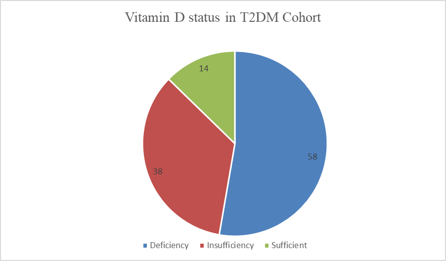
Figure 1: This pie chart represents vitamin D status in T2DM Cohort Study with deficient (52.7%), insufficient (34.5%) and sufficient (12.7%) that emphasize the high prevalence of suboptimal levels (87.3% combined deficiency and insufficiency)
To aim the primary objective, associations between sociodemographic factors and vitamin D status were examined. There were no such significant differences in vitamin D levels were observed across gender (mean in males: 22.1 ± 11.5 ng/mL compared with females: ` ± 11.0 ng/mL; p=0.45, independent t-test). Age showed a weak negative correlation with vitamin D levels (r = -0.12, p=0.21), suggesting minimal age-related decline in this cohort [Figure 2a]. Although, educational qualification did not significantly influence vitamin D status (ANOVA, p=0.32), with similar mean levels across groups (master's: 21.8 ± 11.4 ng/mL; bachelor's: 21.2 ± 10.9 ng/mL; secondary/higher secondary: 21.0 ± 11.6 ng/mL). [Figure 2b].
Marital status and social history (e.g., alcohol use) also showed no associations (chi-square test for categorical vitamin D status: p=0.68 for marital status; p=0.41 for alcohol use). Among females, menstrual status was not linked to vitamin D levels (ongoing cycle: 20.9 ± 10.8 ng/mL vs. postmenopausal: 20.4 ± 11.3 ng/mL; p=0.78). [Figure 2c].
These findings suggest that sociodemographic factors alone do not strongly predict vitamin D status in this population, potentially due to confounding urban lifestyle factors such as limited outdoor activity. [Figure 2].
|
Figure 2a |
|
Figure 2b |
|
|
Figure 2c Figure 2: Association of sociodemographic factor with vitamin D status. (a) Comparison of vitamin D levels across gender and correlation with age (r=-0.12, p=0.21). (b) Vitamin D levels across educational qualification groups (p=0.32). (c) Vitamin D levels in females according to menstrual status (p=0.78). No significant differences were observed, indicating that sociodemographic factors alone are not strong predictors of vitamin D status in this cohort, |
Occupational status was estimate and its impact on vitamin D levels, given its potential influence on sun exposure. Sedentary workers had a mean vitamin D level of 21.3 ± 11.2 ng/mL, homemakers/residents 21.7 ± 11.4 ng/mL, and field workers 21.4 ± 11.1 ng/mL. Spearman’s rank correlation revealed no significant association between occupation and vitamin D deficiency (ρ = -0.031, p=0.744). Prevalence of deficiency was comparable across groups: 53.0% in sedentary, 52.5% in homemakers, and 52.6% in field workers (chi-square, p=0.89). [Figure 3]. Self-reported work type included with day shift compared with night shift, and sun exposure duration did not correlate with vitamin D levels (Pearson’s r = -0.066, p=0.512 for sun exposure). This lack of association may reflect inconsistent sun exposure patterns or inadequate UVB penetration in urban environments.
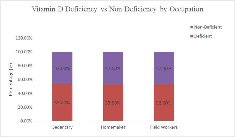
Figure 3: Vitamin D deficiency vs. non-deficiency across occupational categories. Bar chart shows deficiency prevalence among sedentary workers (53.0%), homemakers/residents (52.5%), and field workers (52.6%), compared with the remaining non-deficient group. No significant differences were observed across occupations (Spearman’s ρ = -0.031, p = 0.744; chi-square, p = 0.89).
Symptoms were self-reported and evaluated in connection with vitamin D status. Common symptoms among deficient participants included fatigue/lethargy (reported in ~35% of cases extracted from qualitative review), bone/joint pain (~28%), headache (~25%), and muscle weakness (~20%). Non-specific symptoms such as gastritis, hair fall, and numbness were also noted but not exclusively linked to deficiency. No significant correlation was found between symptom burden and vitamin D levels (Spearman’s ρ = 0.09, p=0.35), underscoring the non-specific nature of vitamin D deficiency manifestations [Figure 4].
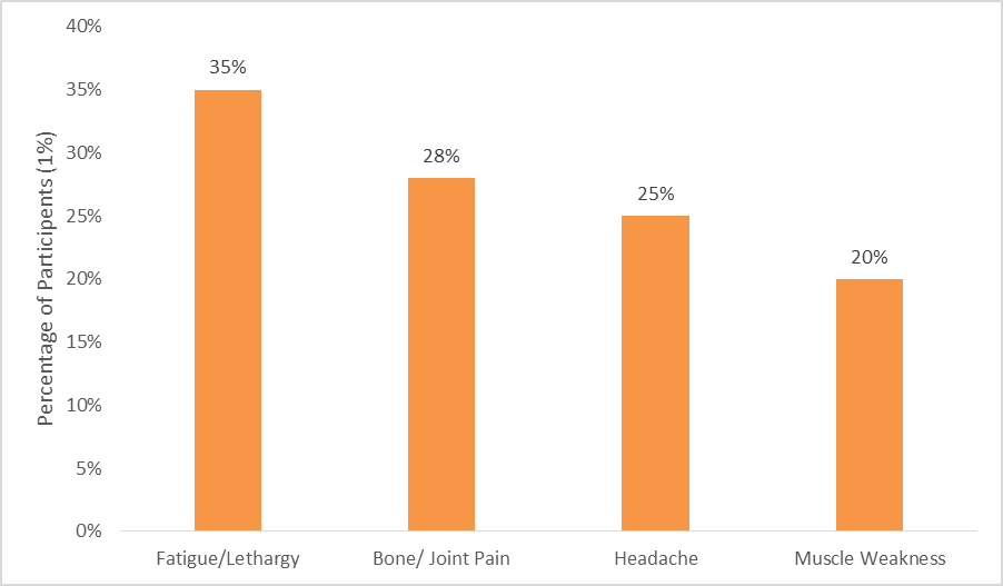
Figure 4: Common self-reported symptoms among participant with vitamin D deficiency. Fatigue/lethargy (35%) was the most frequently reported complaint followed by bone/joint pain (28%), headache (25%) and muscle weakness (20%).
Pearson’s correlation analysis ensures that no significant relationship between BMI and vitamin D levels (r = -0.004, p=0.970), nor between T2DM duration and vitamin D (r = -0.05, p=0.60). Family history of metabolic disorders (e.g., diabetes, hypertension) was prevalent but not associated with vitamin D status (chi-square, p=0.52). Exploratory non-parametric tests on social history (e.g., alcohol) reiterated no correlations (ρ = -0.085, p=0.382). Multidimentional linear regression, adjusting for age, gender, BMI, and sun exposure, explained only 8% of variance in vitamin D levels (R²=0.08, p=0.15), indicating other unmeasured factors (e.g., diet, genetics) may predominate.
In summary, this study reveals a high burden of vitamin D deficiency in T2DM patients attending a quaternary care hospital, with limited influence from sociodemographic and occupational factors. These findings align with the study's objectives and highlight opportunities for intervention. All analyses were performed using SPSS software, with p<0.05 considered significant.
DISCUSSION :
Vitamin D deficiency is widely recognized as a global health problem despite the availability of abundant sunlight in many regions. It plays an essential role in calcium and bone metabolism but also contributes to immune function, glucose regulation, and cardiovascular health. Deficiency arises from a combination of factors such as reduced outdoor activity, poor dietary intake, cultural clothing practices, and genetic influences. The reviewed studies provide important insights into how vitamin D deficiency interacts with metabolic and clinical outcomes across different populations(Kaur et al., 2025).
In patients with type 2 diabetes, nearly one third were found to be vitamin D deficient, yet no significant association was observed with cardiovascular disease or glycemic control (Al Zarooni et al., 2022; Kwiendacz et al., 2023). However, these patients were more frequently treated with sodium glucose cotransporter 2 inhibitors, raising the possibility that such medications could influence vitamin D status and bone health, which merits further exploration. In the Abu Dhabi population, deficiency was far more widespread, affecting close to eighty percent of individuals. Lifestyle factors such as low sun exposure, conservative clothing, poor diet, and physical inactivity were identified as major contributors, with younger people being more at risk due to modern indoor lifestyles and reliance on fast food (Mejaddam et al., 2023).
Deficiency in early life was demonstrated in Bangladeshi children, where despite sufficient sunlight, a large proportion were affected. Sun exposure, breastfeeding, and protective genetic variants were associated with better vitamin D status, while diet alone was insufficient to prevent deficiency. This highlights the need for public health initiatives promoting safe sun exposure and targeted supplementation (Das et al., 2022). In a South Indian cohort, vitamin D levels did not differ significantly between diabetic foot ulcer patients, diabetics without ulcers, and healthy controls. Furthermore, polymorphisms in the vitamin D receptor gene (FokI, TaqI, and ApaI) showed no direct association with ulcer development, though age and sex were linked to certain genetic variants. These findings suggest that ulcer risk is multifactorial and not solely determined by vitamin D status or receptor polymorphisms (Kurian et al., 2024).
Among elderly care home residents, over one third were deficient. While no effect was seen on muscle strength or mobility, deficiency was associated with higher bone turnover, increasing the risk of osteoporosis and fractures. This supports the need for routine vitamin D and calcium supplementation in this population to maintain skeletal health (Feehan et al., 2022). In Saudi Arabia, although mothers expressed generally favorable attitudes toward vitamin D, major gaps were evident in knowledge and practices regarding supplementation and sun exposure. Educational and socioeconomic status strongly influenced these behaviors, emphasizing the importance of culturally tailored public health strategies (Alharbi & Ahmad Rezq, 2025).
In relation to obesity and metabolic syndrome, several key findings emerged. Among obese worksite employees, nearly three fourths had metabolic syndrome. Low levels of eicosapentaenoic acid, an omega 3 fatty acid, and a high arachidonic acid to eicosapentaenoic acid ratio were significantly associated with insulin resistance and inflammation, whereas vitamin D levels showed no meaningful association. This suggests that fatty acid imbalance may be a stronger determinant than vitamin D in this context (Negi et al., 2022). In contrast, another study reported that deficiency was more prevalent among prediabetics and was strongly associated with fasting glucose, HbA1c, fasting insulin, and insulin resistance, especially in overweight and obese individuals. This supports the role of deficiency in impaired glucose metabolism, even though its effect on lipid profiles remained unclear (Atia et al., 2023).
In youth with obesity, branched chain and aromatic amino acids were significantly higher than in normal weight peers and were strongly linked to inflammation, visceral adiposity, and insulin resistance. A novel negative association between these amino acids and vitamin D was identified, pointing toward complex nutritional and metabolic interactions. Importantly, lifestyle intervention improved body composition but did not lower amino acid levels, indicating the need for more intensive or combined approaches to address these metabolic abnormalities (Cosentino et al., 2021).
Taken together, these studies demonstrate that vitamin D deficiency is highly prevalent across diverse populations but its impact differs depending on age, lifestyle, genetic factors, and comorbidities. It is strongly implicated in glucose metabolism, insulin resistance, and bone health, while its links to cardiovascular disease and lipid regulation appear less consistent. Preventive and therapeutic strategies should therefore adopt a population specific approach that integrates supplementation, safe sun exposure, dietary modification, and culturally appropriate education to reduce the burden of vitamin D deficiency and its associated risks.
In this study, more than half of the patients with type 2 diabetes were vitamin D deficient and another one third had insufficient levels, showing that the problem is highly prevalent in this population. Unlike some earlier reports, no significant link was found between vitamin D levels and factors such as age, gender, education, occupation, sun exposure, or lifestyle habits. This suggests that vitamin D status in these patients is not explained by common demographic or behavioral factors alone, but may be more strongly shaped by diet, genetics, or underlying metabolic mechanisms. Compared with other studies that showed associations with insulin resistance, bone health, or obesity-related inflammation, our findings point to a different picture where deficiency is common but not clearly tied to measurable social or lifestyle patterns. This highlights the complexity of vitamin D metabolism and the need for deeper exploration of hidden influences, such as genetic variations and dietary intake, in the Indian diabetic population. Overall, the study reinforces the importance of regular screening and preventive strategies even when obvious risk factors are not apparent.
CONCLUSION :
This study shows that vitamin D deficiency is very common in people with type 2 diabetes, with most participants having low or insufficient levels. The lack of strong links with lifestyle or social factors suggests that other influences, such as diet, genetics, and body metabolism, may play a bigger role. These results highlight the importance of regular vitamin D checks, better awareness, and practical steps like safe sun exposure, improved diet, and supplementation. More research is needed to understand exactly how vitamin D affects diabetes and to design strategies that can prevent deficiency and improve long-term health.
Acknowledgements
The authors express their sincere gratitude to all hospital staff and laboratory personnel for their invaluable support in conducting this study and ensuring accurate data collection. We also extend our special thanks to the Climed Care Internship Program for their assistance in manuscript development and drafting support.
BIBLIOGRAPHY:
1. Al Zarooni, A. A. R., Nagelkerke, N., Al Marzouqi, F. I., & Al Darmaki, S. H. (2022). Risk factors for vitamin D deficiency in Abu Dhabi Emirati population. PloS One, 17(2). https://doi.org/10.1371/JOURNAL.PONE.0264064
2. Alharbi, H. F., & Ahmad Rezq, K. (2025). Vitamin D supplementation in Saudi Arabia: mothers’ knowledge, practices, and attitudes. PeerJ, 13, e19781. https://doi.org/10.7717/PEERJ.19781/SUPP-3
3. Atia, T., Abdelzaher, M. H., Nassar, S. A., Gafar, H. H., Husseini, M. A. M., Kaabi, A. M. Y., & Sakr, H. I. (2023). Investigating the relationship between vitamin-D deficiency and glycemia status and lipid profile in nondiabetics and prediabetics in Saudi population. Medicine, 102(47), E36322. https://doi.org/10.1097/MD.0000000000036322
4. Bucurica, S., Prodan, I., Pavalean, M., Taubner, C., Bucurica, A., Socol, C., Calin, R., Ionita-Radu, F., & Jinga, M. (2023). Association of Vitamin D Deficiency and Insufficiency with Pathology in Hospitalized Patients. Diagnostics, 13(5), 998. https://doi.org/10.3390/DIAGNOSTICS13050998/S1
5. Cosentino, R. G., Churilla, J. R., Josephson, S., Molle-Rios, Z., Hossain, M. J., Prado, W. L., & Balagopal, P. B. (2021). Branched-chain Amino Acids and Relationship with Inflammation in Youth with Obesity: A Randomized Controlled Intervention Study. Journal of Clinical Endocrinology and Metabolism, 106(11), 3129–3139. https://doi.org/10.1210/clinem/dgab538
6. Cui, A., Xiao, P., Ma, Y., Fan, Z., Zhou, F., Zheng, J., & Zhang, L. (2022). Prevalence, trend, and predictor analyses of vitamin D deficiency in the US population, 2001–2018. Frontiers in Nutrition, 9, 965376. https://doi.org/10.3389/FNUT.2022.965376/BIBTEX
7. Das, S., Hasan, M. M., Mohsin, M., Jeorge, D. H., Rasul, M. G., Khan, A. R., Gazi, M. A., & Ahmed, T. (2022). Sunlight, dietary habits, genetic polymorphisms and vitamin D deficiency in urban and rural infants of Bangladesh. Scientific Reports, 12(1). https://doi.org/10.1038/S41598-022-07661-Y
8. Feehan, O., Armstrong, D. J., Magee, P. J., Pourshahidi, K. L., Strain, J. J., Beggan, L., Cobice, D. F., & McSorley, E. M. (2022). Vitamin D and Bone Health of Older Adults within Care Homes: An Observational Study. Nutrients, 14(13). https://doi.org/10.3390/NU14132680
9. Gordon, C. M., DePeter, K. C., Feldman, H. A., Grace, E., & Emans, S. J. (2004). Prevalence of vitamin D deficiency among healthy adolescents. Archives of Pediatrics & Adolescent Medicine, 158(6), 531–537. https://doi.org/10.1001/ARCHPEDI.158.6.531
10. Kaur, J., Khare, S., Sizar, O., & Givler, A. (2025). Vitamin D Deficiency. Endocrinology and Diabetes: A Problem Oriented Approach: Second Edition, 245–256. https://doi.org/10.1007/978-3-030-90684-9_23
11. Kurian, S. J., Baral, T., Benson, R., Munisamy, M., Saravu, K., Rodrigues, G. S., Sunil Krishna, M., Shetty, S., Kumar, A., & Miraj, S. S. (2024). Association of vitamin D status and vitamin D receptor polymorphism in diabetic foot ulcer patients: A prospective observational study in a South-Indian tertiary healthcare facility. International Wound Journal, 21(8). https://doi.org/10.1111/IWJ.70027
12. Kwiendacz, H., Nabrdalik, K., Wijata, A. M., Bartman, W., Tabor, E., Olejarz, A., Krol, J., Piaśnik, J., Nalepa, J., Stompor, T., Gumprecht, J., & Lip, G. Y. H. (2023). Relationship of vitamin D deficiency with cardiovascular disease and glycemic control in patients with type 2 diabetes mellitus: the Silesia Diabetes-Heart Project. Polish Archives of Internal Medicine, 133(6). https://doi.org/10.20452/PAMW.16445
13. Mejaddam, A., Höskuldsdóttir, G., Lenér, F., Wallenius, V., Trimpou, P., Fändriks, L., Mossberg, K., Eliasson, B., & Landin-Wilhelmsen, K. (2023). Effects of medical and surgical treatment on vitamin D levels in obesity. PLOS ONE, 18(12), e0292780. https://doi.org/10.1371/JOURNAL.PONE.0292780
14. Melguizo-Rodríguez, L., Costela-Ruiz, V. J., García-Recio, E., Luna-Bertos, E. De, Ruiz, C., & Illescas-Montes, R. (2021). Role of Vitamin D in the Metabolic Syndrome. Nutrients, 13(3), 1–17. https://doi.org/10.3390/NU13030830
15. Nair, P., Venkatesh, B., & Center, J. R. (2018). Vitamin D deficiency and supplementation in critical illness-the known knowns and known unknowns. Critical Care (London, England), 22(1). https://doi.org/10.1186/S13054-018-2185-8
16. Negi, P. C., Sharma, C. K., Nihjawan, R., Sharma, R., & Asotra, S. (2022). Role of omega 3 and omega 6 poly unsaturated fatty acids (PUFA) and vitamin D deficiency as risk determinants of metabolic syndrome in obesity: Worksite based case-control observational study. Diabetes & Metabolic Syndrome, 16(4). https://doi.org/10.1016/J.DSX.2022.102467
17. P, A., S, M., B, N., & SK, G. (2018). Vitamin D deficiency in India. Journal of Family Medicine and Primary Care, 7(2), 41-44+55. https://doi.org/10.4103/JFMPC.JFMPC_78_18
18. Park, J. E., Pichiah, P. B. T., & Cha, Y. S. (2018). Vitamin D and Metabolic Diseases: Growing Roles of Vitamin D. Journal of Obesity & Metabolic Syndrome, 27(4), 223–232. https://doi.org/10.7570/JOMES.2018.27.4.223
19. Siddiqee, M. H., Bhattacharjee, B., Siddiqi, U. R., & MeshbahurRahman, M. (2021). High prevalence of vitamin D deficiency among the South Asian adults: a systematic review and meta-analysis. BMC Public Health, 21(1). https://doi.org/10.1186/S12889-021-11888-1