A Comparative Study To Evaluate The Efficacy of Primary Closure Without Subcutaneous Drain Versus with Subcutaneous Drain In Preventing Surgical Site Infection In Hollow Viscus Perforation Undergoing Exploratory Laparotomyt
- Manogya Khanna , Department of General Surgery, Maharishi Markandeshwar Institute Of Medical Sciences & Research, Mullana, Ambala, India
Article Information:
Abstract:
Surgical site infection (SSI) is a common postoperative complication following exploratory laparotomy for hollow viscus perforation and is associated with increased morbidity, prolonged hospitalization, and higher healthcare costs. However, the ideal method of abdominal wall closure in contaminated surgical wounds remains a subject of debate.Aim & Objective:To evaluate and compare the outcomes of primary skin closure with and without placement of a subcutaneous suction drain in patients undergoing emergency exploratory laparotomy for hollow viscus perforation, with respect to SSI, seroma formation, wound dehiscence, and duration of hospital stay.Materials & Methods:This prospective case-control study included 50 patients who underwent emergency exploratory laparotomy for hollow viscus perforation. Patients were divided into two groups: Group A (n=25), in whom a subcutaneous suction drain was placed, and Group B (n=25), who underwent primary skin closure without drain placement. Baseline demographic and clinical characteristics were comparable between the groups. Postoperative outcomes, including SSI, seroma formation, wound dehiscence, and length of hospital stay, were recorded and statistically analyzed.Results: Patients in the drain group demonstrated lower incidences of SSI (12.0% vs. 36.0%; p=0.095), seroma formation (8.0% vs. 48.0%; p=0.002), and wound dehiscence (4.0% vs. 20.0%; p=0.189) compared to the no-drain group. The mean duration of hospital stay was also shorter in the drain group (10.32 vs. 11.88 days; p=0.153). Reduction in seroma formation was statistically significant, while decreases in SSI and wound dehiscence showed favorable trends without reaching statistical significance. Conclusion: The use of a subcutaneous suction drain after exploratory laparotomy for hollow viscus perforation significantly decreases seroma formation and may contribute to lower rates of SSI and wound dehiscence, thereby potentially improving postoperative outcomes.
Keywords:
Article :
INTRODUCTION:
Hollow viscus perforation is one of the most serious surgical emergencies encountered in general surgery because leakage of enteric contents into the peritoneal cavity rapidly produces peritonitis, sepsis, fluid imbalance, and organ dysfunction. Early resuscitation, antibiotics, source control, and timely surgery are therefore essential for survival [1]. In many patients with diffuse peritonitis or hemodynamic instability, exploratory laparotomy remains the standard life-saving procedure through which the perforation is identified, repaired, and the contaminated peritoneal cavity is irrigated [1].
The condition is particularly important in low- and middle-income settings, where patients often present late with severe sepsis, dehydration, malnutrition, anemia, and extensive peritoneal contamination. As a result, laparotomy wounds in hollow viscus perforation are created in contaminated or dirty operative fields, making postoperative wound healing difficult from the outset [2].
Unlike elective abdominal surgery, emergency laparotomy involves significant bacterial contamination, tissue edema, inflammatory exudate, and physiologic stress, all of which increase the risk of postoperative complications, especially surgical site infection (SSI) [3].
SSI is one of the most common and clinically significant complications after exploratory laparotomy for hollow viscus perforation. It may involve superficial tissues, deep fascial layers, or organ-space infection and can result in pain, purulent discharge, wound gaping, wound dehiscence, burst abdomen, delayed recovery, prolonged antibiotic use, repeated dressings, and extended hospital stay [4]. The WHO and CDC continue to emphasize SSI prevention as a major component of surgical quality and patient safety because contaminated emergency abdominal surgeries remain at particularly high risk despite advances in antibiotics and operative care [5].
The pathogenesis of SSI in perforation peritonitis is multifactorial. The subcutaneous plane of emergency midline laparotomy may accumulate blood, serum, liquefied fat, and inflammatory exudate, creating dead space that favors bacterial proliferation and impaired wound healing [6].
In addition, systemic factors such as sepsis, hypoalbuminemia, anemia, diabetes, obesity, delayed presentation, and prolonged operative time further compromise tissue healing. These factors have led surgeons to explore technical modifications in wound closure aimed at reducing postoperative wound complications [7].
One such strategy is placement of a subcutaneous suction drain before skin closure. The rationale is that the drain evacuates blood, serum, and contaminated fluid from the subcutaneous tissue, thereby reducing dead space, wound tension, and bacterial growth [7]. Closed suction drainage systems may also improve wound edge apposition and reduce the incidence of seroma, SSI, and wound dehiscence. Consequently, many surgeons use subcutaneous drains as a simple and low-cost adjunct in contaminated abdominal surgeries [8].
However, the routine use of drains remains controversial because drains do not eliminate the underlying causes of infection such as heavy contamination, tissue ischemia, malnutrition, and systemic sepsis. Some studies have also questioned their universal benefit and emphasized that drains should not replace meticulous surgical technique and standard SSI prevention measures [9].
The question becomes especially relevant in patients undergoing primary closure after exploratory laparotomy for hollow viscus perforation. While primary closure offers advantages such as faster healing, easier wound care, and earlier mobilization, the surgeon must decide whether closure should be performed with or without a subcutaneous drain in contaminated wounds. Even small reductions in SSI can significantly decrease hospital stay, dressing burden, antibiotic use, and healthcare costs, particularly in resource-limited settings [10].
Recent studies in perforation peritonitis and emergency laparotomy have reported lower rates of SSI, seroma, and shorter hospital stay with subcutaneous suction drainage, although the effect on wound dehiscence has not always been statistically significant [11].
Therefore, a focused comparative evaluation of primary closure with and without subcutaneous suction drainage is clinically important. Since hollow viscus perforation patients constitute a high-risk group for postoperative wound complications, identifying the more effective wound closure strategy may improve postoperative recovery, reduce SSI and wound-related morbidity, shorten hospital stay, and help standardize operative practice in emergency abdominal surgery [10,12].
Hollow viscus perforation is associated with a high incidence of postoperative SSI because exploratory laparotomy is performed in contaminated or dirty operative fields in patients already compromised by sepsis, malnutrition, anemia, and fluid imbalance. SSI in such patients contributes to wound discharge, seroma formation, wound dehiscence, prolonged hospitalization, repeated dressings, and increased healthcare costs [13].
Although subcutaneous suction drainage is commonly used to reduce dead space and evacuate contaminated collections, available evidence regarding its effectiveness remains inconclusive. Therefore, this study was undertaken to compare primary skin closure with and without subcutaneous suction drain in patients undergoing exploratory laparotomy for hollow viscus perforation, with the aim of evaluating their impact on SSI, wound dehiscence, and duration of hospital stay [10,14].
MATERIALS AND METHODS:
This prospective case-control study was conducted in patients admitted to the Department of General Surgery with a clinical diagnosis of hollow viscus perforation requiring emergency exploratory laparotomy. Eligible patients were recruited consecutively during the study period until the required sample size was achieved. A total of 50 adult patients (>18 years) of both sexes who fulfilled the inclusion criteria were enrolled and divided into two groups based on the abdominal wall closure technique used. Group A included 25 patients who underwent primary closure with placement of a subcutaneous suction drain, while Group B included 25 patients who underwent primary closure without a subcutaneous drain. Patients undergoing emergency exploratory laparotomy for hollow viscus perforation were included in the study, whereas pediatric patients and those undergoing elective abdominal surgeries were excluded.
RESULT:
Table 1: Distribution of patients according to age group (n = 50)
|
Age group (years) |
Frequency |
Percentage |
|
18–27 |
7 |
14.0% |
|
28–37 |
9 |
18.0% |
|
38–47 |
8 |
16.0% |
|
48–57 |
8 |
16.0% |
|
58–67 |
11 |
22.0% |
|
68–77 |
6 |
12.0% |
|
78–87 |
1 |
2.0% |
|
Total |
50 |
100% |
Table 1 analyses that the majority of patients belonged to the 58–67 years age group (22.0%), followed by the 28–37 years age group (18.0%). Patients were relatively evenly distributed across other age groups, with the least number of patients in the 78–87 years category (2.0%). Overall, the study population represented a wide age distribution ranging from young adults to elderly individuals.
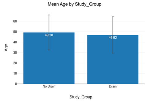
Figure 1: Comparison of age distribution between drain and no-drain groups
Figure 1. The mean age was 46.92 ± 16.78 years in the drain group and 49.28 ± 17.41 years in the no-drain group. There was no significant difference in age distribution between the groups (p = 0.628), with homogeneity of variances confirmed by Levene’s test (p = 0.915). The small effect size (Cohen’s d = 0.14) indicated that both groups were comparable with respect to age.
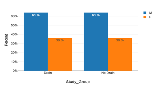
Figure 2: Sex distribution in the drain and no-drain groups.
Figure 2 evaluates a sex distribution was identical in both groups, with males comprising 64.0% and females 36.0% of patients in each group. No significant association was observed between study group and sex (p = 1.000), indicating complete comparability between the drain and no-drain groups.
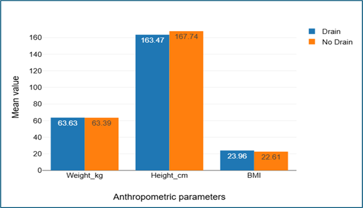
Figure 3: Bar diagram depicts the comparison of mean weight, height, and body mass index between the drain and no-drain groups.
Figure 3 examines the mean body weight and BMI were comparable between the drain and no-drain groups, with no statistically significant differences in weight (63.63 vs 63.39 kg; p = 0.907) or BMI (23.96 vs 22.61 kg/m²; p = 0.144). However, mean height was significantly lower in the drain group compared to the no-drain group (163.47 vs 167.74 cm; p = 0.026). Overall, the anthropometric characteristics of both groups were broadly comparable, with only height showing a significant difference.
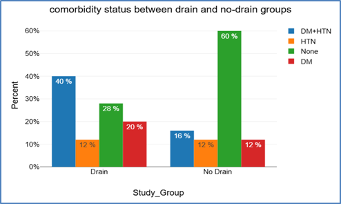
Figure 4: Comparison of comorbidity status between drain and no-drain groups
Figure 4 focuses specifically on diabetes mellitus with hypertension was more common in the drain group, while more patients without comorbidities were observed in the no-drain group. However, the overall distribution of comorbidities was broadly comparable between the groups, with no statistically significant association found on chi-square or Fisher’s exact testing (p > 0.05).
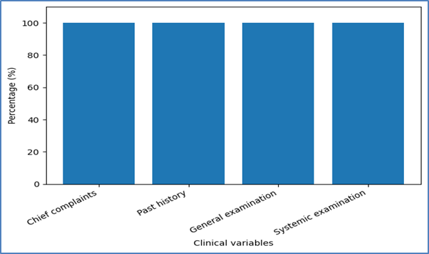
Figure 5: Clinical presentation and examination findings of the study population.
Figure 5 shows that all patients in both groups presented with abdominal pain, distension, vomiting, toxic dehydration, and generalized abdominal tenderness, with no significant past medical or surgical illness. As these findings were uniform across both groups, no comparative statistical analysis was performed.
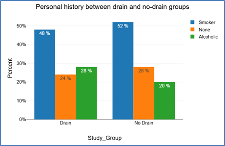
Figure 6: Comparison of personal history between drain and no-drain groups
Smoking was the most common personal habit in both groups, followed by alcohol consumption, while some patients reported no habits. The distribution of personal history was comparable between the drain and no-drain groups, with no statistically significant association observed (p = 0.798; Cramér’s V = 0.09) (Figure 6).
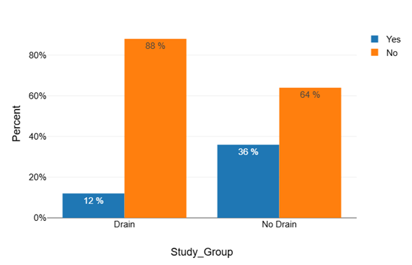
Figure 7: Comparison of surgical site infection (SSI) between drain and no-drain groups
Figure 7 shows that surgical site infection occurred in 12.0% of patients in the drain group compared to 36.0% in the no-drain group, indicating a clinically lower SSI rate with subcutaneous suction drainage. Although the chi-square test showed statistical significance (p = 0.047), Fisher’s exact test did not demonstrate a statistically significant association (p = 0.095).
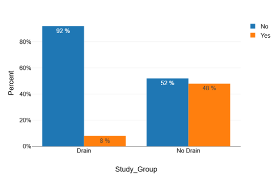
Fig 8: Comparison of seroma formation between drain and no-drain groups
Figure 8 shows that seroma formation was significantly lower in the drain group (8.0%) compared to the no-drain group (48.0%). Both chi-square (p = 0.002) and Fisher’s exact test (p = 0.004) demonstrated a statistically significant association, indicating that subcutaneous suction drainage significantly reduced postoperative seroma formation.
Table 2: Comparison of wound dehiscence between drain and no-drain groups
|
Wound dehiscence |
Drain n (%) |
No Drain n (%) |
Total n (%) |
|
No |
24 (96.0%) |
20 (80.0%) |
44 (88.0%) |
|
Yes |
1 (4.0%) |
5 (20.0%) |
6 (12.0%) |
|
Total |
25 (100%) |
25 (100%) |
50 (100%) |
|
chi-square test, p = 0.082 |
|||
Wound dehiscence occurred in 4.0% of patients in the drain group compared to 20.0% in the no-drain group, showing a lower incidence with subcutaneous suction drainage. However, Fisher’s exact test did not demonstrate a statistically significant association (p = 0.189), although a moderate association was observed (Cramér’s V = 0.25) (Table 2).
Table 3: Distribution of HHH status between drain and no-drain groups
|
HHH status |
Drain n (%) |
No drain n (%) |
Total n (%) |
|
Negative |
24 (96.0%) |
22 (88.0%) |
46 (92.0%) |
|
HCV positive |
1 (4.0%) |
1 (4.0%) |
2 (4.0%) |
|
HIV positive |
0 (0.0%) |
1 (4.0%) |
1 (2.0%) |
|
HBsAg positive |
0 (0.0%) |
1 (4.0%) |
1 (2.0%) |
|
Total |
25 (100%) |
25 (100%) |
50 (100%) |
|
χ²-test, p = 0.555 |
|||
Most patients in both groups were HHH negative, with only a few patients positive for HCV, HIV, or HBsAg. The distribution of HHH status was comparable between the drain and no-drain groups, with no statistically significant association observed (p > 0.05) (Table 3).
Table 4: Distribution of drain duration among patients in the drain group
|
Frequency |
Mean |
Std. Deviation |
Minimum |
Maximum |
||
|
Drain_Duration_days |
Drain |
25 |
5.44 |
0.77 |
4 |
7 |
|
No Drain |
25 |
- |
- |
- |
- |
The mean duration of subcutaneous suction drain placement was 5.44 ± 0.77 days, with a range of 4 to 7 days, indicating relatively uniform drain usage among patients in the drain group. As drain duration was applicable only to the drain group, no comparative statistical analysis was performed (Table 4).
Table 5: Comparison of duration of hospital stay between drain and no-drain groups
|
Study group |
Frequency |
Mean (days) |
Std. Deviation |
Minimum |
Maximum |
|
Drain |
25 |
10.32 |
3.28 |
3 |
16 |
|
No drain |
25 |
11.88 |
4.26 |
3 |
19 |
|
t-test, p = 0.153 |
|||||
The mean hospital stay was shorter in the drain group (10.32 days) compared to the no-drain group (11.88 days). However, the difference was not statistically significant (p = 0.153), indicating that duration of hospital stay was broadly comparable between the two groups (Table 5).
Table 6: Distribution of type of incision wound between drain and no-drain groups
|
Type of incision wound |
Drain n (%) |
No Drain n (%) |
Total n (%) |
|
Clean-contaminated |
4 (16.0%) |
4 (16.0%) |
8 (16.0%) |
|
Contaminated |
13 (52.0%) |
11 (44.0%) |
24 (48.0%) |
|
Dirty |
8 (32.0%) |
10 (40.0%) |
18 (36.0%) |
|
Total |
25 (100%) |
25 (100%) |
50 (100%) |
|
chi-square test, p = 0.823 |
|||
The distribution of incision wound types was comparable between the drain and no-drain groups, with contaminated wounds being the most common, followed by dirty and clean-contaminated wounds. No statistically significant association was observed between study group and wound type (p = 0.823; Cramér’s V = 0.09) (Table 6).
Table 7: Association between type of incision wound and surgical site infection
|
Type of Incision Wound |
SSI in Group A (Drain) |
SSI in Group B (No Drain) |
z-proportion |
p-value |
||
|
Present |
Absent |
Present |
Absent |
|||
|
Clean-Contaminated |
1 |
3 |
0 |
4 |
1.07 |
.285 |
|
Contaminated |
1 |
12 |
6 |
5 |
2.06 |
.039* |
|
Dirty |
1 |
7 |
3 |
7 |
0.89 |
.373 |
|
Total |
3 |
22 |
9 |
16 |
1.99 |
.047* |
Surgical site infection was less frequent in the drain group compared to the no-drain group across contaminated and dirty wounds. Overall, SSI occurred in 3 patients in the drain group and 9 patients in the no-drain group, with the difference being statistically significant (p = 0.047), indicating a lower incidence of infection with subcutaneous drain placement (Table 7).
Table 8: Association between type of incision wound and seroma formation
|
Type of Incision Wound |
Seroma in Group A (Drain) |
Seroma in Group B (No Drain) |
z-proportion |
p-value |
||
|
Present |
Absent |
Present |
Absent |
|||
|
Clean-Contaminated |
0 |
4 |
1 |
3 |
0.73 |
.463 |
|
Contaminated |
2 |
11 |
6 |
5 |
1.78 |
.075 |
|
Dirty |
0 |
8 |
5 |
5 |
2.31 |
.021* |
|
Total |
2 |
23 |
12 |
13 |
3.15 |
.002 ** |
Seroma formation was markedly lower in the drain group across all wound types. Overall, seroma occurred in 2 patients in the drain group compared to 12 patients in the no-drain group, with the difference being statistically highly significant (p = 0.002), indicating that subcutaneous drain placement significantly reduced postoperative seroma formation (Table 8).
Table 9: Association between type of incision wound and wound dehiscence
|
Type of Incision Wound |
Wound Dehiscence in Group A (Drain) |
Wound Dehiscence in Group B (No Drain) |
z-proportion |
p-value |
||
|
Present |
Absent |
Present |
Absent |
|||
|
Clean-Contaminated |
0 |
4 |
0 |
4 |
— |
— |
|
Contaminated |
1 |
12 |
3 |
8 |
0.87 |
.383 |
|
Dirty |
0 |
8 |
2 |
8 |
1.47 |
.141 |
|
Total |
1 |
24 |
5 |
20 |
1.56 |
.118 |
Wound dehiscence was less frequent in the drain group than in the no-drain group across contaminated and dirty wounds. Overall, wound dehiscence occurred in 1 patient in the drain group and 5 patients in the no-drain group; however, the difference was not statistically significant (p = 0.118) (Table 9).
DISCUSSION :
Surgical site infection (SSI) remains one of the most frequent and clinically significant complications following abdominal surgery because it increases postoperative morbidity, prolongs hospitalization, increases healthcare expenditure, and delays recovery [17]. According to the World Health Organization, approximately 11% of surgical patients in low- and middle-income countries develop SSI, while the CDC reports that SSI constitutes nearly 20% of healthcare-associated infections and is associated with increased mortality and prolonged hospital stay [20]. The problem becomes particularly important in emergency gastrointestinal surgery, where patients commonly present late with diffuse peritonitis, severe contamination, sepsis, dehydration, and poor nutritional status. Such conditions make wound-related complications especially common after exploratory laparotomy for hollow viscus perforation.
Hollow viscus perforation is a life-threatening surgical emergency resulting from disruption of gastrointestinal tract integrity with leakage of bowel contents into the peritoneal cavity, leading to secondary peritonitis and systemic sepsis [21]. Emergency surgical guidelines emphasize urgent operative intervention and effective source control because delayed management significantly worsens outcomes [22]. In many patients, exploratory laparotomy remains the standard life-saving procedure, particularly in cases with generalized peritonitis and gross contamination [23]. However, closure of laparotomy wounds in contaminated or dirty abdominal fields remains a major surgical challenge because postoperative SSI, seroma formation, and wound dehiscence are common complications [24].
A key technical issue during abdominal wall closure is management of the subcutaneous plane. After fascial closure, residual dead space may accumulate blood, serum, liquefied fat, and inflammatory exudate, providing a favorable environment for bacterial proliferation and impaired wound healing [15]. Subcutaneous suction drainage has therefore been proposed as a method to reduce fluid accumulation, decrease tissue tension, and minimize superficial wound infection. Nevertheless, evidence regarding its effectiveness has remained inconsistent. Some studies and meta-analyses have questioned the routine use of subcutaneous drains in abdominal surgery [25], whereas more recent evidence has demonstrated lower SSI rates and shorter hospital stay with drainage, particularly in gastrointestinal and emergency contaminated surgeries [26]. Therefore, the role of subcutaneous drainage in hollow viscus perforation remains clinically relevant and warrants focused evaluation.
The present study compared primary skin closure with and without subcutaneous suction drainage in patients undergoing exploratory laparotomy for hollow viscus perforation. The findings demonstrated that the drain and no-drain groups were well matched in terms of baseline demographic characteristics, thereby reducing confounding factors. Mean age was comparable between the two groups (46.92 ± 16.78 vs 49.28 ± 17.41 years; p=0.628), and both groups showed identical male predominance (64%; p=1.000). Similar demographic comparability has been reported by Mallaiah et al. and Nawab et al., who also observed no significant baseline differences between drain and no-drain groups in emergency laparotomy patients [10,27]. This demographic balance strengthens the reliability of our comparison and suggests that postoperative outcome differences were likely related to the intervention itself rather than baseline variation.
Anthropometric and clinical characteristics were also largely comparable between groups. Weight and BMI showed no significant difference, although height was greater in the no-drain group (p=0.026). Comparable BMI and comorbidity profiles between study groups have similarly been reported by Farooq et al. and Mallaiah et al. [10,28]. Our cohort also demonstrated a clinically severe but homogeneous presentation, with generalized abdominal pain, distension, vomiting, dehydration, and diffuse tenderness present in most patients. Previous studies by Chandran et al. and Neupane et al. have similarly described hollow viscus perforation as a high-burden emergency condition commonly associated with severe peritonitis and postoperative wound complications [16,22].
The most important finding of our study was the significantly lower incidence of postoperative wound morbidity in patients managed with subcutaneous suction drainage. SSI occurred in only 12% of patients in the drain group compared with 36% in the no-drain group (p=0.047). Seroma formation was also significantly reduced in the drain group (8% vs 48%; p=0.002). These findings are consistent with recent studies supporting the role of subcutaneous drainage in contaminated laparotomy wounds. Mallaiah et al. reported significantly lower SSI, seroma, and wound discharge rates with subcutaneous suction drainage in emergency laparotomy wounds [10]. Similarly, Ishinuki et al., in a systematic review and meta-analysis of randomized controlled trials, found significantly lower overall SSI rates in drained patients compared with controls [18]. Although their review did not demonstrate a significant reduction in seroma formation overall, the marked seroma reduction observed in our study may reflect the particularly heavy contamination associated with hollow viscus perforation, where evacuation of infected exudate becomes especially beneficial.
Our findings also demonstrated a lower incidence of wound dehiscence in the drain group compared with the no-drain group (4% vs 20%), although the difference did not reach statistical significance (p=0.082). Similar trends have been reported in contemporary literature. Mallaiah et al. observed wound dehiscence rates of 4% in the drain group compared with 16% in controls [10]. Likewise, Tagar et al. reported fewer wound dehiscence cases with subcutaneous drainage, although statistical significance was not consistently achieved [19]. These findings suggest that while subcutaneous drainage may improve wound healing and reduce tension-related complications, larger sample sizes may be required to demonstrate statistically significant differences in wound dehiscence outcomes.
The duration of postoperative hospital stay was shorter in the drain group (10.32 ± 3.28 days) than in the no-drain group (11.88 ± 4.26 days), although this difference was not statistically significant (p=0.153). This trend is consistent with studies by Mallaiah et al. and Tagar et al., both of whom reported reduced hospital stay in patients managed with subcutaneous drainage [10,19]. Improved wound condition, reduced seroma formation, and lower SSI rates likely contribute to earlier recovery and discharge in drained patients.
An important observation in our study was that the protective effect of drainage was most evident in contaminated wounds. Contaminated wounds constituted the largest wound category overall, and SSI in this subgroup was significantly lower in patients with drainage compared to those without drainage (1/13 vs 6/11; p=0.039). Similar findings have been reported by Mallaiah et al. and Khan et al., who concluded that subcutaneous suction drainage was particularly beneficial in class III and class IV abdominal wounds characterized by high bacterial contamination [10,19]. The mechanism likely relates to evacuation of inflammatory exudate, reduction of dead space, and prevention of bacterial proliferation within the subcutaneous plane.
The present study further demonstrated that the greatest benefit of subcutaneous drainage was observed in prevention of seroma formation. Seroma was significantly reduced overall and particularly in dirty wounds, where no patient with drainage developed seroma compared with multiple cases in the no-drain group. Comparable findings have been described by Mallaiah et al. and Tagar et al., who also found significant reductions in seroma formation with subcutaneous drainage, while reductions in wound dehiscence were less consistent [10,19]. These observations suggest that the principal advantage of subcutaneous suction drainage lies in effective evacuation of postoperative fluid collections and obliteration of dead space, thereby improving local wound conditions and indirectly reducing SSI risk.
Overall, the findings of the present study support the use of subcutaneous suction drainage following exploratory laparotomy for hollow viscus perforation. The study demonstrated significantly lower rates of SSI and seroma formation, along with favorable trends toward reduced wound dehiscence and shorter hospital stay in the drain group. These results are consistent with recent evidence suggesting that subcutaneous drainage may be particularly beneficial in contaminated and dirty emergency laparotomy wounds. Although larger multicentric studies are required for further validation, the present study indicates that subcutaneous suction drainage is a simple, cost-effective, and clinically useful adjunct in reducing postoperative wound complications after emergency laparotomy for hollow viscus perforation [10,18,28].
CONCLUSION :
The present prospective case-control study titled “A Comparative Study to Evaluate the Efficacy of Primary Closure Without Subcutaneous Drain Versus With Subcutaneous Drain in Preventing Surgical Site Infection in Hollow Viscus Perforation Undergoing Exploratory Laparotomy” was conducted to evaluate the role of subcutaneous suction drainage in reducing postoperative wound complications in contaminated emergency laparotomy wounds. A total of 50 patients with hollow viscus perforation undergoing exploratory laparotomy were divided into two equal groups: patients managed with subcutaneous suction drain placement and those undergoing primary closure without drain. Both groups received comparable perioperative management. The study demonstrated that placement of a subcutaneous suction drain significantly reduced seroma formation and was associated with lower rates of surgical site infection, wound dehiscence, and shorter hospital stay compared with primary closure without drain. These findings suggest that subcutaneous suction drainage is a simple, safe, and cost-effective adjunct that may improve postoperative wound outcomes in patients undergoing emergency exploratory laparotomy for hollow viscus perforation.
BIBLIOGRAPHY:
1. Tarasconi A, Coccolini F, Biffl WL, Tomasoni M, Ansaloni L, Picetti E, et al. Perforated and bleeding peptic ulcer: WSES guidelines. World J Emerg Surg. 2020;15(1):1-24.
2. Rickard J, Beilman G, Forrester J, Sawyer R, Stephen A, Weiser TG, et al. Surgical infections in low- and middle-income countries: a global assessment of the burden and management needs. Surg Infect (Larchmt). 2020;21(6):478-494.
3. Singh M, Kumar A, Singh KP, Singh R, Singh K, Singh R. Prospective randomized control study to evaluate the effectiveness of hypochlorous acid as a peritoneal lavage agent to prevent surgical site infection after exploratory laparotomy for perforation peritonitis. Cureus. 2025;17(11):1-16.
4. Bozzay JD, Walker PF, Schechtman DW, Shaikh F, Stewart L, Carson ML, et al. Risk factors for abdominal surgical site infection after exploratory laparotomy among combat casualties. J Trauma Acute Care Surg. 2021;91(2):247-255.
5. Berríos-Torres SI, Umscheid CA, Bratzler DW, Leas B, Stone EC, Kelz RR, et al. Centers for Disease Control and Prevention guideline for the prevention of surgical site infection, 2017. JAMA Surg. 2017;152(8):784-791.
6. Tweedle E. Postoperative complications. In: Ellis and Calne's Lecture Notes in General Surgery. 14th ed. Hoboken: Wiley-Blackwell; 2023. p. 27-48.
7. Francis DM. Surgical techniques. In: Textbook of Surgery. 3rd ed. London: Elsevier; 2006. p. 27-36.
8. Harish R, Kazi FN, Sharma JP. Efficacy of subcutaneous closed suction drain in reduction of postoperative surgical site infection. Surg J (N Y). 2021;7(4):275-280.
9. Khan S, Rai P, Misra G. Is prophylactic drainage of peritoneal cavity after gut surgery necessary? A non-randomized comparative study from a teaching hospital. J Clin Diagn Res. 2015;9(10):1-3.
10. Mallaiah B, Ramachandra J. A comparative study on subcutaneous negative suction drain vs no drain in emergency laparotomy wounds for peritonitis. Cureus. 2025;17(5):1-9.
11. Singh S, Tandup C, Singh H, Kumar H, Khare S, Sahu S, et al. A prospective, non-randomized study to determine the role of intraperitoneal drain placement in perforation peritonitis. Ulus Travma Acil Cerrahi Derg. 2022;28(10):1397-1403.
12. Leaper D, Edmiston C. World Health Organization: global guidelines for the prevention of surgical site infection. J Hosp Infect. 2017;95(2):135-136.
13. Russo V, Leaptrot D, Otis M, Smith H, Hebden JN, Wright MO. Health care-associated infections studies project: an American Journal of Infection Control and National Healthcare Safety Network data quality collaboration case study—chapter 9 surgical site infection event case study. Am J Infect Control. 2022;50(7):799-800.
14. Solomkin J, Gastmeier P, Bischoff P, Latif A, Berenholtz S, Egger M, et al. WHO global guidelines for the prevention of surgical site infection. Lancet Infect Dis. 2017;17(3):262-264.
15. Huang Q, Li J, Lau WY. Techniques for abdominal wall closure after damage control laparotomy: from temporary abdominal closure to early/delayed fascial closure—a review. Gastroenterol Res Pract. 2016;2016:1-15.
16. Agrawal SN, Singh K. A prospective study of single layer abdominal wall closure in the tertiary care hospital. Int Surg J. 2019;6(5):1693-1697.
17. Neupane S, Koirala DP, Kharel S, Silwal S, Yadav KK. Clinical profile and management of perforation peritonitis in Bharatpur Hospital, Nepal: a prospective study. Ann Med Surg (Lond). 2022;82:1-5.
18. Aga E, Keinan-Boker L, Eithan A, Mais T, Rabinovich A, Nassar F. Surgical site infections after abdominal surgery: incidence and risk factors. Infect Dis (Lond). 2015;47(11):761-767.
19. Ishinuki T, Shinkawa H, Kouzu K, Shinji S, Goda E, Ohyanagi T, et al. Recent evidence for subcutaneous drains to prevent surgical site infections after abdominal surgery: a systematic review and meta-analysis. World J Gastrointest Surg. 2023;15(12):2879-2891.
20. Tagar E, Kpolugbo J, Dongo AE, Osime C, Eshiobo I, Irabor D. Abdominal wound closure in the presence of sepsis: our experience with the use of subcutaneous drain. Ghana Med J. 2024;58(1):26-32.
21. Monahan M, Jowett S, Pinkney T, Brocklehurst P, Morton DG, Abdali Z, et al. Surgical site infection and costs in low- and middle-income countries: a systematic review of the economic burden. PLoS One. 2020;15(6):1-9.
22. Chandran M, Shankar A, Krishnan K, Sundar M, Sundar SRM, KG M. A study on hollow viscus perforation in a tertiary care hospital in South India. Cureus. 2024;16(10):e00000.
23. Sartelli M, Chichom-Mefire A, Labricciosa FM, Hardcastle T, Abu-Zidan FM, Adesunkanmi AK, et al. The management of intra-abdominal infections from a global perspective: 2017 WSES guidelines for management of intra-abdominal infections. World J Emerg Surg. 2017;12(1):29-63.
24. Coccolini F, Sartelli M, Sawyer R, Rasa K, Viaggi B, Abu-Zidan F, et al. Source control in emergency general surgery: WSES, GAIS, SIS-E, SIS-A guidelines. World J Emerg Surg. 2023;18(1):41-58.
25. Mogoanta SS, Paitici S, Mogoanta CA. Postoperative follow-up and recovery after abdominal surgery. In: Abdominal Surgery-A Brief Overview. London: IntechOpen; 2021. p. 1-15.
26. Grönroos-Korhonen M. Failure to rescue for severe complications in colorectal surgery [dissertation]. Helsinki: University of Helsinki; 2025:1-111.
27. Nawab U, Ashraf I, Ayub A, Tahir M, Nawab M, Javed H. Comparison of frequency of surgical site infection in patients of peritonitis undergoing laparotomy with closed wound and drain placed vs open wound without drain. Pak Armed Forces Med J. 2025;75:763-767.
28. Farooq H, Shami AI, Haq BU. Subcutaneous suction drain to reduce surgical site infection in contaminated cases of emergency laparotomy: a randomized controlled trial. Ann PIMS-Shaheed Zulfiqar Ali Bhutto Med Univ. 2025;21(4):683-688.