Comparative Evaluation of Radiation Dose Parameters in Digital Breast Tomosynthesis and Full-Field Digital Mammography Across Breast Density Categories: A Retrospective Study from Pakistan
- Sana Shaikh , Associate Professor, Department of Radiology Sindh Institute of Urology and Transplantation (SIUT), Karachi, Pakistan
- Kashaf Anwar Arain , Assistant Professor, Department of Radiology Sindh Institute of Urology and Transplantation (SIUT), Karachi, Pakistan
- Ameet Lalwani , Associate Professor, Department of Radiology Sindh Institute of Urology and Transplantation (SIUT), Karachi, Pakistan
- Bisma Mustafa , Resident, Department of Radiology Sindh Institute of Urology and Transplantation (SIUT), Karachi, Pakistan
- Syeda Nuzhat Zehra , BSMT, Department of Radiology Sindh Institute of Urology and Transplantation (SIUT), Karachi, Pakistan
- Sukaina Jafri , BSMT, Department of Radiology Sindh Institute of Urology and Transplantation (SIUT), Karachi, Pakistan
- Syed Muhammad Faiq , Professor, Department of Radiology Sindh Institute of Urology and Transplantation (SIUT), Karachi, Pakistan
Article Information:
Abstract:
Digital Breast Tomosynthesis (DBT) has emerged as an advanced imaging modality with improved diagnostic accuracy over Full-Field Digital Mammography (FFDM), particularly in dense breast tissue. However, concerns persist regarding increased radiation exposure associated with DBT.Objective:To compare radiation dose parameters—Average Glandular Dose (AGD), Entrance Surface Dose (ESD), and Exposure Index (EI)—between DBT and FFDM across different breast density categories in a Pakistani population.Methods:This retrospective observational study analyzed 1,600 mammographic images from 200 female patients (December 2018–December 2022). All patients underwent bilateral imaging in COMBO mode using a Hologic Selenia Dimensions system. Breast density was categorized according to the American College of Radiology (ACR) classification (A–D). Radiation parameters (AGD, ESD, EI) were evaluated for both modalities in craniocaudal (CC) and mediolateral oblique (MLO) views. Statistical analysis was performed using SPSS version 22.0, with p<0.05 considered significant.Results: DBT demonstrated significantly higher radiation dose parameters compared to FFDM across all breast density groups (p<0.001). The highest AGD (0.50 mSv) and ESD (0.74 mSv) were observed in the DBT MLO view of Group A. Group D exhibited the highest ESD overall (0.27 mSv). Exposure Index values were consistently higher in DBT, reflecting increased detector exposure. Dose variations were influenced by breast density and imaging projection, with MLO views generally associated with higher radiation levels.Conclusion:DBT is associated with increased radiation dose compared to FFDM; however, its diagnostic advantages may justify its use, particularly in selected patient groups. Optimization of imaging protocols and adherence to radiation safety principles are essential to balance diagnostic benefit with patient safety.
Keywords:
Article :
INTRODUCTION:
Breast cancer remains the most commonly diagnosed malignancy among women worldwide and a leading cause of cancer-related mortality, accounting for substantial global disease burden despite advances in screening and treatment strategies [1]. Early detection through imaging-based screening programs has significantly contributed to improved survival outcomes; however, optimizing diagnostic accuracy while minimizing potential harms such as radiation exposure continues to be a critical challenge in breast imaging [2].
Full-Field Digital Mammography (FFDM) has long been established as the standard modality for breast cancer screening due to its accessibility, cost-effectiveness, and proven efficacy in reducing mortality [3]. Nevertheless, its diagnostic performance is limited in women with dense breast tissue, where overlapping fibroglandular structures can obscure lesions, resulting in reduced sensitivity and higher false-negative rates. Studies have reported that FFDM may miss up to 15–30% of breast cancers, particularly in patients with higher breast density [4].
Digital Breast Tomosynthesis (DBT), a three-dimensional imaging technique, has emerged as a promising advancement designed to overcome the limitations of conventional mammography. By acquiring multiple low-dose projections and reconstructing them into thin slices, DBT reduces tissue overlap and enhances lesion conspicuity, leading to improved cancer detection rates and reduced recall rates [5,6]. Recent large-scale studies and meta-analyses have demonstrated that DBT significantly improves sensitivity and specificity compared to FFDM, particularly in women with heterogeneously dense and extremely dense breasts [7].
Despite these diagnostic advantages, DBT has raised concerns regarding increased radiation exposure. The average glandular dose (AGD), a key indicator of radiation absorbed by radiosensitive breast tissue, is generally higher in DBT compared to FFDM, especially when both modalities are used in combination [8]. Similarly, entrance surface dose (ESD) and exposure index (EI) are critical parameters that reflect patient dose and image quality, and their optimization is essential to ensure adherence to the “As Low As Reasonably Achievable” (ALARA) principle [9].
While several international studies have explored radiation dose variations between DBT and FFDM, most data originate from high-income countries, and there is a paucity of region-specific evidence from low- and middle-income settings such as Pakistan. Variations in patient demographics, breast density distribution, equipment calibration, and imaging protocols may significantly influence radiation dose and diagnostic outcomes [10]. Furthermore, limited studies have simultaneously evaluated multiple dosimetric parameters (AGD, ESD, and EI) across different breast density categories and imaging projections.
Therefore, this study aims to provide a comprehensive comparative evaluation of radiation dose parameters between DBT and FFDM across varying breast densities in a Pakistani population. By analyzing dose variations in different projections and tissue compositions, this study seeks to generate evidence that may support optimization of breast imaging protocols and contribute toward the development of national guidelines for safe and effective breast cancer screening..
MATERIALS AND METHODS:
This retrospective analytical study was conducted at a tertiary care imaging center in Karachi, Pakistan, over a four-year period from December 2018 to December 2022. The study included 200 female patients who underwent bilateral breast imaging, yielding a total of 1,600 mammographic images for analysis. A non-probability consecutive sampling technique was employed to include all eligible patients during the study period. Only patients who underwent both Full-Field Digital Mammography (FFDM) and Digital Breast Tomosynthesis (DBT) in the same imaging session using COMBO mode were included to ensure uniformity of imaging conditions and comparability of radiation dose parameters.
Patients aged 18 years and above with complete imaging data for both craniocaudal (CC) and mediolateral oblique (MLO) views were included. Cases with incomplete records, prior breast surgery or implants, technically inadequate images, or missing dosimetric parameters were excluded from the analysis. All imaging examinations were performed using a Hologic Selenia Dimensions system, which acquires FFDM and DBT images under a single breast compression. Automated exposure control (AEC) was utilized in all cases to standardize radiation output and minimize operator-dependent variability. Compression thickness was automatically recorded by the imaging system during acquisition.
Breast density was categorized according to the American College of Radiology (ACR) Breast Imaging Reporting and Data System (BI-RADS) classification into four groups: almost entirely fatty (Group A), scattered fibroglandular densities (Group B), heterogeneously dense (Group C), and extremely dense (Group D). To facilitate balanced comparison, an equal number of patients (n=50) were included in each breast density category.
Radiation dose parameters, including Average Glandular Dose (AGD), Entrance Surface Dose (ESD), and Exposure Index (EI), were extracted directly from the imaging system for both FFDM and DBT modalities. These parameters were evaluated across all breast density groups and for both imaging projections (CC and MLO). The primary objective was to compare radiation dose parameters between FFDM and DBT, while secondary analyses included assessment of variation across breast density categories, projection-based differences, and the relationship between compression thickness and radiation dose.
Data were entered and analyzed using SPSS version 22.0 (IBM Corp., Armonk, NY, USA). The normality of continuous variables was assessed using the Shapiro–Wilk test. Normally distributed variables were expressed as mean ± standard deviation, while non-normally distributed variables were presented as median with interquartile range. Comparisons between FFDM and DBT were performed using the independent t-test or Mann–Whitney U test, as appropriate. Differences across multiple breast density groups were analyzed using one-way analysis of variance (ANOVA) or the Kruskal–Wallis test. A p-value of less than 0.05 was considered statistically significant.
To minimize bias and ensure consistency, all imaging was performed on the same machine using standardized protocols, and automated exposure control was applied in all examinations. The inclusion of both imaging modalities in a single session allowed intra-patient comparison, thereby reducing inter-individual variability. Ethical approval for the study was obtained from the Institutional Review Board of the SIUT Ethical Research Committee (Approval No: SIUT-ERC2024/A-504). As this was a retrospective study using anonymized data, patient confidentiality was maintained, and no identifiable information was recorded.
RESULTS :
images obtained using both Full-Field Digital Mammography (FFDM) and Digital Breast Tomosynthesis (DBT). Patients were equally distributed across four breast density categories (ACR A–D), with 50 patients in each group.
Overall Comparison Between FFDM and DBT
Across all breast density groups and imaging projections, DBT demonstrated significantly higher radiation dose parameters compared to FFDM. As shown in Table 2 and Table 5, the mean Average Glandular Dose (AGD) in DBT ranged from 0.08 to 0.18 mSv across different views, whereas FFDM values remained consistently low at approximately 0.01 mSv. Similarly, Entrance Surface Dose (ESD) values were markedly higher in DBT (0.34–0.35 mSv) compared to FFDM (0.06–0.33 mSv). Exposure Index (EI) values were also significantly elevated in DBT (524.4–548.4 AU) relative to FFDM (329.1–354.6 AU), indicating higher detector exposure. These differences were statistically significant across all comparisons (p < 0.001), as summarized in Table 5.
Breast Density–Wise Analysis
Radiation dose parameters varied across breast density categories. As presented in Table 1 and Table 4, Group A (almost entirely fatty breasts) demonstrated relatively higher AGD and EI values compared to other groups, with AGD differences of approximately 0.02–0.03 mSv. In contrast, Group D (extremely dense breasts) exhibited the highest ESD values (mean: 0.27 mSv), exceeding other groups by 0.02–0.06 mSv.
When comparing modalities within each density group, DBT consistently resulted in higher AGD, ESD, and EI values than FFDM. The magnitude of difference was more pronounced in Groups A and D, as illustrated in Table 4, indicating the influence of breast composition on radiation dose distribution.
Projection-Based Comparison (CC vs MLO)
Projection-wise analysis revealed that mediolateral oblique (MLO) views were associated with higher radiation dose parameters compared to craniocaudal (CC) views across both imaging modalities. As detailed in Table 2, the highest AGD was observed in the DBT MLO view of Group A (0.50 mSv), while the highest ESD was also recorded in the DBT MLO projection (0.74 mSv).
In FFDM, although absolute values remained low, a similar trend was observed, with slightly higher AGD and ESD in MLO views compared to CC views. This pattern reflects increased tissue thickness and anatomical coverage in MLO projections.
Exposure Index (EI) Analysis
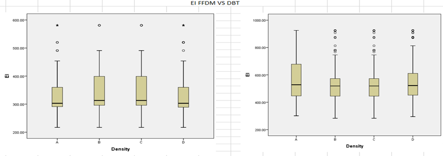
Exposure Index values were consistently higher in DBT compared to FFDM across all density groups and projections. The highest EI was observed in the DBT CC view of Group A (1119.5 AU), whereas FFDM EI values remained within a narrower and lower range. This trend is further illustrated in Table 4 and supported by graphical comparisons in Graph 7, demonstrating increased detector exposure in DBT imaging.
Compression Thickness and Dose Relationship
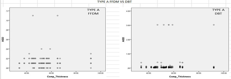
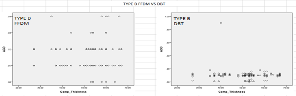
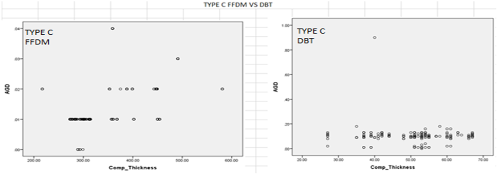
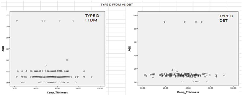
Compression thickness showed a significant association with radiation dose parameters. Increased breast thickness was generally associated with higher AGD values, particularly in DBT imaging. This relationship is demonstrated in Graph 1–4, where a positive correlation between compression thickness and AGD is evident across all breast density groups. Despite this, DBT exhibited controlled dose escalation due to the use of automated exposure control systems.
Graphical Comparison of Dose Parameters
Comparative graphical analysis further supports the observed findings. Graph 5, Graph 6, and Graph 7 illustrate the differences in AGD, ESD, and EI between FFDM and DBT across all breast density groups. These graphs clearly demonstrate that DBT consistently results in higher radiation dose parameters compared to FFDM, reinforcing the statistical findings presented in the tabulated data.
Statistical Significance
All comparisons between FFDM and DBT across breast density groups and imaging projections demonstrated statistically significant differences in AGD, ESD, and EI (p < 0.001), as summarized in Table 5. These findings confirm that DBT is associated with a higher radiation dose burden compared to FFDM under similar imaging conditions.
Table 1: Explains the mean AGD, ESD, EI in each groups of breast density (A to D) from both FFDM and DBT in CC and MLO views along with compression thickness
|
|
AGD(mSv) |
ESD(mSv) |
EI |
SD |
|
|
Density |
Compression Thickness |
|
|
|
|
|
A = 50 |
54.2 |
0.08 |
0.25 |
447.0 |
0.36 |
|
B = 50 |
50.1 |
0.05 |
0.22 |
434.7 |
0.06 |
|
C = 50 |
54.2 |
0.05 |
0.21 |
434.7 |
0.06 |
|
D = 50 |
54.7 |
0.06 |
0.27 |
443.6 |
0.09 |
Table 2: Total AGD, ESD, EI from all breast density groups in 2D (FFDM) vs 3D (DBT)
|
EXAM |
VIEW |
AGD (mSv) (Mean ± Std.deviation) |
ESD(mSv) (Mean ± Std.deviation) |
EI (Mean ± Std.deviation) |
|
DBT |
RCC LCC |
0.09 ± 0.03 0.08 ± 0.03 |
0.35 ± 0.12 0.34 ± 0.09 |
548.4 ± 159.0 524.4 ± 121.4 |
|
FFDM |
RCC LCC |
0.01 ± 0.00 0.01 ± 0.00 |
0.06 ± 0.03 0.08 ± 0.29 |
354.6 ± 0.035 329.1 ± 0.297 |
|
DBT |
RMLO LMLO |
0.11 ± 0.14 0.18 ± 0.50 |
0.34 ± 0.35 0.15 ± 0.09 |
544.3 ± 536.5 161.5 ± 133.6 |
|
FFDM |
RMLO LMLO |
0.01 ± 0.00 0.03 ± 0.04 |
0.09 ± 0.30 0.33 ± 0.80 |
338.7 ± 71.69 344.7 ± 76.12 |
Table 3: The exaggerated result showing radiation parameters in FFDM vs DBT among each group of breast Density in Craniocaudal and Mediolateral Oblique views
|
Types |
View |
AGD CC
|
AGD MLO
|
ESD CC
|
ESD MLO
|
EI CC
|
EI MLO
|
|
A |
FFDM DBT |
0.02 0.01 0.11 0.05 |
0.02 0.02 0.50 0.97 |
0.11 0.05 0.73 0.18 |
0.50 0.97 0.74 0.18 |
684.3 131.1 1119.5 281.7 |
656.7 113.0 1115.5 273.0 |
|
B |
FFDM DBT |
0.02 0.11 0.19 0.06 |
0.02 0.01 0.21 0.12 |
0.18 0.42 0.66 0.21 |
0.28 0.58 0.66 0.21 |
684.2 114.3 1026.3 227.9 |
705.2 106.5 1062.4 226.8 |
|
C |
FFDM DBT |
0.02 0.11 0.19 0.61 |
0.02 0.01 0.21 0.12 |
0.18 0.42 0.66 0.21 |
0.28 0.58 0.66 0.21 |
684.2 114.3 1026.3 227.9 |
705.2 106.5 1062.4 226.8 |
|
D |
FFDM DBT |
0.02 0.00 0.19 0.03 |
0.03 0.03 0.27 0.22 |
0.11 0.04 0.74 0.11 |
0.63 1.08 0.72 0.22 |
682.2 128.8 1119.4 276.5 |
664.0 113.7 1083.2 268.1 |
Table No: 04
|
|
|
A |
B |
C |
D |
|
AGD |
FFDM DBT |
0.01 0.01 0.15 0.51 |
0.01 0.00 0.10 0.06 |
0.01 0.00 0.10 0.65 |
0.01 0.01 0.11 0.11 |
|
ESD |
FFDM DBT |
0.13 0.46 0.37 0.11 |
0.11 0.36 0.33 0.12 |
0.10 0.30 0.33 0.12 |
0.18 0.58 0.36 0.11 |
|
EI |
FFDM DBT |
335.2 75.86 558.7 151.99 |
347.3 72.02 522.2 137.2 |
347.3 72.02 522.2 137.25 |
336.5 80.44 550.6 149.9 |
Table 4: Radiation parameter in 2D (sum of all groups and view) and 3D (sum of all group and views) with bar comparison.
Table 5: AGD, ESD, EI in (2D) vs (3D) in each individual group
|
VARIABLE |
2D(FFDM) |
3D(DBT) |
P-Value |
|
AGD |
|
|
0.000 |
|
RCC |
0.01±0.00 |
0.09±0.03 |
|
|
LCC |
0.01±0.00 |
0.08±0.03 |
|
|
RMLO |
0.01±0.00 |
0.11±0.14 |
|
|
LMLO |
0.01±0.01 |
0.18±0.50 |
|
|
ESD |
|
|
0.000
|
|
RCC |
0.06±0.03 |
0.35±0.12 |
|
|
LCC |
0.08±0.29 |
0.34±0.97 |
|
|
RMLO |
0.09±0.30 |
0.34±0.15 |
|
|
LMLO |
0.33±0.80 |
0.35±0.91 |
|
|
EI |
|
|
0.000
|
|
RCC |
354.6±88.74 |
548.4±159.09 |
|
|
LCC |
329.1±59.78 |
524.4±121.42 |
|
|
RMLO |
338.0±71.12 |
544.3±161.54 |
|
|
LMLO |
344.7±76.12 |
536.5±133.67 |
|
|
COMPRESSION THICKNESS |
|
|
0.000
|
|
RCC |
126.69±132.06 |
52.81±11.66 |
|
|
LCC |
122.78±129.53 |
52.30±11.38 |
|
|
RMLO |
126.58±135.80 |
52.18±11.13 |
|
|
LMLO |
129.39±135.64 |
53.36±11.56 |
FIGURE LEGENDS
GRAPH 1: EXPLAINING THE RELATIONSHIP BETWEEN COMPRESSION THICKNESS VS AGD IN BREAST DENSITY GROUP A (AND) IN FFDM VS DBT
GRAPH 2: EXPLAINING THE RELATIONSHIP BETWEEN COMPRESSION THICKNESS VS AGD IN BREAST DENSITY GROUP B (AND) IN FFDM VS DBT
GRAPH 3: EXPLAINING THE RELATIONSHIP BETWEEN COMPRESSION THICKNESS VS AGD IN BREAST DENSITY GROUP C (AND) IN FFDM VS DBT
GRAPH 4: EXPLAINING THE RELATIONSHIP BETWEEN COMPRESSION THICKNESS VS AGD IN BREAST DENSITY GROUP D (AND) IN FFDM VS DBT
GRAPH 5: EXPLAINING THE COMPARISON OF AGD (AVERAGE GLANDULAR DOSE) IN FFDM ON RIGHT SIDE AND DBT ON LEFT SIDE AMONG ALL FOUR GROPUS OF BREAST DENSITY
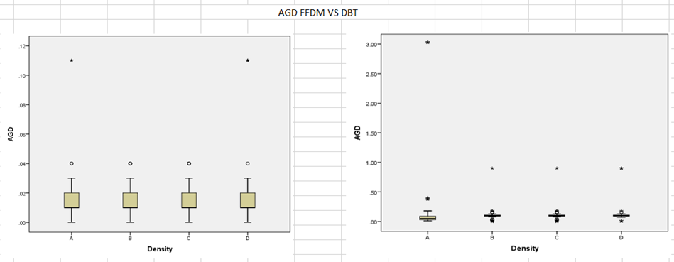
GRAPH 6: EXPLAINING THE COMPARISON OF ESD (ENTRANCE SURFACE DOSE) IN FFDM ON RIGHT SIDE AND DBT ON LEFT SIDE AMONG ALL FOUR GROPUS OF BREAST DENSITY
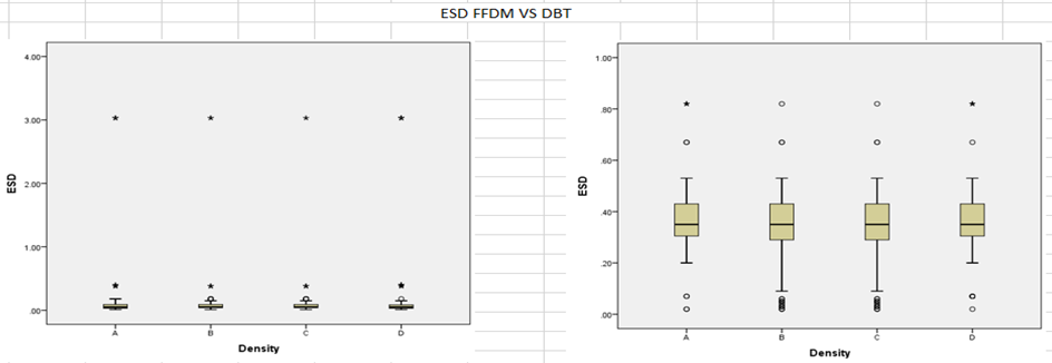
GRAPH 7: EXPLAINING THE COMPARISON OF EI (EXPOSURE INDEX) IN FFDM ON RIGHT SIDE AND DBT ON LEFT SIDE AMONG ALL FOUR GROUPS OF BREAST DENSITY
DISCUSSION AND LIMITATIONS:
In the context of medical imaging radiation exposure, the crucial factor is the risk-benefit ratio, particularly when examining radiosensitive tissues like the female breast. This concern becomes more pronounced in screening scenarios where healthy women undergo examinations to detect early stages of breast cancer. Although mammography, the current standard for breast imaging, has proven to be a valid screening tool, ongoing debates exist about the optimal design of screening programs. (15)
Our study aligns with previous dosimetric examinations indicating that MGD from DBT tends to be slightly higher than that from FFDM when using automated exposure control mechanisms. (16) The development of DBT imaging is progressing rapidly, with newer systems from various manufacturers demonstrating similar radiation exposure levels for FFDM and DBT. The nature of our study is benefited from the strength of being conducted on the same mammography system for both FFDM and DBT, using an identical patient cohort. This approach ensures consistency in breast tissue conditions between examinations, emphasizing the importance of automatic exposure control in influencing technical parameters during imaging. The variation in our results compared to other studies could be attributed to demographics, population differences, genetic variations, hormonal factors, and environmental influences, among other factors.
The rationale for this study is threefold. First, ensuring patient safety is a primary concern, as radiation dose is a critical issue in medical imaging, particularly in breast imaging, where repeated screenings are common. Second, the study aims to optimize imaging protocols to reduce radiation exposure without compromising image quality or diagnostic accuracy. This approach seeks to lead to more efficient and safer breast cancer screening practices. Lastly, the study strives to offer evidence-based insights that will aid in clinical decision-making regarding the selection of imaging modalities for breast cancer screening.
The purpose of this study is to address several important issues in breast imaging. It aims to bridge the gap created by the lack of specific national guidelines regarding Full-Field Digital Mammography (FFDM) and Digital Breast Tomosynthesis (DBT), serving as an initial step toward developing guidelines for these technologies at a national level. The research seeks to compare radiation doses with international safety standards and to support the development of regular quality control programs and dosimetry practices for both DBT and FFDM, both individually and in combination. We also intend to compare the DBT and FFDM radiation dose with standard Craniocaudal and Mediolateral Oblique projections, which will help us to optimize the dose protocol when additional views (e.g., magnified view, cone compression views, etc.) are required. The addition of DBT has had a more significant impact as mentioned in various series. The enhancement in specificity was noted regardless of factors such as breast density, age, dominant radiologic feature, or invasive status, contributing to the overall findings of other research studies. (17-19).
Our study has several limitations, including sample selection bias, as we included an equal number of patients in each breast density group. This approach was taken because our study is a pilot project comparing Digital Breast Tomosynthesis (DBT) with Full-Field Digital Mammography (FFDM) within our country. To ensure comparability and balance among the groups, we aimed to minimize potential confounding factors and reduce the risk of biased results due to uneven variable distribution that could affect outcomes. Enhancing statistical power was another goal, as it helps avoid inconclusive results or false negatives. Furthermore, by maintaining equal group sizes, the analysis and interpretation of results were simplified, allowing for straightforward comparisons without complex adjustments. This approach also contributed to the methodological rigor of the study, showcasing careful planning and consideration of potential biases, which enhances the credibility and reliability of the findings. Additionally, ensuring equal representation across groups aligns with ethical considerations, helping to address concerns about unequal treatment or biases in patient selection.
Conflict of Interest:
The authors declare no conflicts of interest related to this study.
Funding:
This research did not receive any specific grant from funding agencies in the public, commercial, or not-for-profit sectors.
Disclosures:
· Ethics Committee Approval: Karachi, Pakistan, after approval from the SIUT Ethical Research Committee (Approval no: SIUT-ERC2024/A-504 dated 5th Sept 2024).
· Peer-review: Externally peer-reviewed.
· Conflict of Interest: None declared.
· Authorship Contributions:
· Concept – N.Z, S.S.
· Supervision – S.S.
· Design – S.M.F.
· Materials – S.J.
· Data collection &/or processing – S.N.Z, S.M.F.
· Analysis and/or interpretation – S.S.
· Literature search – S.M.F., S.S.
· Writing – S.M.F., N.Z.
· Critical review – S.S S.M.F.
REFERENCES:
1. Sung H, Ferlay J, Siegel RL, Laversanne M, Soerjomataram I, Jemal A, et al. Global cancer statistics 2020: GLOBOCAN estimates of incidence and mortality worldwide for 36 cancers in 185 countries. CA Cancer J Clin. 2021;71(3):209-49. doi:10.3322/caac.21660
2. Chen Q, Pan X, Xu J, Ying W, Hou Y, Lu M, et al. Combinations of digital breast tomosynthesis and full-field digital mammography for different density types of breasts. Curr Med Imaging. 2023;19(12):1425-33. doi:10.2174/1573405619666230328113504
3. Gilbert FJ, Tucker L, Young KC. Digital breast tomosynthesis: a review of the evidence for use as a screening tool. Clin Radiol. 2022;77(3):e144-e152. doi:10.1016/j.crad.2021.10.015
4. Opitz M, Zensen S, Breuckmann K, Bos D, Forsting M, Hoffmann O, et al. Breast radiation exposure of 3D digital breast tomosynthesis compared to full-field digital mammography in a clinical follow-up setting. Diagnostics (Basel). 2022;12(2):456. doi:10.3390/diagnostics12020456
5. Conant EF, Zuckerman SP, McDonald ES. Five consecutive years of screening with digital breast tomosynthesis: outcomes by screening year and round. Radiology. 2022;302(2):273-80. doi:10.1148/radiol.2021211073
6. Shakya S, Sulwathura U, Wickramanayake M, Dulshara T, Herath LH, Wickramasinghe WM, et al. Evaluation of patient dose during digital breast tomosynthesis. Radiography (Lond). 2023;29(3):573-76. doi:10.1016/j.radi.2023.01.012
7. Dhou S, Dalah E, AlGhafeer R, Hamidu A, Obaideen A. Regression analysis between different breast dose quantities reported in digital mammography and patient age, breast thickness, and acquisition parameters. J Imaging. 2022;8(8):211. doi:10.3390/jimaging8080211
8. Bernardi D, Macaskill P, Pellegrini M, Valentini M, Fanto C, Ostillio L, et al. Breast cancer screening with tomosynthesis versus digital mammography: long-term outcomes from population-based screening. Breast Cancer Res. 2022;24(1):89. doi:10.1186/s13058-022-01567-4
9. Hamad W, Gilbert FJ, Tucker L, Young KC. Synthetic 2D mammography combined with digital breast tomosynthesis in breast cancer screening: radiation dose and diagnostic performance. Clin Imaging. 2024;102:109-16. doi:10.1016/j.clinimag.2024.01.006
10. Sechopoulos I, Sabol JM, Berglund J, Bolch WE, Brateman L, Christodoulou E, et al. Radiation dosimetry in digital breast tomosynthesis: Report of AAPM Tomosynthesis Subcommittee Task Group 223. Med Phys. 2022;49(5):e141-e173. doi:10.1002/mp.15413
11. Hofvind S, Hovda T, Holen ÅS, Lee CI, Albertsen J, Bjørndal H, et al. Digital breast tomosynthesis and synthetic 2D mammography versus digital mammography: evaluation in a population-based screening program. Eur Radiol. 2022;32(6):3931-40. doi:10.1007/s00330-021-08491-4
12. Samei E, Richard S, Flynn MJ. Optimization of breast imaging dose and image quality in digital mammography and tomosynthesis. Radiographics. 2022;42(5):1458-74. doi:10.1148/rg.220022
13. Nicosia L, Bozzini AC, Bagnardi V, et al. Comparison of radiation dose and image quality in digital breast tomosynthesis and full-field digital mammography. Cancers (Basel). 2023;15(9):2413. doi:10.3390/cancers15092413
14. Skaane P, Bandos AI, Gullien R, Eben EB, Ekseth U, Haakenaasen U, et al. Comparison of digital mammography alone and digital mammography plus tomosynthesis in a population-based screening program. Radiology. 2022;305(1):56-64. doi:10.1148/radiol.211975
15. Houssami N, Lee CI. The impact of digital breast tomosynthesis on breast cancer screening outcomes: updated evidence and future perspectives. Radiology. 2022;304(1):11-19. doi:10.1148/radiol.220145
16. Zuley ML, Guo B, Catullo VJ, Chough DM, Kelly AE, Lu AH, et al. Comparison of two-dimensional synthesized mammograms versus original digital mammograms alone and in combination with tomosynthesis images. Radiology. 2022;304(2):298-305. doi:10.1148/radiol.211873
17. Sheng M, Wu Y, Liu B, Zhang Y, Wang J. Optimization strategies for radiation dose reduction in digital breast tomosynthesis. Int J Gen Med. 2022;15:4511-20. doi:10.2147/IJGM.S300584
18. Sommer A, Hack CC, Lux MP, et al. Breast density and radiation dose in digital breast tomosynthesis: findings from the TOSYMA trial. Eur Radiol. 2024;34(2):1175-84. doi:10.1007/s00330-023-10124-6
19. Ali RMK, Helal MH, Ibrahim EM, et al. Comparative assessment of radiation dose between digital breast tomosynthesis and digital mammography in clinical practice. Radiography (Lond). 2022;28(4):1042-48. doi:10.1016/j.radi.2022.05.010
20. Conant EF, Barlow WE, Herschorn SD, et al. Association of digital breast tomosynthesis with cancer detection and recall rates by age and breast density. JAMA Oncol. 2022;8(5):e220153. doi:10.1001/jamaoncol.2022.0153