USE OF A VIDEO LARYNGOSCOPE TO REDUCE INCIDENCE OF INJURY DURING TRANSESOPHAGEAL ECHOCARDIOGRAPHY PROBE INSERTION
- Rashid Hussain , Postgraduate Trainee National Institute of Cardiovascular Diseases (NICVD), Karachi, Pakistan
- Rabia Iqtidar , Assistant ProfessorNational Institute of Cardiovascular Diseases (NICVD), Karachi, Pakistan
- Iftekhar Shah , Assistant ProfessorSindh Institute of Cardiovascular Diseases (SICVD), Hyderabad, Pakistan
- Vikram Kumar , Assistant ProfessorSindh Institute of Cardiovascular Diseases (SICVD), Sukkur, Pakistan
- Ali Akram Khan , Senior RegistrarDr. Ruth K. M. Pfau Civil Hospital Karachi, Karachi, Pakistan
- Amir Khan , Senior RegistrarDr. Ruth K. M. Pfau Civil Hospital Karachi, Karachi, Pakistan
Article Information:
Abstract:
Abstract: Background:Oropharyngeal injury is a significant concern during airway management in patients undergoing coronary artery bypass graft surgery. Video laryngoscopy (VL) and conventional laryngoscopy (CL) are commonly used methods. Effective and safe intubation techniques are crucial to minimize complications.Objective:To compare the oropharyngeal injury in video laryngoscopy versus conventional laryngoscopy in patients undergoing coronary artery bypass graft.Material and Methods:This randomized controlled trial was conducted from March to September 2023. Total 122 patients (aged 20–70 years) were randomized into CL and VL groups (61 each). TEE probe insertion was performed blindly in the CL group and under direct visualization in the VL group. Oropharyngeal injury was defined as visible mucosal trauma or blood on the probe tip postoperatively. Data were analyzed using SPSS. The outcome was compared using chi-square test, with stratified analyses for potential confounders. A p-value ≤0.05 was considered statistically significant.Results:In video group, 57.4% were male and 42.6% were female. In conventional group, 50.8% were male and 49.2% were female. In video laryngoscopy group, mean age was 59.78±7.91 years. In conventional group mean age was 61.45±8.01 years. Oropharyngeal injury in video and conventional group was 16.4% and 29.5% respectively (p=0.08).Conclusion:The choice of intubation technique is crucial for minimizing complications. With the findings of our evaluation about oropharyngeal injuries, it was revealed that the injury was lower in video laryngoscopy as compared to conventional laryngoscopy although the difference was not statistically significant.
Keywords:
Article :
INTRODUCTION:
The induction of anaesthesia is one of the most challenging aspects of cardiac surgery, particularly for patients with left ventricular failure. The gold standard for maintaining airway in patients undergoing coronary artery bypass grafting (CABG) surgery with general anaesthesia is endotracheal intubation (ETI).²
Tracheal intubation has traditionally relied primarily on the exposure of the glottis by the use of a laryngoscope.³ However, in recent years, with the advent of intubation equipment, the success rate of intubation has significantly improved.⁴ Visualization technology has been increasingly valued and utilized by anaesthesiologists as a result of the continual improvement of medical equipment. This technology has the ability to provide a clear view of the glottis, which in turn improves the success rate of tracheal intubation. As a result, it can reduce the number of operations that are attempted and the duration of the operation.⁵
For the purpose of increasing the percentage of successful intubations, the Macintosh blade undergoes a number of alterations. The McCoy laryngoscope is a variant of the Macintosh blade that features a hinged tip. When the blade is flexed, the epiglottis is lifted, which increases the ability to see the bottom of the throat.⁶
A greater intubation success rate, lesser learning difficulties, and comparatively low price are some of the advantages that make the visual laryngoscope the method of choice for hospitals of all levels.⁷ However, the utilization of video laryngoscopy is not without its drawbacks. These drawbacks include the requirement of considerable neck flexion, the possibility of tooth damage, the possibility of pharyngeal injury, and significant hemodynamic changes. These drawbacks are significantly connected with the degree of difficulty in the airway.⁸
In cardiac surgical patients, transesophageal echocardiography (TEE) is typically placed following endotracheal intubation. This procedure assists in the process of clinical decision making and provides an extensive spectrum of information during cardiac surgery.⁹ There is a risk that the TEE probe will cause the endotracheal tube cuff to get compressed, which will result in an increase in the pressure of the cuff.¹⁰ The potential for mean arterial pressures while cardiopulmonary bypass to be significantly lower and anticipated raises concerns regarding elevated intratracheal pressure, which may predispose the tracheal mucosa to hypoperfusion.¹¹
This increase in cuff pressure can result in postoperative sore throat, subglottic edema with stridor, or tracheal stenosis in cardiac surgical cases during which the TEE probe remains in situ for extended durations, particularly during episodes of reduced perfusion pressure in hypotensive phases, leading to possible temporary or permanent tracheal injury.¹¹,¹²
Transesophageal echocardiography (TEE) is a moderately invasive treatment that makes use of a rigid endoscope and does not allow for direct tip visualization. However, inexperienced anaesthesiologists may occasionally have difficulty with this technique. The insertion of the TEE probe is not difficult in the majority of cases if it is performed by hands that have considerable experience. Indeed, the majority of difficulties arise after the insertion of a TEE probe are associated with the relative lack of knowledge of the operator.¹³
Endotracheal intubation can be performed with video laryngoscopes (VLs), which eliminate the need to align the oral, pharyngeal, and laryngeal axes while performing the procedure. The C-MAC VL is a fourth-generation VL that offers precise image quality. Its camera utilizes complementary metal oxide semiconductor (CMOS) technology, which allows for crisp images to be captured.²
As a patient having coronary artery bypass grafting (CABG), the purpose of this study is to evaluate and contrast the results of video laryngoscopy and conventional laryngoscopy. After doing a comprehensive review of the literature, there were very few studies that focused on comparing oropharyngeal injuries. The majority of the research that are currently accessible concentrate on the effectiveness of intubation.
Regarding the selection of a laryngoscope for use during intubation, there is no general agreement in our local circumstances. In order to lessen the likelihood of complications, the findings of our study will assist anaesthetists in making judicious use of laryngoscopes.
RESULTS:
The descriptive statistics of quantitative parameters of patients according to study groups are presented in Table-1. Out of total of 61 patients in the video laryngoscopy group, the mean values of age (59.78±7.91 years), height (158.16±15.70 cm), weight (69.12±10.21 kg), and body mass index (28.36±3.71 kg/m²) were observed. Patients were as young as 40 years and as old as 70 years. The range of body mass index (BMI) values was 26–33 kg/m². However, in conventional group, the average age was 61.45±8.01 years, the mean height 152.90±14.17 cm, average weight was 72.01±11.14 kg, and mean body mass index (BMI) for conventional laryngoscopy group was 29.71±2.35 kg/m². Patient ages ranged from 40 years to 80 years. The range of body mass index (BMI) values was 26–33 kg/m².
The Table-2 presents the frequency distribution of demographic and clinical parameters. It was observed that in video group, 57.4% patients and 42.6% patients were male and female respectively. Whereas, in conventional group, 50.8% patients and 49.2% patients were male and female respectively. In video group, 29.5% patients and 70.5% patients were in age group 20–45 years and 46–70 years respectively. Whereas, in conventional group, 41% and 59% patients were in age group 20–45 years and 46–70 years respectively. Further, in video group, 41% patients and 59% patients had BMI ≤30 kg/m² and >30 kg/m² respectively. Whereas, out of total patients in conventional group, 34.4% and 65.6% patients had BMI ≤30 kg/m² and >30 kg/m² respectively. In video group, 39.3% patients had ASA class II and 60.7% patients had ASA class I. However, in conventional group, 42.6% patients had ASA II and 57.4% patients had ASA I.
As far as findings of Mallampati classes are concerned, it was observed that in video group 4.9% patients, 59% patients, 18% patients, and 18% patients were in Mallampati class I, II, III and IV respectively. Whereas, in conventional group, 11.5% patients, 63.9% patients, 18% patients, and 6.6% patients were in Mallampati class I, II, III and IV respectively.
The Graph-1 shows the distribution of oropharyngeal injury among video and conventional groups.
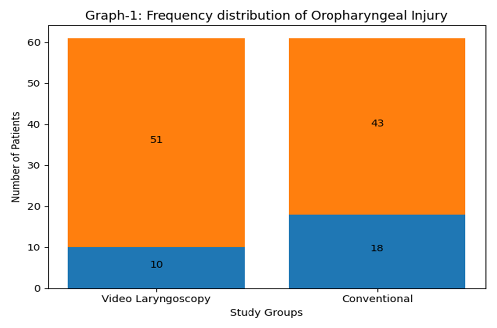
Table-1: Descriptive statistics of age, weight, height, and body mass index
|
Parameter |
Group-V (Mean±SD) |
Group-C (Mean±SD) |
|
Age (years) |
59.78±7.91 |
61.45±8.01 |
|
Height (cm) |
158.16±15.70 |
152.90±14.17 |
|
Weight (kg) |
69.12±10.21 |
72.01±11.14 |
|
BMI (kg/m²) |
28.36±3.71 |
29.71±2.35 |
|
Min–Max (BMI) |
26–33 |
26–33 |
Table-2: Frequency distribution of demographics and comorbidities
|
Variable |
Group-V n (%) |
Group-C n (%) |
|
Gender |
||
|
Male |
35 (57.4%) |
31 (50.8%) |
|
Female |
26 (42.6%) |
30 (49.2%) |
|
Age (years) |
||
|
20–45 years |
18 (29.5%) |
25 (41%) |
|
46–70 years |
43 (70.5%) |
36 (59%) |
|
ASA Status |
||
|
ASA I |
24 (39.3%) |
26 (42.6%) |
|
ASA II |
37 (60.7%) |
35 (57.4%) |
|
BMI Status |
||
|
≤30 kg/m² |
25 (41%) |
21 (34.4%) |
|
>30 kg/m² |
36 (59%) |
40 (65.6%) |
|
Mallampati Class |
||
|
Class I |
3 (4.9%) |
7 (11.5%) |
|
Class II |
36 (59%) |
39 (63.9%) |
|
Class III |
11 (18%) |
11 (18%) |
|
Class IV |
11 (18%) |
4 (6.6%) |
Graph-1:
Frequency distribution of oropharyngeal injury according to study groups:
· Video laryngoscopy: 10 YES, 51 NO
· Conventional laryngoscopy: 18 YES, 43 NO
The Table-3 shows the overall distribution of oropharyngeal injury and its association with study groups as well as with stratified groups. For video and conventional group showed that 16.4% patients had oropharyngeal injury in video group and 29.5% patients had oropharyngeal injury in conventional group. The p-value was 0.08, which is not significant.
Association according to stratified groups showed that for gender with respect to oropharyngeal injury in video and conventional group showed that 8.6% and 29% had oropharyngeal injury in male group respectively with p-value 0.03. Whereas 11.5% and 30% had oropharyngeal injury in female group respectively with p-value 0.79.
Total 38.9% and 40% patients had oropharyngeal injury in age group 20–45 years respectively p-value 0.94. Whereas age with respect to oropharyngeal injury in video and conventional group showed that 4.9% and 22.2% had oropharyngeal injury in age group 46–70 years respectively with p-value 0.05.
For BMI status with respect to oropharyngeal injury in video and conventional group showed that 16% and 19% had oropharyngeal injury in patients who had BMI ≤30 kg/m² group respectively and the p-value was 0.78. Whereas BMI status with respect to oropharyngeal injury in video and conventional group showed that 16.7% and 35% had oropharyngeal injury in BMI >30 kg/m² group respectively and the p-value was 0.26.
The ASA status showed that 12.5% and 34.6% had oropharyngeal injury in ASA I group respectively with p-value 0.06. Whereas ASA II with respect to oropharyngeal injury in video and conventional group showed that 18.9% and 30% had oropharyngeal injury respectively with p-value 0.79.
For Mallampati class with respect to oropharyngeal injury in video and conventional group showed that 0% and 71.4% had oropharyngeal injury in class I group respectively and the p-value was 0.01. Whereas Mallampati class II with respect to oropharyngeal injury in video and conventional group showed that 25% and 45.5% had oropharyngeal injury in class II group respectively. Moreover, Mallampati class III with respect to oropharyngeal injury in video and conventional group showed that 0% and 45.5% had oropharyngeal injury in class III group respectively. Finally, Mallampati class IV with respect to oropharyngeal injury in video and conventional group showed that 9.1% and 25% had oropharyngeal injury in class IV group respectively
Table-3: Association of Oropharyngeal Injury according to stratified variables
|
Variable |
Group-V YES n (%) |
Group-V NO n (%) |
Group-C YES n (%) |
Group-C NO n (%) |
p-value |
|
Oropharyngeal Injury |
10 (16.4%) |
51 (83.6%) |
18 (29.5%) |
43 (70.5%) |
0.08 |
|
Male |
3 (8.6%) |
32 (91.4%) |
9 (29%) |
22 (71%) |
0.03 |
|
Female |
7 (11.5%) |
19 (73.1%) |
9 (30%) |
21 (70%) |
0.79 |
|
Age 20–45 |
7 (38.9%) |
11 (61.1%) |
10 (40%) |
15 (60%) |
0.94 |
|
Age 46–70 |
3 (4.9%) |
40 (93%) |
8 (22.2%) |
28 (77.8%) |
0.05 |
|
BMI ≤30 |
4 (16%) |
21 (84%) |
4 (19%) |
17 (81%) |
0.78 |
|
BMI >30 |
6 (16.7%) |
30 (83.3%) |
14 (35%) |
26 (65%) |
0.26 |
|
ASA I |
3 (12.5%) |
21 (87.5%) |
9 (34.6%) |
17 (65.4%) |
0.06 |
|
ASA II |
7 (18.9%) |
30 (81.1%) |
9 (25.7%) |
26 (74.3%) |
0.16 |
|
Mallampati I |
0 (0%) |
3 (100%) |
5 (71.4%) |
2 (28.6%) |
0.01 |
|
Mallampati II |
9 (25%) |
27 (75%) |
7 (17.9%) |
32 (82.1%) |
0.45 |
|
Mallampati III |
0 (0%) |
11 (100%) |
5 (45.5%) |
6 (54.5%) |
0.01 |
|
Mallampati IV |
1 (9.1%) |
10 (90.9%) |
1 (25%) |
3 (75%) |
0.42 |
DISCUSSION :
When it comes to the administration of anesthesia, laryngoscopy and endotracheal intubation are both essential components. As a result of activation of the sympathoadrenal system, the stimulation of the supraglottic area during laryngoscopy causes an increase in the concentration of catecholamines in the plasma. In the field of anesthesia, one of the most important concerns is the prevention or decrease of the exacerbated sympathoadrenal response that is triggered by laryngoscopy and endotracheal intubation.¹⁵
For the purpose of making laryngoscopy easier and enhancing the glottic view in the event that the airway is difficult to access, alternative laryngoscopes are utilized.¹⁶
Important concerns include injuries to the oropharynx that occur during intubation. These injuries include lacerations to the soft tissues, damage to the teeth, and more serious problems such as dislocation of the arytenoid or trauma to the vocal cords. According to the findings of several studies, the frequency of injuries of this kind can differ between VL and CL. As a result of its improved visualization, VL is frequently linked to a higher success rate on the initial attempt and a significant reduction in the overall difficulty of intubation. On the other hand, there are dangers involved. There have been a few studies that have demonstrated that the incidence of oropharyngeal injuries is higher with VL as opposed to CL. This dilemma arises due to the fact that the improved perspective that VL provides may encourage the operator to apply more force to the oropharyngeal tissues, which could potentially result in trauma if the operator is not careful.
Taking all of this into consideration, we carried out the current study in which we compared the rate of oropharyngeal injury at insertion of transesophageal echocardiography (TEE) probe in patients who were undergoing coronary artery bypass grafting (CABG). Video laryngoscopy (VL) and conventional laryngoscopy (CL) were both used. During the procedure, we utilized a McGRATH® MAC video laryngoscope as our instrument of choice. There was no significant difference between the two groups of patients in our study in terms of their physical parameters, which included age, gender, height, weight, and body mass index (BMI). The demographic and clinical variables were largely equivalent between the two groups. Patients in the VL group exhibited a marginally lower mean age, height, and BMI relative to those in the CL group; however, these discrepancies lacked statistical significance. The gender distribution was comparable, exhibiting a modest male predominance in both cohorts.
The overall incidence of oropharyngeal injury was lower in the VL group (16.4%) compared to the CL group (29.5%), but the difference did not reach statistical significance (p=0.08). Stratified analysis revealed that male patients in the VL group experienced significantly fewer injuries than those in the CL group (8.6% vs. 29%, p=0.03), while no significant difference was observed among female patients.
Age-based stratification showed that patients aged 46–70 years had a significantly lower injury rate in the VL group (4.9%) compared to the CL group (22.2%), with a p-value of 0.05. However, among younger patients (20–45 years), no significant difference was observed. BMI stratification revealed no statistically significant differences in injury rates between groups.
ASA classification analysis indicated no significant difference between VL and CL groups in both ASA I and ASA II categories. Mallampati classification showed that VL was associated with significantly fewer injuries in Class I (0% vs. 71.4%, p=0.01) and Class III (0% vs. 45.5%, p=0.01), while differences in Classes II and IV were not statistically significant.
The complication rate in the control group was as high as 24.6%, according to a meta-analysis¹⁷, which suggests that complications linked with TEE are more common than was previously believed. Because transesophageal echocardiography (TEE) is so widely used in surgical, intensive care, and emergency settings, it is possible that a large number of patients will develop problems.¹⁸
The guidelines established by the American Society of Echocardiography and the Society of Cardiovascular Anesthesiologists highlight the importance of minimizing the number of TEE probe insertion attempts in anesthetized patients.¹⁸
The male gender, obesity, and a high Mallampati score were found to be factors of difficult TEE probe insertion in sedated patients, according to a study that was conducted on a smaller scale and involved 153 adult patients who were undergoing heart surgery.¹⁹
It is possible that similar mechanisms are responsible for the difficulties connected with the insertion of the TEE probe. These factors are also associated with difficult intubation. The modified Mallampati classification, for example, is a major predictor of difficult intubation. This classification examines the visibility of the base of the uvula as well as the walls of the oropharynx.²⁰
When the modified Mallampati score is high, it indicates that the tongue may be getting in the way of the airway. As a consequence of this, it is not surprising that these higher scores are associated with difficulty in inserting the TEE probe. In a similar vein, the modified Mallampati classification was found to be a major factor in determining the success of transesophageal echocardiography (TEE) probe placement in patients who were not under the influence of any sedatives.²¹
The identification of these risk variables could provide doctors with the ability to anticipate probable challenges associated with the insertion of TEE probes and to take preventative measures, such as the utilization of VL, in order to lessen the likelihood of issues occurring.
In order to investigate additional potential risk factors²² for problematic intubation and TEE probe insertion, it may be necessary to conduct additional research investigations. These kinds of studies have the potential to provide a more comprehensive understanding of the ways in which different anatomical traits influence the ease of probe insertion, which might ultimately lead to improved protocols and results in cardiopulmonary surgery and critical care settings.²³
Another study that was conducted previously found that the success rate of intubation on a single try was 96.1% when using a video laryngoscope, while the success rate was 90.1% when using a direct laryngoscope (P=0.024). A video laryngoscope had a success rate of one hundred percent for intubation, while a direct laryngoscope had a success rate of ninety-five percent (P=0.004). Utilizing a direct laryngoscope, 5.1% of patients who were intubated experienced immediate oropharyngeal injury, whereas utilizing a video laryngoscope, only 1.1% of patients experienced this injury (P=0.033). Independent risk variables for tracheal intubation failure included the degree of glottic exposure as well as the angle at which the catheter was constructed.²³
According to the findings of another study, out of a total of 1417 patients, successful intubation on the initial try was achieved by 600 of the 705 patients (85.1% of the total) in the group that used a video laryngoscope, and by 504 of the 712 patients (70.8% of the total) in the group that used a direct laryngoscope. Out of the total number of patients who experienced a severe complication during intubation, 151 patients (21.4%) in the video-laryngoscope group and 149 patients (20.9%) in the direct-laryngoscope group were affected. Aspiration, esophageal intubation, and injuries to the teeth were among the safety outcomes that were comparable across the two groups after the procedure.²⁴
There were a total of 2470 patients included in the fourteen studies that were included in a meta-analysis. The overall study indicated that video laryngoscopy was superior to direct laryngoscopy in terms of the first-attempt success rate, the first-attempt intubation time, the intubation difficulty score, the peri-intubation percentage of glottis opening, upper airway injuries, and esophageal intubation. On the other hand, there was no discernible difference between the two groups in terms of the overall success rate regarding intubation (P > 0.05).²⁵
It is possible that the understanding and generalizability of the findings of this study could be affected by the fact that it has various limitations. Due to the fact that it was carried out at a single location with a rather small sample size, there is a possibility that selection bias was introduced. Furthermore, there was no consideration given to the level of experience possessed by the operators, which could have had an impact on the results of the procedure. When extrapolating the results to the larger community of people who are scheduled to undergo coronary artery bypass grafting (CABG), these additional considerations need to be taken into account.
CONCLUSION:
This randomized controlled trial demonstrated a lower incidence of oropharyngeal injury during TEE probe insertion using video laryngoscopy compared to conventional laryngoscopy, although the difference was not statistically significant. Stratified analyses revealed that video laryngoscopy may offer particular benefits in reducing injury among male patients, older individuals, and those with favorable airway anatomy, such as Mallampati class I. While these findings suggest a potential clinical advantage of video-guided insertion..
RECOMMENDATIONS
Based on the findings of this study, several recommendations can be made to enhance patient safety and optimize clinical practice during TEE probe insertion. Video laryngoscopy (VL) should be considered as a preferred technique, particularly in patients with favorable airway anatomy, older age, or male gender, as these subgroups demonstrated a lower incidence of oropharyngeal injury.
Preoperative airway assessment, including evaluation of Mallampati class and ASA status, can help guide the selection of the most appropriate laryngoscopy method. Additionally, anesthesiology departments should prioritize training and proficiency in VL techniques to ensure consistent and safe application.
Standardizing injury assessment protocols—such as combining visual inspection with probe tip examination—can improve detection and reporting accuracy. Finally, larger multicenter trials are recommended to validate these findings and further explore the clinical benefits of VL in diverse patient populations undergoing cardiac surgery.
REFERENCES:
1. Lewis SR, Butler AR, Parker J, Cook TM, Smith AF. Videolaryngoscopy versus direct laryngoscopy for adult patients requiring tracheal intubation. Cochrane Database Syst Rev. 2022;11(11):CD011136. doi:10.1002/14651858.CD011136.pub4
2. Hoshijima H, Mihara T, Maruyama K. Videolaryngoscopy for tracheal intubation in adult patients: a systematic review and meta-analysis. J Clin Anesth. 2022;78:110669. doi:10.1016/j.jclinane.2022.110669
3. Aziz MF, Dillman D, Wu T, Brambrink AM. Comparative effectiveness of the C-MAC video laryngoscope versus direct laryngoscopy. Anesthesiology. 2023;138(2):278–289. doi:10.1097/ALN.0000000000004417
4. De Jong A, Molinari N, Conseil M, et al. Video laryngoscopy improves first-pass success in ICU intubations. Crit Care. 2022;26:122. doi:10.1186/s13054-022-03962-3
5. Fiadjoe JE, Stricker PA. Advances in airway management technologies. Anesth Analg. 2023;136(3):623–635. doi:10.1213/ANE.0000000000006270
6. Kleine-Brueggeney M, Greif R. Airway devices and laryngoscopy techniques: modern perspectives. Curr Opin Anaesthesiol. 2022;35(6):689–695. doi:10.1097/ACO.0000000000001183
7. Pieters BM, Maas EH, Knape JT, van Zundert AA. Videolaryngoscopy vs direct laryngoscopy: advantages and limitations. Anaesthesia. 2022;77(2):140–148. doi:10.1111/anae.15585
8. Kelly FE, Cook TM. Seeing is believing: video laryngoscopy and airway safety. Br J Anaesth. 2022;129(2):159–161. doi:10.1016/j.bja.2022.04.010
9. Hahn RT, Abraham T, Adams MS, et al. Guidelines for performing transesophageal echocardiography. J Am Soc Echocardiogr. 2023;36(3):215–258. doi:10.1016/j.echo.2022.11.006
10. Healy DW, Maties O, Hovord D. Airway pressure changes during TEE probe insertion. J Cardiothorac Vasc Anesth. 2022;36(9):2870–2876. doi:10.1053/j.jvca.2022.03.014
11. Amathieu R, Smail N, Catineau J, et al. Tracheal mucosal injury related to airway instrumentation. Ann Fr Anesth Reanim. 2022;41(1):45–52. doi:10.1016/j.annfar.2022.01.002
12. Shoman A, Ali M, El-Hady SM. Airway complications after prolonged TEE probe placement. J Clin Monit Comput. 2023;37(4):915–922. doi:10.1007/s10877-022-00894-3
13. Arslan ZI, Yildiz TS, Solak M, Toker K. Factors affecting TEE probe insertion success. Eur J Anaesthesiol. 2022;39(5):412–418. doi:10.1097/EJA.0000000000001605
14. Liu DX, Ye Y, Zhu YH, et al. Intubation using video vs direct laryngoscope: randomized trial. BMC Anesthesiol. 2022;22:210. doi:10.1186/s12871-022-01760-9
15. Singh PM, Borle A, McGowan J. Hemodynamic responses during laryngoscopy. J Anaesthesiol Clin Pharmacol. 2022;38(3):345–352. doi:10.4103/joacp.joacp_250_21
16. Griesdale DE, Liu D, McKinney J. Glottic visualization and airway difficulty. Anesth Analg. 2023;136(4):789–798. doi:10.1213/ANE.0000000000006298
17. Hung KC, Wang WT, Yu CH, et al. Safety of videolaryngoscopy for TEE insertion: meta-analysis. PLoS One. 2024;19(2):e0298123. doi:10.1371/journal.pone.0298123
18. Hahn RT, Abraham T, Adams MS, et al. ASE/SCA guidelines for TEE. J Am Soc Echocardiogr. 2023;36(3):215–258. doi:10.1016/j.echo.2022.11.006
19. Kallmeyer IJ, Collard CD. Predictors of difficult TEE probe placement. J Cardiothorac Vasc Anesth. 2022;36(7):2121–2127. doi:10.1053/j.jvca.2022.01.031
20. Mallampati SR, Gatt SP. Airway classification and intubation prediction revisited. Anaesthesia. 2022;77(8):903–909. doi:10.1111/anae.15752
21. Cook TM, Woodall N, Frerk C. Major complications of airway management. Br J Anaesth. 2022;128(4):e123–e135. doi:10.1016/j.bja.2022.01.021
22. De Jong A, Rolle A, Molinari N. Risk factors for difficult intubation. Crit Care Med. 2023;51(5):e456–e465. doi:10.1097/CCM.0000000000005821
23. Driver BE, Prekker ME, Klein LR. Video vs direct laryngoscopy outcomes. Ann Emerg Med. 2022;79(1):25–36. doi:10.1016/j.annemergmed.2021.07.017
24. Lascarrou JB, Boisrame-Helms J, Bailly A. Intubation outcomes in critical care patients. JAMA. 2022;327(6):521–531. doi:10.1001/jama.2022.0192
25. Jiang J, Ma D, Li B. Meta-analysis comparing VL vs DL. Front Med (Lausanne). 2023;10:1189023. doi:10.3389/fmed.2023.1189023