Diagnostic Accuracy of Transvaginal Sonography for Ectopic Pregnancy: A Validation against Histopathology
- Alina Farooq , RadiologyDepartment, Ayub Teaching Hospital (ATH), Abbottabad, Pakistan
- Faiza Akram , RadiologyDepartment, Ayub Teaching Hospital (ATH), Abbottabad, Pakistan
- Muhammad Hadi Farooq , 3rd Year MBBS, Ayub Medical College , Abbottabad, Pakistan
- Nuzhat Raza , Professor of Physiology, Ayub Medical College Abbottabad, Pakistan
Article Information:
Abstract:
Background: Ectopic pregnancy (EP) is a significant obstetric emergency with high morbidity and mortality if undiagnosed. Transvaginal sonography (TVS) has emerged as a superior diagnostic modality due to its high resolution and ability to detect pelvic abnormalities. This study aimed to evaluate the diagnostic accuracy of TVS in detecting ectopic pregnancies, using histopathology as the gold standard. Methods: A cross-sectional validation study was conducted at the Radiology Department of Ayub Teaching Hospital, Abbottabad. A total of 228 women aged 20–40 years with suspected ectopic pregnancy were enrolled during October 2023 to December 2024. TVS findings were compared with histopathological results, and diagnostic accuracy metrics, including sensitivity, specificity, positive predictive value (PPV), and negative predictive value (NPV), were calculated. Results: TVS demonstrated a sensitivity of 92.9%, specificity of 81.4%, PPV of 93.4%, and NPV of 80.0% for diagnosing ectopic pregnancy. Significant associations were observed between ectopic pregnancy and risk factors such as a history of pelvic inflammatory disease and previous ectopic pregnancies (p < 0.05). Conclusion: TVS is a reliable and accurate diagnostic tool for ectopic pregnancy, offering high sensitivity and specificity. Its integration into clinical practice can enhance early diagnosis and reduce complications, particularly in resource-limited settings.
Keywords:
Article :
INTRODUCTION:
Ectopic pregnancy (EP) is a significant challenge in obstetrics, often considered one of the most critical gynecological emergencies. It occurs when a fertilized egg implants outside the uterine cavity, most commonly in the fallopian tube, though it can also occur in the ovary, cervix, or abdominal cavity. This condition is not only life-threatening if undiagnosed but also a major contributor to maternal morbidity and mortality, particularly in resource-limited settings like Pakistan, where timely diagnostic tools and treatments are often unavailable 1,2.
Despite advances in medical diagnostics, ectopic pregnancy remains notoriously difficult to diagnose due to its nonspecific and variable clinical presentations. Studies suggest that up to 40% of ectopic pregnancies are initially missed, highlighting the limitations of relying solely on clinical history and physical examination for diagnosis 3,4. Delayed or missed diagnoses can lead to ruptured ectopic pregnancies, posing grave risks to maternal health, including severe morbidity and even death 5-7.
A variety of diagnostic tools are available for ectopic pregnancy, ranging from serum beta-hCG levels and posterior fornix puncture to laparoscopy and histopathological examination 8. However, these approaches often require advanced resources and expertise, which may not be accessible in many healthcare settings. Among imaging techniques, transabdominal ultrasound (TAS) has been widely used for early pregnancy assessments. However, TAS often has limitations, such as reduced resolution due to factors like bladder filling, abdominal adiposity, and bowel gas interference, making it suboptimal for diagnosing ectopic pregnancies in certain cases 9.
Transvaginal sonography (TVS), on the other hand, has emerged as a superior diagnostic modality due to its high resolution and close proximity to pelvic structures. TVS has demonstrated high sensitivity and specificity, with an overall diagnostic accuracy of 93%, making it a highly reliable tool in the timely diagnosis of ectopic pregnancies 10. These attributes are particularly crucial in settings with limited access to invasive diagnostic methods.
This study aims to evaluate the diagnostic accuracy of TVS for ectopic pregnancy, using histopathology as the gold standard. By validating TVS as a non-invasive and dependable diagnostic tool, we hope to contribute to improved outcomes for women presenting with suspected ectopic pregnancies, especially in resource-constrained healthcare environments. Early and accurate diagnosis is not only essential for initiating timely management but also for reducing the devastating complications associated with ruptured ectopic pregnancies.
MATERIALS AND METHODS:
The study was conducted as a cross-sectional validation at the Department of Radiology, Ayub Teaching Hospital, Abbottabad. To ensure methodological rigor and transparent reporting, the study adhered to the Standards for Reporting of Diagnostic Accuracy (STARD) guidelines. A total of 228 participants were included, with the sample size calculated based on statistical requirements for sensitivity and specificity. We used a 95% confidence level, assuming an estimated prevalence of ectopic pregnancy of 25.4% 4, a sensitivity of 92%10 and a specificity of 93.3%10 with a 7% precision. Participants were enrolled consecutively over a period of fifteen months from October 2023 to December 2024, ensuring all eligible women referred for ultrasound with suspected ectopic pregnancy during the study period were included. The study was conducted in compliance with the Declaration of Helsinki. Ethical approval was obtained from the Ayub Teaching Hospital Ethical Review Board. All participants were thoroughly informed about the study’s purpose, and confidentiality was maintained throughout.
The study focused on women aged 20 to 40 years, referred from the Gynecology and Obstetrics outpatient department. Those with intrauterine abortion, ovarian cyst torsion, acute tubal infection, vaginal inflammation, known gynecological malignancies, urogenital anomalies, or contraindications to vaginal examination were excluded. After obtaining written informed consent, we collected demographic and clinical data, including age, body mass index (BMI), parity, socioeconomic status, and medical history, such as prior ectopic pregnancy or pelvic inflammatory disease. Ultrasound examinations were performed by a consultant radiologist with more than five years of post-fellowship experience, using a Canon Aplio a ultrasound machine with a 3.0–10.0 MHz probe. To ensure consistency, the procedure was standardized, with patients positioned supine and examined with an empty bladder for optimal imaging. The scans were carefully reviewed to identify adnexal masses, free fluid, or other findings suggestive of ectopic pregnancy. Histopathological examination of surgical specimens was used as the gold standard for confirming the diagnosis, providing an objective basis for comparison.
Data analysis was conducted using SPSS version 27.0. Continuous variables, such as age and BMI, were summarized as means and standard deviations, while categorical variables, like parity and history of pelvic inflammatory disease, were expressed as frequencies and percentages. The diagnostic performance of TVS was evaluated using sensitivity, specificity, positive predictive value (PPV), and negative predictive value (NPV). To explore potential confounding factors, such as variations in age or BMI, stratified analyses were performed, with a p-value of less than 0.05 considered statistically significant.
RESULTS:
Table 1: Descriptive statistics of demographic and clinical characteristics of the study participants
|
Continuous Parameters |
Mean ± Standard Deviation |
|
|
Age (Years) |
31.6 ± 4.96 |
|
|
Weight (Kg) |
65 ± 8.29 |
|
|
Height (cm) |
163.2 ± 9.14 |
|
|
BMI (Kg/m2) |
24.4 ± 3.26 |
|
|
Categorical Parameters |
N (%) |
|
|
Residence |
Rural |
110 (48.2%) |
|
Urban |
118 (51.8%) |
|
|
Duration since last menstruation cycle (weeks) |
5 |
65 (28.5%) |
|
6 |
60 (26.3%) |
|
|
7 |
59 (25.9%) |
|
|
8 |
44 (19.3%) |
|
|
Socioeconomic Status |
Upper Middle class |
70 (30.7%) |
|
Lower class |
75 (32.9%) |
|
|
Middle Class |
83 (36.4%) |
|
|
Parity |
Nulliparous |
7 (3.1%) |
|
Multipara |
166 (72.8%) |
|
|
Grand Multipara |
55 (24.1%) |
|
|
History of Ectopic Pregnancy |
Yes |
37 (16.2%) |
|
No |
191 (83.8%) |
|
|
History of PID |
Yes |
32 (14.0%) |
|
No |
196 (86.0%) |
|
The table 1 summarizes the demographic data of study participants. The mean age is 31.6 ± 4.96 years, with an average BMI of 24.4 ± 3.26 Kg/m². Categorical variables detail the distribution of residence (48.2% rural), most prevalent average time since last menstrual cycle was 5 weeks for 65 (28.5%) participants, socioeconomic status was middle class 83 (36.4%), and most participants were multiparous (72.8%), reflecting diverse reproductive histories within this cohort. History of ectopic pregnancy was positive in only 37 (16.2%) and Pelvic Inflammatory Disease in 32 (14.0%) participants.
Table 2: Comparison of demographic and clinical metrics between ectopic pregnancy and other pathology cases
|
Diagnosed via Histopathology |
N |
Mean |
Std. Deviation |
P Value |
|
|
Age (Years) |
Ectopic Pregnancy |
169 |
31.57 |
4.841 |
0.796 |
|
Other Pathology |
59 |
31.76 |
5.344 |
||
|
Weight (Kg) |
Ectopic Pregnancy |
169 |
65.207 |
8.3408 |
0.523 |
|
Other Pathology |
59 |
64.403 |
8.1762 |
||
|
Height (cm) |
Ectopic Pregnancy |
169 |
163.35 |
8.985 |
0.745 |
|
Other Pathology |
59 |
162.90 |
9.656 |
||
|
BMI (Kg/m2) |
Ectopic Pregnancy |
169 |
24.180 |
3.2923 |
0.045 |
|
Other Pathology |
59 |
25.166 |
3.0665 |
||
The table 2 compares demographic and clinical metrics (age, weight, height, and BMI) between patients diagnosed with ectopic pregnancy and those with other pathologies, as confirmed by histopathology. Mean values for age, weight, and height are similar across both groups, with no significant differences (P > 0.05). However, a statistically significant difference in BMI was observed between the groups (P = 0.045), indicating a lower average BMI in patients with ectopic pregnancy compared to those with other pathologies.
Table 3: Cross-tabulation of ectopic pregnancy diagnosis by clinical factors
|
Cross-tabulation |
Diagnosis by Histopathology |
Total |
P value |
|
||
|
Other Pathology n (%) |
Ectopic Pregnancy n (%) |
|||||
|
N (%) |
|
|||||
|
History of Ectopic Pregnancy |
Yes |
0 (00%) |
36 (21.3%) |
36 (15.8%) |
<0.001 |
|
|
No |
59 (100%) |
133 (78.7%) |
192 (84.2%) |
|
||
|
History of PID |
Yes |
1 (1.7%) |
31 (18.3%) |
32 (14%) |
0.002 |
|
|
No |
58 (98.3%) |
138 (81.7%) |
196 (86%) |
|
||
|
Parity |
Nulliparous |
1 (1.7%) |
6 (3.6%) |
7 (3.1%) |
0.761 |
|
|
Multipara |
43 (72.9%) |
123 (72.8%) |
166 (72.8%) |
|
||
|
Grand Multipara |
15 (25.4%) |
40 (23.7%) |
55 (24.1%) |
|
||
The cross-tabulation presented in table 3 examines the relationship between ectopic pregnancy (as confirmed by histopathology) and various clinical factors, including a history of ectopic pregnancy, PID (Pelvic Inflammatory Disease), and parity. A statistically significant association was found between ectopic pregnancy diagnosis and both history of ectopic pregnancy (P < 0.001) and PID (P = 0.002). No significant difference was observed with parity (P = 0.761), indicating parity distribution is similar between groups with ectopic pregnancy and other pathologies.
Table 4: Cross-tabulation comparing diagnosis of ectopic pregnancy via TVS with histopathology
|
Cross-tabulation |
Diagnosis by Histopathology |
Total |
P value |
|||
|
Other Pathology n (%) |
Ectopic Pregnancy n (%) |
|||||
|
N (%) |
||||||
|
Diagnosis by TVS |
No |
48 (81.4%) |
12 (7.1%) |
60 (26.3%) |
<0.001 |
|
|
Yes |
11 (18.6%) |
157 (92.9%) |
168 (73.7%) |
|||
|
Total |
59 (100%) |
169 (100%) |
228 (100%) |
|||
The table presents a cross-tabulation comparing TVS diagnosis of ectopic pregnancy with histopathology results in 228 patients. TVS demonstrated a high sensitivity (92.9%) and specificity (81.4%), with an overall accuracy of 89.9% for the diagnosis of ectopic pregnancy. The positive predictive value was 93.4%, and the negative predictive value was 80.0%, showing TVS's strong diagnostic reliability (P < 0.001).
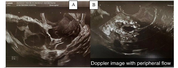
Figure 1: Grey scale and Color Doppler Transvaginal Ultrasound images of the Ectopic Pregnancy
Figure 1 shows the TVS images (A- Greyscale, B- Doppler image) showing anteverted normal sized uterus measuring 7.4x2.6x4 cm with endometrial thickness of 2.5mm. No intra-uterine focal lesion was seen. The left adnexa were bulky measuring approximately 3.7x3.3x3.2 cm having anechoic/ cystic component with increased peripheral flow giving ring of fire sign suggesting ectopic pregnancy.
DISCUSSION :
Our study evaluated the diagnostic accuracy of transvaginal sonography (TVS) in identifying ectopic pregnancies, using histopathology as the definitive gold standard. The findings demonstrate that TVS is a highly reliable diagnostic tool, with a sensitivity of 92.9% and specificity of 81.4%. These results affirm TVS’s critical role in early detection and management of ectopic pregnancies, particularly in clinical settings where histopathology or invasive diagnostic procedures may not be immediately accessible. Similar findings have been reported in studies by Hu et al. and Malik et al., who observed comparable sensitivity and specificity values for TVS 10, 11.
When compared to existing literature, our results align closely with previously reported sensitivity and specificity values for TVS. Hu et al. 10 noted that TVS outperformed transabdominal sonography (TAS) due to its superior resolution and ability to closely visualize pelvic structures. Malik et al. reported a sensitivity of 92% for TVS in ectopic pregnancy diagnosis, corroborating our findings 11. However, our specificity (81.4%) was slightly lower than the 93.3% reported by Verma et al., possibly due to variations in study populations, operator expertise, or equipment quality 3. Notably, the high positive predictive value (PPV) of 93.4% observed in our study further underscores the reliability of TVS in confirming ectopic pregnancies when clinical suspicion is high.
The association of ectopic pregnancy with risk factors such as a history of pelvic inflammatory disease (PID) and previous ectopic pregnancies was also significant in our study. Similar findings were highlighted by Obajimi et al., who reported PID and prior tubal surgeries as major contributors to ectopic pregnancy risk 4. Bouyer et al. also emphasized the importance of these clinical risk factors in their population-based study, further supporting our results 12. Additionally, we observed a statistically significant association between ectopic pregnancy and lower body mass index (BMI). Although this finding is consistent with other studies linking BMI to reproductive health, the mechanisms underlying this association remain unclear and warrant further investigation 13, 14. Compared to alternative diagnostic modalities, TVS continues to demonstrate clear advantages. TAS, though widely used, is often limited by factors such as bowel gas and bladder filling, which can obscure imaging. Baker and dela Cruz highlighted that TAS is less effective in visualizing small adnexal masses or subtle pelvic findings compared to TVS 5. Similarly, the diagnostic performance of TAS was found to be inferior in large-scale analyses, particularly in early-stage ectopic pregnancies 15. By contrast, TVS provides high-resolution images, offering a reliable method for detecting adnexal masses, free fluid, and other key indicators of ectopic pregnancy.
Despite these strengths, our study acknowledges certain limitations. Being a single-center study, the results may not be fully generalizable to broader populations. Additionally, the non-probability sampling method may have introduced selection bias, although efforts were made to minimize this through consecutive sampling. Operator dependency in ultrasound interpretation is another inherent limitation, as noted in similar studies emphasizing the role of radiologist expertise 13, 14. our findings affirm that TVS is a highly sensitive and specific modality for diagnosing ectopic pregnancies, with diagnostic performance comparable to existing literature. The integration of TVS into routine diagnostic protocols can facilitate early diagnosis, reducing the incidence of ruptured ectopic pregnancies and associated morbidity and mortality. Future research may explore the utility of advanced imaging techniques, to further enhance diagnostic accuracy in complex cases 16.
CONCLUSION :
TVS is a highly accurate and reliable diagnostic tool for detecting ectopic pregnancies, with strong sensitivity and specificity. Its non-invasive nature and accessibility make it an essential modality, particularly in resource-limited settings. Integrating TVS into routine diagnostic protocols can enable early detection and timely management, significantly reducing complications and improving maternal outcomes.
BIBLIOGRAPHY:
1. Tonick S, Conageski C. Ectopic pregnancy. Obstet Gynecol Clin North Am. 2022;49(1):537-49.
2. Committee on Practice Bulletins-Gynecology. ACOG Practice Bulletin No. 191: Tubal Ectopic Pregnancy. Obstet Gynecol. 2018;131:65-77.
3. Verma ML, Singh U, Solanki V, Sachan R, Sankhwar PL. Spectrum of ectopic pregnancies at a tertiary care center of Northern India: A retrospective cross-sectional study. Gynecol Minim Invasive Ther. 2022;11(1):36-40.
4. Obajimi GO, Smart AE, Adekanmi AJ, Adeniji-Sofoluwe A, Jinadu FO. Correlation between transabdominal ultrasound features of ectopic gestation and surgical findings at the University College Hospital, Ibadan: A preliminary review. Trop J Obstet Gynaecol. 2019;36:265-70.
5. Baker M, dela Cruz J. Ectopic pregnancy, ultrasound. [Updated 2023 Jan 16]. In: StatPearls [Internet]. Treasure Island (FL): StatPearls Publishing; 2023 Jan. Available from: https://www.ncbi.nlm.nih.gov/books/NBK482192.
6. Beals T, Naraghi L, Grossestreuer A, Schafer J, Balk D, Hoffman B. Point of care ultrasound is associated with decreased ED length of stay for symptomatic early pregnancy. Am J Emerg Med. 2019;37(6):1165-8.
7. Wang PS, Rodgers SK, Horrow MM. Ultrasound of the first trimester. Radiol Clin North Am. 2019;57(3):617-33.
8. Mukherjee R, Samanta S. Surgical emergencies in pregnancy in the era of modern diagnostics and treatment. Taiwan J Obstet Gynecol. 2019;58(2):177-82.
9. Shen Z, Li C, Zhao L, Xu L, Peng B, Chen Z, et al. Minimally-invasive management of intramural ectopic pregnancy: An eight-case series and literature review. Eur J Obstet Gynecol Reprod Biol. 2020;253(1):180-6.
10. Hu HJ, Sun J, Feng R, Yu L. Comparison of the application value of transvaginal ultrasound and transabdominal ultrasound in the diagnosis of ectopic pregnancy. World J Clin Cases. 2023;11(13):2945-55.
11. Malik SA, Malik S, Maqbool A. Comparison of transabdominal and transvaginal sonography in the diagnosis of ectopic pregnancy. Pak J Med Health Sci. 2010;4(1):22-7.
12. Bouyer J, Coste J, Shojaei T, Pouly JL, Fernandez H, Gerbaud L, Job-Spira N. Risk factors for ectopic pregnancy: a comprehensive analysis based on a large case-control, population-based study in France. American journal of epidemiology. 2003 Feb 1;157(3):185-94.
13. Young L, Barnard C, Lewis E, Jones M, Furlan J, Karatasiou A, Necas M. The diagnostic performance of ultrasound in the detection of ectopic pregnancy. The New Zealand Medical Journal (Online). 2017 Mar 24;130(1452):17.
14. Ectopic pregnancy: Epidemiology, risk factors, and anatomic sites (2024) UpToDate. Available at: https://www.uptodate.com/contents/ectopic-pregnancy-epidemiology-risk-factors-and-anatomic-sites (Accessed: 11 January 2025).
15. Condous G, Okaro E, Khalid A, Lu C, Van Huffel S, Timmerman D, Bourne T. The accuracy of transvaginal ultrasonography for the diagnosis of ectopic pregnancy prior to surgery. Human reproduction. 2005 May 1;20(5):1404-9. https://doi.org/10.1093/humrep/deh770
16. Hendriks E, Rosenberg R, Prine L. Ectopic pregnancy: diagnosis and management. American family physician. 2020 May 15;101(10):599-606. Available from: https://www.reproductiveaccess.org/wp-content/uploads/2020/05/Ectopic-Pregnancy-Diagnosis-and-Management.pdf.