POST-MORTEM AND HISTO PATHOLOGICAL ANALYSIS OF SUBMERGED WISTAR RAT LIVER
- Arun Ganesh M K , Department of Pathology,Saveetha Dental College and Hospitals,Saveetha Institute of Medical and Technical Sciences,Saveetha University, Chennai-600 077, India.
- R Priyadharshini , Department of Pathology,Saveetha Dental College and Hospitals,Saveetha Institute of Medical and Technical Sciences, Saveetha University, Chennai-600 077,India.
Article Information:
Abstract:
The act of suffering with breathing impairments as a result of submersion or immersion in liquid is called drowning. Aim is to evaluate the post mortem, histopathological and electron microscopic findings of submerged wistar rat liver. Materials & methods : Wistar rat was sacrificed by submerging in water for three days and autopsy was done on liver tissue. Result : There was loss of morphological structure and nuclear cytoplasmic detail, sinusoids were constructed, coagulative necrosis, homogenous eosinophilic hyaline like appearance of hepatocyte cells. Hepatocyte cells are seen as clusters, the central vein were constricted because of oedema with CD34 & SMA showing positivity. Conclusion: From the study we may conclude that the aspiration of water into the airways causes drowning which results in death. If the findings are further explored, forensic pathology could benefit as a highly reliable technique at low cost.
Keywords:
Article :
INTRODUCTION:
Finding the cause of death (Eg: Drowning, Natural ailment) and the manner of death (Eg: Murder, Suicide, or Accident) when a body is discovered in water is frequently difficult for forensic pathologists (1). India accounts for 17% of all accidental drowning deaths worldwide, making it a major contributor to the global burden of drowning. India's population is frequently exposed to water due to its enormous inland freshwater systems and more than 8000 km of coastline (2).
Young children are more likely to die from drowning, with 27% of unintentional injury deaths in United States occurring before the age of 1-4 years. More often than women, men are involved. The location will obviously depend on the nation and region, with seawater drownings being more common in coastal areas. Coastal areas also have a higher rate of boating accidents and drownings due to water sports like diving. Suicide is a more frequent cause of drowning in adults and is frequently linked to a preexisting psychiatric condition (3). Most at risk in terms of access to suicide tools as well, while it is also easy to commit suicide by drowning in a bathtub or other such home amenities (4).
According to data through 2021, there were 2,36,000 drowning deaths registered worldwide (5). According to the National Crime Records Bureau from 2019, there are 82 drowning deaths each day in India. Of the overall number of drowning deaths, nearly 11,884 instances (39.74%) were caused by accidental falls into water, while 7426 cases (5.6%) were caused by suicidal drowning. Drowning was the second most common cause of unnatural death after traffic accidents (53.4%), accounting for 29,903 cases (9.4%) out of 316,828 cases of unnatural deaths. Of these, 23,166 cases (77.47%) were male, 6736 cases (22.52%) were female, and 1 case (0.0033%) was transgender (6).
The difficulty of making the postmortem diagnosis of drowning is one factor contributing to the confusion surrounding the number of drowning deaths. An autopsy, toxicological and microscopic tests, as well as a thorough investigation of the circumstances are all part of the inquiry of remains found in water. The main issue is whether the person died as a result of the submersion or whether it happened after death. Sadly, "real" drownings are not always easy to identify (7,8). There are no pathognomonic postmortem findings that identify drowning (9,10). The diagnosis is instead based on two variables. First, the circumstances and history that supports drowning. Second, a sickness or physical injury whose severity or extent is irreconcilable with continuing life is not revealed by the autopsy. Numerous non-specific ("soft") anatomical data can also be taken into account. As drowning is an excluded diagnosis, a full autopsy is necessary, including a toxicological study (11).
Hypoxia is a significant pathophysiological aspect of drowning. A tissue is analysed by post-mortem, histopathology to elucidate the cause and mechanism of death and injury (12). The factors that affect the post-mortem autolytic process are temperature, air humidity and environment (12,13). This research is needed to get data from gross, histopathology analysis and electron microscopic findings of submerged wistar rat liver. We will be able to have data about the changes that occur in a liver tissue of wistar rats which occurs due to submerging. Our team has extensive knowledge and research experience that has translate into high quality publications(14–28). The aim of the study is to understand Post-mortem, histopathological and electron microscopic findings of submerged wistar rat liver.
Materials and methods:
The Male wistar rats used in this study were from BRULAC animal house of Saveetha Dental College and Hospitals in Chennai's. With ethical clearance given by Saveetha Scientific Review Board and ethical clearance number IHEC/SDC/FACULTY/22/GPATH/451, this experiment was carried out in June 2022. The rat was housed in an airtight cage at a temperature between 24 and 27 ℃elsius prior to being submerged. The rats were allowed to consume as much food and liquid as they pleased.
A healthy wistar rat weighing between 274g and 375g was used. The sacrificed anaesthetized rat was immersed in water. After two days, the submerged rat were removed from water. The swollen submerged rat liver after sacrificing were grossed. The study comprised healthy submerged healthy rat as well as non-submerged healthy rat. Improperly sectioned and stained slides, damaged archival tissues, artefact slides and drugged rats and rats suffering from systemic disorders were all ruled out.
After grossing, the liver tissue was fixed in formalin for 24 hours, processed in alcohol, and embedded. Then, 3–4 micrometre section of the tissues were made with a semi-automated microtome, and hematoxylin and eosin staining was performed on them.
After being submerged in xylene for ten minutes, the tissues were rinsed under running tap water. The tissues were subjected to three xylene changes for three minutes each, and is rinsed with distilled water, and then subjected to three propanol changes for three minutes. The tissues were stained for one minute with hematoxylin, rinsed with a drop of 1% acid alcohol, and then left in tap water for five minutes. The tissue was then stained with Eosin for a minute before being cleaned with a drop of alcohol (ethanol). The stained tissue was then re-immersed for five minutes in tap water for bluing. The tissue was then submerged in propanol and xylene, followed by drying and DPX mounting.
Result
On autopsy, the liver was enlarged, brown in colour, with the size of around 9-9.5cm in transverse diameter (Figure 1). While sacrificing areas of liver was hard intermixed with soft consistency, the size was about 2-3cm and margins were well demarcated. Submerged rat liver histological findings demonstrated loss of morphological structures, loss of nuclear cytoplasmic detail, constricted sinusoids, hepatocyte cells with homogenous eosinophilic hyaline like appearance, clustered hepatocyte cells, and constricted central vein due to oedematous liver (Figure 2) along with Coagulative necrosis. Immunohistochemical findings were positive for CD34 and SMA (Figure 3).
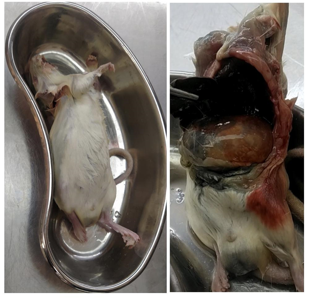
Figure 1: Grossing of submerged rat liver
Figure 2: The above hematoxylin and eosin lung sections show 2A) loss of morphological structure, 2B) loss of nuclear cytoplasmic detail, 2C) constricted sinusoids, 2D) homogenous eosinophilic hyaline like appearance of hepatocyte cells, 2E) clustered hepatocyte cells
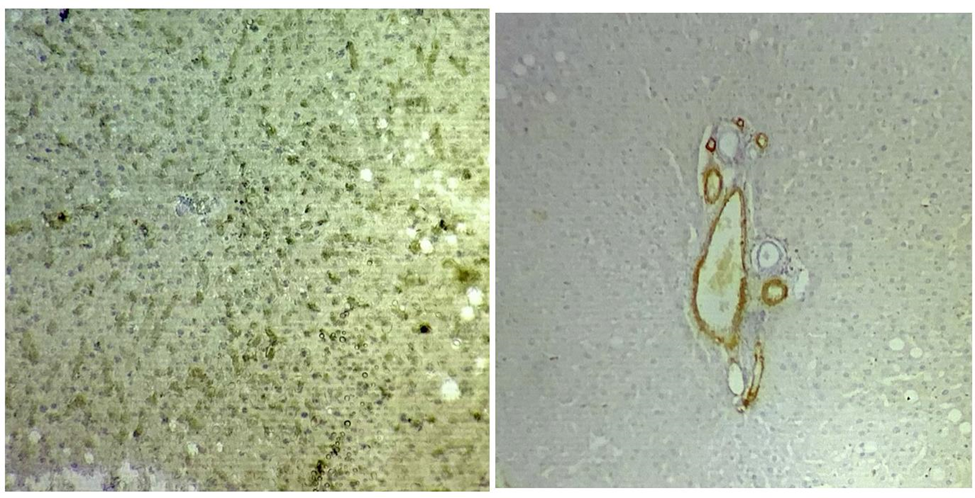
Figure 3: Immunohistochemistry findings were positive for CD34 and SMA
DISCUSSION :
Our present study evaluated, the Histological findings of two days of submerged rat liver which revealed loss of morphological structures, loss of nuclear cytoplasmic detail, constricted sinusoids, homogenous eosinophilic hyaline-like hepatocyte cells, clustered hepatocyte cells, a constricted central vein due to oedema and areas of coagulative necrosis.
A similar study was done by Yukari Tomita on Ultrastructural changes during in situ early postmortem autolysis in kidney, pancreas, liver, heart and skeletal muscle of rats. Cell edema (cell swelling), nuclear chromatin clumping, and the loss of mitochondrial cristae were observed in the hepatocytes one hour after death; the extent of mitochondrial cristae loss increased over time. The hepatocytes showed amorphous thick deposits in mitochondria fifteen hours after death.
Another study by Yukari Tomita on Histology of postmortem changes in rat livers to ascertain hour of death has the following results on liver postmortem autolysis. Male rats held at room temperature (23 ℃), body temperature (37.5 ℃), and cool temperature (5 ℃) after death had their postmortem hepatic tissue changes studied in relation to the hour of death. The temperature at which the body was stored was found to affect the timing and severity of histological postmortem alterations in the liver (5℃ 23 ℃ 37.5 ℃). After 48 hours at 5 ℃, there was decreased stainability of erythrocytes, atrophy of hepatocytes, sinusoidal dilatation, and karyopyknosis of endothelium or Kupffer cells. In contrast to 12 hours at 23 ℃, these observations were made in rat tissues held at 37.5 ℃ after 3-5 hours (29). Hepatocyte atrophy and sinusoidal dilatation were both noticeable in the subcapsular area. There were swollen nuclei and karyopyknosis with an eosinophilic cytoplasm in the epithelia of the interlobular bile ducts (IBD) (30).
According to electron microscopy, the first shift involved nucleoplasmic heterogeneity and peripheral distribution of nuclear chromatin with coagulative cytoplasmic organelles, whereas atrophy of the nuclei with cytoplasm, including rupture and organelle elimination were also noted (31,32). According to the timing of these observations in dead rats housed at various temperatures, postmortem alterations of epithelial nuclei associated with swelling to karyorrhexis (disappearance) via karyopyknosis (33).
Further studies on drowning will be highly effective by interpreting the time of death with liver histopathological findings by conducting it at various intervals of time and varying temperature in submerged rat liver which can enable a better interpretation for forensic identification.
CONCLUSION :
From the study we may conclude that the aspiration of water into the airways causes drowning which results in death. If the findings are further explored, forensic pathology could benefit as a highly reliable technique at low cost.
Acknowledgement
The authors are thankful to Saveetha Institute of Medical and Technical Sciences, Saveetha Dental College and Hospitals, Saveetha University for giving a platform to conduct the study.
Conflict of interest
The authors would like to declare no conflict of interest in the present study.
The present project is funded by
• Saveetha Institute of Medical and Technical Sciences,
• Saveetha Dental College and Hospitals and Saveetha University
• Saveetha University
• Shanmuga Dental Clinic
BIBLIOGRAPHY:
1. A fatal drowning filmed in a private pool: Analysis of the sequences of submersion. Forensic Science International: Reports. 2021 Jul 1;3:100189.
2. Lukaszyk C, Ivers RQ, Jagnoor J. Systematic review of drowning in India: assessment of burden and risk. Inj Prev. 2018 Dec;24(6):451–8.
3. Abelairas-Gómez C, Tipton MJ, González-Salvado V, Bierens JJ. Drowning: epidemiology, prevention, pathophysiology, resuscitation, and hospital treatment. Emergencias. 2019 Aug;31(4):270–80.
4. Haw C, Hawton K. Suicide and Self-Harm by Drowning: A Review of the Literature. Arch Suicide Res. 2016;20(2):95–112.
5. Scarr JP, Buse K, Norton R, Meddings DR, Jagnoor J. Tracing the emergence of drowning prevention on the global health and development agenda: a policy analysis. Lancet Glob Health. 2022 Jul;10(7):e1058–66.
6. Liu L, Villavicencio F, Yeung D, Perin J, Lopez G, Strong KL, et al. National, regional, and global causes of mortality in 5-19-year-olds from 2000 to 2019: a systematic analysis. Lancet Glob Health. 2022 Mar;10(3):e337–47.
7. Rony Varughese RM, Pathoor NN, Ranganathan P, Ganesh PS. Efficacy of Rhamnus frangula extract against Acinetobacter baumannii biofilms: Histopathological evidence from ex vivo goat models. World Academy of Sciences Journal. 2025 Feb 12;7(3):36.
8. Stephenson L, Van den Heuvel C, Byard RW. The persistent problem of drowning - A difficult diagnosis with inconclusive tests. J Forensic Leg Med. 2019 Aug;66:79–85.
9. Armstrong EJ, Erskine KL. Investigation of Drowning Deaths: A Practical Review. Acad Forensic Pathol. 2018 Mar;8(1):8–43.
10. Saukko P, Knight B. Knight’s Forensic Pathology [Internet]. 2015. Available from: http://dx.doi.org/10.1201/b13266
11. Priyadharshini Ranganathan. Clinico-pathological comparison of Salivary TNF-α among Menopause Women with and without Periodontitis - A short study. Bulletin of Stomatology and Maxillofacial Surgery. 2025;21(3). 49-54.doi: 10.58240/1829006X-2025.3-49
12. Yu QC, Lipsky M, Trump BF, Marzella L. Response of human hepatocyte lysosomes to postmortem anoxia [Internet]. Vol. 19, Human Pathology. 1988. p. 1174–80. Available from: http://dx.doi.org/10.1016/s0046-8177(88)80149-7
13. Penttilå A, Ahonen A. Electron microscopical and enzyme histochemical changes in the rat myocardium during prolonged autolysis. Beitr Pathol. 1976 Apr;157(2):126–41.
14. Lakshmi T, Krishnan V, Rajendran R, Madhusudhanan N. Azadirachta indica: A herbal panacea in dentistry - An update. Pharmacogn Rev. 2015 Jan;9(17):41–4.
15. Jain RK, Kumar SP, Manjula WS. Comparison of intrusion effects on maxillary incisors among mini implant anchorage, j-hook headgear and utility arch. J Clin Diagn Res. 2014 Jul;8(7):ZC21–4.
16. Johnson J, Lakshmanan G, M B, R M V, Kalimuthu K, Sekar D. Computational identification of MiRNA-7110 from pulmonary arterial hypertension (PAH) ESTs: a new microRNA that links diabetes and PAH. Hypertens Res. 2020 Apr;43(4):360–2.
17. Keerthana B, Thenmozhi MS. Occurrence of foramen of huschke and its clinical significance. Research Journal of Pharmacy and Technology. 2016;9(11):1835–6.
18. Mootha A, Malaiappan S, Jayakumar ND, Varghese SS, Toby Thomas J. The Effect of Periodontitis on Expression of Interleukin-21: A Systematic Review. Int J Inflam. 2016 Feb 22;2016:3507503.
19. Marofi F, Motavalli R, Safonov VA, Thangavelu L, Yumashev AV, Alexander M, et al. CAR T cells in solid tumors: challenges and opportunities. Stem Cell Res Ther. 2021 Jan 25;12(1):81.
20. Thejeswar EP, Thenmozhi MS. Educational research-iPad system vs textbook system. J Adv Pharm Technol Res. 2015;8(8):1158.
21. Krishna RN, Nivesh Krishna R, Yuvaraj Babu K. Estimation of stature from physiognomic facial length and morphological facial length [Internet]. Vol. 9, Research Journal of Pharmacy and Technology. 2016. p. 2071. Available from: http://dx.doi.org/10.5958/0974-360x.2016.00423.6
22. Menon A, Thenmozhi MS. Correlation between thyroid function and obesity. J Adv Pharm Technol Res. 2016 Oct;9(10):1568.
23. Sriram N, Thenmozhi, Yuvaraj S. Effects of Mobile Phone Radiation on Brain: A questionnaire based study. J Adv Pharm Technol Res. 2015;8(7):867.
24. Subashri A, Thenmozhi MS. Occipital Emissary Foramina in Human Adult Skull and Their Clinical Implications [Internet]. Vol. 9, Research Journal of Pharmacy and Technology. 2016. p. 716. Available from: http://dx.doi.org/10.5958/0974-360x.2016.00135.9
25. Rubika J, Felicita AS, Sivambiga V. Gonial angle as an indicator for the Prediction of Growth Pattern. World J Dent. 2015 Sep;6(3):161–3.
26. Viswanath A, Ramamurthy J, Dinesh SPS, Srinivas A. Obstructive sleep apnea: awakening the hidden truth. Niger J Clin Pract. 2015 Jan;18(1):1–7.
27. Nandhini JST, Babu KY, Mohanraj KG. Size, shape, prominence and localization of gerdy’s tubercle in dry human tibial bones’. J Adv Pharm Technol Res. 2018;11(8):3604.
28. Pratha AA, Thenmozhi MS. A study of occurrence and morphometric analysis on meningo orbital foramen. J Pharm Res [Internet]. 2016; Available from: https://www.indianjournals.com/ijor.aspx?target=ijor:rjpt&volume=9&issue=7&article=024
29. Tomita Y, Nihira M, Ohno Y, Sato S. Ultrastructural changes during in situ early postmortem autolysis in kidney, pancreas, liver, heart and skeletal muscle of rats. Leg Med . 2004 Mar;6(1):25–31.
30. Sri ED, Dhivya Sri E, Priyadharshini R, Sinduja P, Meghashree V. Comparison of Efficacy of the Pap and H & E in Identifying the Various Changes in Liver Samples of High Fat Diet Induced Rat - In vitro Study [Internet]. Journal of Pharmaceutical Research International. 2021. p. 364–80. Available from: http://dx.doi.org/10.9734/jpri/2021/v33i54a33757
31. Jayaraman S, Eswaran A, Rajagopal P, Palanisamy CP, Veerakumar P, Veeraraghavan VP, et al. Neuroendocrine-associated epigenetic factors in cellular senescence: mechanisms and therapeutic implications. Biogerontology [Internet]. 2026 Feb 26;27(2). Available from: http://dx.doi.org/10.1007/s10522-026-10402-7
32. Eswaran A, Natarajan SR, Jayaraman S, Khan JM, Jasmine S, Veeraraghavan VP. Jervine-induced suppression of triple-negative breast cancer (TNBC) cells growth through the regulation of Wnt signaling pathway- an in-silico and in-vitro approach. J Comput Aided Mol Des. 2026 Feb 5;40(1):57.
33. Francis DL, Reddy SS. Microplastics in the pathogenesis of periodontal diseases: A narrative review. Annals of Global Health. 2025 Oct 3;91(1):69.