ROLE OF PERIPARTUM CARDIOMYOPATHY ON MATERNAL MORTALITY DURING THE THIRD TRIMESTER OF PREGNANCY
- MAHNOOR , POST GRADUATE FCPS PART 2 RESIDENT, OBSTETRICS AND GYNECOLOGY, MBBS, SHEIKH ZAYED WOMAN HOSPITAL, CMC @ SMBBMU LARKANA, PAKISTAN.
- SHABNAM NAZ , PROFESSOR OBGYN UNIT 3, OBSTETRICS AND GYNAECOLOGY, FCPS, MRCOG, SHEIKH ZAYED WOMEN HOSPITAL , CMC @SMBBMU LARKANA , PAKISTAN.
- AFSHEEN SHAHID SHAIKH , MEDICAL OFFICER, OBSTETRICS AND GYNECOLOGY, LAST DEGREE: RCPI, RUSTAQ GENERAL HOSPITAL, RUSTAQ CITY OMAN.
- FOUZIA SHAIKH , ASSOCAITE PROFESSOR, OBSTETRICS AND GYNAECOLOGY ,UROGYNAECOLOGY AND PELVIC FLOOR RECONSTRUCTIVE SURGERY, DEPARTMENT, OBSTETRICS AND GYNAECOLOGY, FCPS AND POST FELLOWSHIP IN URO GYNECOLOGY, SHEIKH ZAYED WOMEN HOSPITAL, CMC @SMBBMU LARKANA, PAKISTAN.
- RIMSHA SOLANGI , POST GRADUATE FCPS PART 2 RESIDENT, OBSTETRICS AND GYNECOLOGY, MBBS, SHEIKH ZAYED WOMAN HOSPITAL, CMC @ SMMBMU LARKANA, PAKISTAN.
- KOUSAR , POST GRADUATE FCPS PART 2 RESIDENT, OBSTETRICS AND GYNECOLOGY MBBS, SHEIKH ZAYED WOMAN HOSPITAL, CMC @SMBBMU LARKANA, PAKISTAN.
Article Information:
Abstract:
Objective: To determine the frequency of maternal mortality during the third trimester among women diagnosed with peripartum cardiomyopathy (PPCM) at a tertiary care hospital in Larkana and assess its association with maternal characteristics.Methods: This descriptive cross-sectional study was conducted at the Department of Obstetrics and Gynecology, SMBBMU, Larkana, Pakistan, This research was conducted in a duration of Four months from 15th June 2025- to 15th October 2025, following ethical approval from the Institutional Ethical Review Committee and College of Physicians and Surgeons Pakistan (CPSP). A total of 135 women aged 18–40 years with gestational age ≥28 weeks diagnosed with PPCM were enrolled through non-probability consecutive sampling. Data regarding demographic, obstetric, clinical, laboratory, and echocardiographic characteristics were collected using a structured proforma. PPCM was diagnosed based on heart failure symptoms, NT-pro-BNP >500 pg/mL, left ventricular ejection fraction (LVEF) <45%, and echocardiographic evidence of ventricular dysfunction. Data were analyzed using SPSS version 26, and associations were assessed using the Chi-square test and Fisher’s Exact test.Results: The mean age of participants was 29.4 ± 5.8 years. Maternal mortality was observed in 24 (17.8%) women with PPCM. Shortness of breath (89.6%), fatigue (80.7%), and palpitations (62.2%) were the most common presenting symptoms. Maternal mortality was significantly associated with advanced maternal age (p=0.041), obesity (p=0.022), rural residence (p=0.048), and LVEF <35% (p=0.009).Conclusion: Maternal mortality was considerably high among women with PPCM during the third trimester. Advanced maternal age, obesity, rural residence, and severely reduced LVEF were significantly associated with adverse maternal outcomes.
Keywords:
Article :
INTRODUCTION:
Peripartum cardiomyopathy (PPCM) is a rare but potentially fatal entity characterized by new-onset heart failure within the last month of pregnancy, or following delivery, in women with no previously known cardiac disease. It is defined as decreased left ventricular systolic function (LVSD), usually with a left ventricular ejection fraction (LVEF) <45%, resulting in impaired cardiac output and heart failure signs and symptoms (1). Dyspnoea, fatigue, pedal swelling/pedal, such as oedema, palpitations, and orthopnea, are presenting symptoms of women with PPCM that can resemble physiological changes of pregnancy with a delayed diagnosis. PPCM remains a relevant cause of maternal morbidity and mortality in both low and high-resource settings, despite improvements in global maternal health care (2).
The prevalence of PPCM is heterogeneous across different geographic regions and ethnic groups. Additionally, Nigeria has one of the highest incidences, with 1 in every 102 deliveries being affected compared to many other countries, including Japan, who have reported some of the lowest incidences at close to 1 in 15,533 births (3). Other studies have suggested that genetic variance, socioeconomic status, and access to care may contribute to such differences in disease expression and outcomes in African and African-American women. Studies from Oman (4) and Turkey (5) also clinically highlighted the heavy burden, maternal, and fetal consequences of PPCM, which might lead to heart failure, thromboembolic complications, arrhythmias, and maternal death. International studies have demonstrated a rate of maternal mortality from 18 to 21%, highlighting the poor prognosis associated with the delay in diagnosis and management of patients with PPCM (6).
Pregnancy is a significant hemodynamic stress on the cardiovascular system, particularly with increases in blood volume and cardiac output occurring in the third trimester to supply maternal and fetal demands. In the context of the obstetric changes of pregnancy, these physiological adaptations can trigger or aggravate cardiac dysfunction in women with PPCM and result in life-threatening consequences (7). Echocardiography is the mainstay for diagnosis, proving reduced ventricular function and chamber dilatation, while increased NT-pro-BNP levels aid in confirming heart failure. Early detection and timely management could improve maternal survival and fetal outcome (8).
In Pakistan, PPCM has not yet gained recognition as a major contributor to maternal morbidity and mortality despite significant health and financial burden on suffering families. There is limited local literature on maternal mortality. Outcomes were study-specific, with most available studies assessing overall maternal cardiac disease rather than distinguishing PPCM. Abstract Background The current study assesses the incidence of maternal mortality due to peripartum cardiomyopathy during the third trimester of pregnancy at a tertiary care hospital in Pakistan. The results from this study may enhance early identification, risk stratification, and management of high-risk pregnant women, thereby improving maternal outcomes.
METHODOLOGY:
A descriptive cross-sectional study was conducted through a consecutive sampling technique after approval from the Ethical Review Committee of SMBBMU and the College of Physicians and Surgeons, Pakistan (CPSP). All the procedures were performed in accordance with the ethical principles of the Declaration of Helsinki. All participants provided written informed consent before enrolment in this study. Before the beginning of the study, participants were made aware of the investigation and its aims and were assured regarding their privacy. Data were coded with a number for each participant, and data were only accessible to the research team.
A sample of 135 women was calculated using the WHO sample size calculator, assuming the frequency of maternal mortality in women suffering from peripartum cardiomyopathy to be 18.2% (9), with a confidence interval of 95% and a margin of error as 7%. A further 15% was added in case of potential loss to follow-up. Women presenting with cardiomyopathy between the ages of 18–40 years, gestational age ≥28 weeks, and diagnosed with peripartum cardiomyopathy (PPCM), with any parity or gravida, and willing to participate were included in the study.
Peripartum cardiomyopathy was defined as the presence of clinical features of heart failure, such as shortness of breath, palpitations, and fatigue, with NT-pro-BNP levels >500 pg/mL; reduced left ventricular ejection fraction (LVEF 1.5 mg/dL), pregnancy prolonged past 40 weeks of gestation, or those who were unwilling to participate in the study.
Data collection
Collection of data was done using a predesigned and pretested proforma. Age, gestational age, parity, gravida, weight, height, and BMI (kg/m2), as well as residential status, are collected as baseline demographic and clinical variables. Ultrasonography then confirmed gestational age. The height was measured using a stadiometer (without shoes), while the body weight was recorded in kilograms with participants wearing light clothing, and using a calibrated weighing scale. And BMI was calculated as weight in kilograms and height in meters squared.
Hospital protocol for clinical evaluation and investigations was followed. PPCM was diagnosed based on clinical findings, investigations such as NT-pro-BNP, and references to echocardiography. Maternal mortality related to PPCM was traced during follow-up (until delivery) of enrolled women using pre-defined operational definitions. The principal investigator conducted data collection and assessments under supervision to ensure uniformity and reduce observer bias.
Data analysis
IBM SPSS Statistics version 26.0 software was used to analyze data. The Shapiro-Wilk test was used to assess the normality of continuous variables. Data are expressed as mean ± SD or median (IQR) for normally or non-normally distributed variables, respectively. For categorical variables, including residential status and maternal mortality, results are presented as frequency and percentage values. Stratified analysis was done according to age, gestational age, BMI, parity, gravida, and residential status to see their effect on study outcomes. Constituent Measurement Data were used to perform inferential analysis using Chi-square or Fisher's exact test when indicated. Statistical significance was set at p <0.05.
RESULTS:
A total of 135 women diagnosed with peripartum cardiomyopathy during the third trimester of pregnancy were included in this study. The mean maternal age was 29.4 ± 5.8 years, while the median gestational age at presentation was 34 weeks (IQR: 31–37 weeks). Most participants belonged to the age group of 26–32 years and were from rural areas. The majority of women were multiparous and multigravida. The mean body mass index (BMI) of the study population was 27.1 ± 4.3 kg/m². Table 1.
Table 1: Baseline demographic and obstetric characteristics of study participants
(n=135)
|
Variables |
Frequency (%) / Mean ± SD |
|
Age (years) |
29.4 ± 5.8 |
|
18–25 years |
39 (28.9%) |
|
26–32 years |
61 (45.2%) |
|
33–40 years |
35 (25.9%) |
|
Gestational age (weeks) |
34 (31–37)* |
|
28–32 weeks |
42 (31.1%) |
|
33–36 weeks |
58 (43.0%) |
|
37–40 weeks |
35 (25.9%) |
|
Residential status Urban |
49 (36.3%) |
|
Rural |
86 (63.7%) |
|
BMI (kg/m²) |
27.1 ± 4.3 |
|
Underweight |
11 (8.1%) |
|
Normal weight |
44 (32.6%) |
|
Overweight |
53 (39.3%) |
|
Obese |
27 (20.0%) |
|
Parity |
|
|
Primiparous |
37 (27.4%) |
|
Multiparous |
98 (72.6%) |
*Median (IQR)
BMI = Body mass index
The most frequently observed presenting symptoms among women with PPCM were shortness of breath, fatigue, palpitations, and pedal edema. Echocardiographic assessment demonstrated reduced left ventricular systolic function in all participants. The mean left ventricular ejection fraction (LVEF) was 36.2 ± 5.4%, while elevated NT-pro-BNP levels were observed in all women according to operational diagnostic criteria. Table 2.
Table 2: Clinical and echocardiographic characteristics among women with PPCM
(n=135)
|
Variables |
Frequency (%) / Mean ± SD |
|
Shortness of breath |
121 (89.6%) |
|
Fatigue |
109 (80.7%) |
|
Palpitations |
84 (62.2%) |
|
Pedal edema |
77 (57.0%) |
|
Orthopnea |
52 (38.5%) |
|
NT-pro-BNP >500 pg/mL |
135 (100%) |
|
LVEF (%) |
36.2 ± 5.4 |
|
LVEF <35% |
56 (41.5%) |
|
LVEDD >60 mm |
49 (36.3%) |
Maternal mortality due to PPCM during the third trimester or at delivery was observed in 24 (17.8%) women, whereas 111 (82.2%) women survived until delivery. Graph 1.
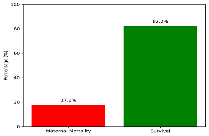
Figure 1 illustrates the frequency of maternal mortality among women diagnosed with PPCM.
Stratification analysis demonstrated that maternal mortality was more frequent among women aged >32 years, obese women, and those with LVEF <35%. Rural residence and multiparity were also associated with relatively higher frequencies of adverse maternal outcomes. Table 3.
Table 3: Association of maternal mortality with maternal characteristics
(n=135)
|
Parameters |
Maternal Mortality n (%) |
Survival n (%) |
p-value |
|
Age 18–25 years |
4 (10.3%) |
35 (89.7%) |
0.041* |
|
Age 26–32 years |
10 (16.4%) |
51 (83.6%) |
|
|
Age 33–40 years |
10 (28.6%) |
25 (71.4%) |
|
|
Urban residence |
6 (12.2%) |
43 (87.8%) |
0.048* |
|
Rural residence |
18 (20.9%) |
68 (79.1%) |
|
|
Obese BMI category |
9 (33.3%) |
18 (66.7%) |
0.022* |
|
LVEF <35% |
15 (26.8%) |
41 (73.2%) |
0.009* |
|
Multiparity |
19 (19.4%) |
79 (80.6%) |
0.173 |
*p-value <0.05 is considered statistically significant
DISCUSSION :
The main objective of this study was to determine the frequency of maternal mortality in 3rd trimester women with peripartum cardiomyopathy. Maternal deaths occurred in 17.8% of women with PPCM in the present study. This finding highlights the gravitas of PPCM as well as its significant contribution to adverse maternal outcomes at the end of pregnancy. Our results are highly comparable to data reported in the international literature. One study in Japan examined maternal mortality related to cardiovascular diseases during pregnancy, reporting this as around 18.2% (10), and another study from the Netherlands documented mortality as nearly 21% in women with severe PPCM (11). Delays in diagnosis and resource-limited health care systems are other contributing factors in observing a higher prevalence of PPCM, as seen elsewhere, particularly in African countries such as Nigeria, where even higher mortality rates have been recorded (12). These similarities reflect that PPCM is still a global public health challenge.
PPCM is also a rare disease with significant regional variation in incidence all over the globe. Incidence is reported to be highest in Nigeria (1:102 deliveries) in studies and lowest in Japan (~1:15533 births) (13). Previous local studies have demonstrated the incidence of close to 1.02 cases per 1,000 deliveries in tertiary care hospitals in Pakistan (14). The relatively high burden seen within developing nations may be attributable to a lack of antenatal care, malnourishment, delayed presentation, and limited access to specialty cardiac services. The maternal mortality rate in our study may be a reflection of similar reasons, including late referrals, poor Economical status, and lack of knowledge regarding cardiac symptoms during pregnancy (15).
The mean maternal age of our study was 29.4±5.8 years, and those aged 33–40 years were significantly at a higher risk for death than younger women. Similar observations were documented globally. From studies, it was found that advanced maternal age is an identifiable precursor of adverse cardiovascular complications and inadequate recovery of ventricular function in PPCM patients from the US as well as Europe (16, 17). Pregnancy among older women has been associated with lower cardiovascular adaptability and comorbid diseases (eg, hypertension, obesity) that are the causes of increased mortality. The same conclusion regarding the predominance of obstetric complications among mothers >30 years of age was derived from Pakistani studies analyzing maternal cardiac ailments (18).
In our study, more than two-thirds of women were multiparous and multigravida, and maternal mortality associated with multiparity was relatively high. Previously published studies also found multiparity to be a significant risk factor for PPCM when analysing data from Indonesia, Nigeria, and South Africa (19, 20). Other studies in Africa reported that multiparous women accounted for >60–70% of PPCM cases (21). Repeated pregnancies are theorized to deliver cumulative hemodynamic stress on the myocardium and put women at risk for heart dysfunction. In contrast, studies from well-developed countries have shown less association between parity and mortality due to earlier diagnosis with a better maternal monitoring system (22). Our study failed to establish a statistically significant association, which may be due to the small sample size.
Area of residence was also an important correlate among maternal outcomes. The risk of maternal mortality among women in rural areas was much higher than that of urban dwellers. Similar findings have been documented in low- and middle-income countries, in which rural populations report reduced access to antenatal care, delayed transport to tertiary health facilities, and lower levels of health literacy (23). The disparities in maternal healthcare in rural Pakistan continue to be a major public health issue (24). A failure to recognize signs of PPCM in rural communities may well account for a considerable proportion of cases with bad maternal outcomes.
Obesity is also a major risk factor in maternal mortality in the current study. Morality was higher in obese women than in those with normal BMI. This finding aligns with international literature that identifies obesity per se as an independent risk factor among women with PPCM (25). Obesity surges cardiac workload and escalates systemic inflammation, endothelial breath is a very short description of system malfunction, causing physical deterioration to renal impairment in hypertension, stimulating excessive stress (requiring physical tranquillising of the nervous system) (26).
In our study, most of them presented with shortness of breath (89.6%), fatigue (80.7%), palpitations (62.2%), and pedal edema (57.0%). This is very similar to international studies that had more than 80% of women with PPCM report dyspnea and fatigue (27). Since many of these symptoms are vague and overlap with the physiological changes associated with pregnancy, diagnosis may be delayed until significant ventricular dysfunction is present. Echocardiography in our cohort demonstrated a mean LVEF of 36.2 ± 5.4%, with mortality being significantly higher among females with LVEF <35%. In addition, studies from South Korea and other international registries showed that severely reduced LVEF is one of the most robust predictors of undesirable maternal outcomes and progressive heart failure (28). Diagnostic accuracy for LVEF <35% in women is 93%, and as such, these women are at significantly greater risk of death, arrhythmias, thromboembolism, and more persistent cardiac dysfunction.
CONCLUSION :
Peripartum cardiomyopathy was associated with a considerable frequency of maternal mortality during the third trimester of pregnancy. Advanced maternal age, obesity, rural residence, and severely reduced left ventricular ejection fraction were important factors associated with adverse maternal outcomes. Early diagnosis, timely echocardiographic evaluation, and multidisciplinary management are essential to reduce maternal mortality and improve outcomes among women with PPCM.
Conflict of Interest: The authors declare no conflict of interest.
Funding: This research received no specific grant from any funding agency in the public, commercial, or not-for-profit sectors.
BIBLIOGRAPHY:
1. Alayed N. Peripartum Cardiomyopathy Complicated by Ventricular Tachycardia during Labor: A Case Report and Literature Review. Open Journal of Obstetrics and Gynecology. 2024.
2. Rodriguez Ziccardi M, Siddique MS. Peripartum Cardiomyopathy. StatPearls. Treasure Island (FL): StatPearls Publishing
3. Copyright © 2026, StatPearls Publishing LLC.; 2026.
4. Isogai T, Kamiya CA. Worldwide Incidence of Peripartum Cardiomyopathy and Overall Maternal Mortality. Int Heart J. 2019;60(3):503-11.
5. Al Riyami N, Al Khayari S, Al Zadjali R, Machado L, Al Madhani A, Al Lawati H. Incidence, Risk Factors, Maternal and Neonatal Outcomes of Peripartum Cardiomyopathy (PPCM) in Oman. Glob Heart. 2023;18(1):23.
6. Kayikcioglu M, Biteker M, Mutluer F, Güzel T, Yılmaz E, Demir E, et al. Baseline Characteristics and Clinical Insights from the ARTEMIS Registry: A Comprehensive Study of Peripartum Cardiomyopathy in Türkiye. Turk Kardiyoloji Dernegi Arsivi-Archives of the Turkish Society of Cardiology. 2024;52:474-83.
7. Iannaccone G, Graziani F, Kacar P, Tamborrino PP, Lillo R, Montanaro C, et al. Diagnosis and management of peripartum cardiomyopathy and recurrence risk. Int J Cardiol Congenit Heart Dis. 2024;17:100530.
8. Yang Y, Lewey J, Arany Z. Cardiovascular complications of pregnancy. J Clin Invest. 2026;136(1).
9. Chen S, Zhou Y, Wu X, Shi S, Wu H, Li P. The Value of Echocardiography Combined with NT-pro BNP Level in Assessment and Prognosis of Diastolic Heart Failure. Comput Math Methods Med. 2022;2022:2102496.
10. Perea Rojas DM, Seni Hernandez CD, Rojas Torres IL, Olivares Olmos M, Garcia Jarava CM, Gaivao Arciniegas DJ, et al. Peripartum Cardiomyopathy: A Case Report of Mortality From a Rare and Potentially Fatal Condition. J Med Cases. 2024;15(8):171-9.
11. Matsushita T, Arakaki T, Sekizawa A, Hasegawa J, Tanaka H, Katsuragi S, et al. Pregnancy-related maternal deaths due to cardiovascular diseases in Japan from 2010 to 2019: an analysis of maternal death exploratory committee data. The Journal of Maternal-Fetal & Neonatal Medicine. 2023;36(1):2175207.
12. Lameijer H, Schutte JM, Schuitemaker NWE, van Roosmalen JJM, Pieper PG. Maternal mortality due to cardiovascular disease in the Netherlands: a 21-year experience. Neth Heart J. 2020;28(1):27-36.
13. Tukeni KN, Asefa ET, Woyimo TG, Gudina EK, Estner H, Haas NA. Peripartum cardiomyopathy: a review of prevalence and treatment trends from an African perspective. Front Cardiovasc Med. 2025;12:1568493.
14. Kitsou V, Dingen H, Lunde T, Engan B, Macsali F, Saeed S. Incidence, characteristics and long-term outcomes of peripartum cardiomyopathy: Insights from the Bergen peripartum cardiomyopathy study. Current Problems in Cardiology. 2026;51(4):103276.
15. Bhatti S, Naz S, Gurmani S, Yasmin H, Atif U, Karim M, et al. Prospective Pakistan Registry of Echocardiographic Screening in Asymptomatic Pregnant Women. JACC: Advances. 2024;3(12_Part_2):101215.
16. Razzaq A, Memon SH, Arain ZI, Khushk SA, Ali A, Bhutto AQ. Outcomes of Newly Diagnosed Patients of Peripartum Cardiomyopathy at Liaquat University Hospital, Hyderabad, Sindh, Pakistan. Journal of Pharmaceutical Research International. 2021;33(31B):10-6.
17. De Viti D, Malvasi A, Busardò F, Beck R, Zaami S, Marinelli E. Cardiovascular Outcomes in Advanced Maternal Age Delivering Women. Clinical Review and Medico-Legal Issues. Medicina (Kaunas). 2019;55(10).
18. Tuvali O, Kuperstein R, Zwas Donna R, Volodarsky I, Vaisbuch E, Shimoni S, et al. Long-Term Outcomes in Women With a History of Peripartum Cardiomyopathy. JACC: Heart Failure. 2026;14(3):102702.
19. Hasan JA, Qureshi A, Ramejo BB, Kamran A. Peripartum cardiomyopathy characteristics and outcome in a tertiary care hospital. J Pak Med Assoc. 2010;60(5):377-80.
20. Dewi TI, Muthiah A, Achmad C, Wirakusumah FF, Anwar R, Nugrahani AD, et al. Association between multiple pregnancy with peripartum cardiomyopathy: a systematic review and meta-analysis. BMC Cardiovasc Disord. 2025;25(1):775.
21. Karaye KM, Ishaq NA, Sa'idu H, Balarabe SA, Talle MA, Isa MS, et al. Incidence, clinical characteristics, and risk factors of peripartum cardiomyopathy in Nigeria: results from the PEACE Registry. ESC Heart Fail. 2020;7(1):235-43.
22. Hammami R, Abdelhedi O, Khanfir F, Shahlaq RSO, Gargouri R, Abid L, et al. Outcomes of peripartum cardiomyopathy in North Africa: insights from a single-center observational study in Tunisia. BMC Pregnancy Childbirth. 2024;24(1):722.
23. Kerpen K, Koutrolou-Sotiropoulou P, Zhu C, Yang J, Lyon J-A, Lima FV, et al. Disparities in death rates in women with peripartum cardiomyopathy between advanced and developing countries: A systematic review and meta-analysis. Archives of Cardiovascular Diseases. 2019;112(3):187-98.
24. Harrington KA, Cameron NA, Culler K, Grobman WA, Khan SS. Rural-Urban Disparities in Adverse Maternal Outcomes in the United States, 2016-2019. Am J Public Health. 2023;113(2):224-7.
25. Hanif M, Khalid S, Rasul A, Mahmood K. Maternal Mortality in Rural Areas of Pakistan: Challenges and Prospects. In: Bacha U, editor. Rural Health. London: IntechOpen; 2021.
26. Cho SH, Leonard SA, Lyndon A, Main EK, Abrams B, Hameed AB, et al. Pre-pregnancy Obesity and the Risk of Peripartum Cardiomyopathy. Am J Perinatol. 2021;38(12):1289-96.
27. Putra ICS, Irianto CB, Raffaello WM, Suciadi LP, Prameswari HS. Pre-pregnancy obesity and the risk of peripartum cardiomyopathy: A systematic review and meta-analysis. Indian Heart Journal. 2022;74(3):235-8.
28. Givertz MM. Peripartum Cardiomyopathy. Circulation. 2013;127(20):e622-e6.
29. Bak M, Youn JC, Bae DH, Lee JH, Lee S, Cho DH, et al. Temporal Trends in Clinical Characteristics and Outcomes for Peripartum Cardiomyopathy: The Nationwide Multicenter Registry Over 20 Years. J Am Heart Assoc. 2024;13(13):e034055.