FORMULATION AND CHARACTERIZATION OF GALLIC ACID LOADED ETHOSOMAL GEL FOR THE ANTI-INFLAMMATORY ACTIVITY
- Sagar pal , NKBR College of Pharmacy and Research Centre, Meerut-Hapur Road, Phaphunda, Meerut, Uttar Pradesh, 245206, India
- Sachin Kumar , NKBR College of Pharmacy and Research Centre, Meerut-Hapur Road, Phaphunda, Meerut, Uttar Pradesh, 245206, India
- Ravi Jayant , NKBR College of Pharmacy and Research Centre, Meerut-Hapur Road, Phaphunda, Meerut, Uttar Pradesh, 245206, India
Article Information:
Abstract:
The present study aimed to formulate and characterize a Gallic acid-loaded ethosomal gel for enhanced anti-inflammatory activity and improved topical drug delivery. Gallic acid-loaded ethosomes were prepared by the cold method followed by probe ultrasonication using soya lecithin, ethanol, and propylene glycol. The prepared ethosomal formulations were evaluated for particle size, polydispersity index (PDI), zeta potential, entrapment efficiency, and stability. The optimized formulation (GE6) exhibited a particle size of 288.9 nm with a PDI of 0.308, indicating uniform vesicle distribution. The zeta potential value of –38.3 mV confirmed good physical stability, while the entrapment efficiency was found to be 88.5 ± 0.26%, demonstrating efficient drug incorporation within the vesicles. The optimized ethosomal suspension was incorporated into a Carbopol 934P gel base and further evaluated for pH, viscosity, gelation temperature, drug content, and in-vitro drug release. The prepared ethosomal gel showed satisfactory physicochemical properties with acceptable pH, viscosity, and spreadability suitable for topical application. In-vitro diffusion studies revealed sustained and controlled drug release from the ethosomal gel, achieving 98.82% cumulative drug release over 24 hours compared to plain gel and drug solution. Release kinetic studies indicated that the formulation followed first-order kinetics. Stability studies demonstrated that the optimized formulation remained stable without significant changes in physical appearance and evaluation parameters. Overall, the developed Gallic acid-loaded ethosomal gel may serve as a promising topical delivery system for prolonged anti-inflammatory activity and enhanced therapeutic effectiveness.
Keywords:
Article :
INTRODUCTION:
Inflammation is a complex biological response triggered by tissue injury, infection, or exposure to harmful stimuli (Medzhitov 2021). Although inflammation serves as a protective mechanism, prolonged or uncontrolled inflammatory responses may lead to various chronic disorders such as psoriasis, rheumatoid arthritis, dermatitis, and other skin-related diseases (Ujiie et al. 2022). Conventional anti-inflammatory therapies are often associated with limitations including poor skin penetration, low bioavailability, frequent administration, and undesirable side effects. Therefore, the development of advanced topical drug delivery systems capable of improving therapeutic efficacy and patient compliance has gained considerable attention in recent years.
Gallic acid is a naturally occurring polyphenolic compound widely distributed in fruits, tea leaves, grapes, and several medicinal plants (Wianowska and Olszowy-Tomczyk 2023). It possesses remarkable pharmacological properties such as anti-inflammatory, antioxidant, antimicrobial, anticancer, and wound-healing activities. The anti-inflammatory activity of gallic acid is mainly attributed to its ability to inhibit inflammatory mediators, suppress oxidative stress, and regulate pro-inflammatory cytokines (Hadidi et al. 2024). Despite its promising therapeutic potential, the clinical application of gallic acid is limited due to its poor permeability across biological membranes, instability, and inadequate retention at the site of application. Hence, an efficient carrier system is required to enhance its topical delivery and therapeutic effectiveness.
Among the various novel vesicular drug delivery systems, ethosomes have emerged as a promising approach for transdermal and dermal drug delivery. Ethosomes are soft, malleable lipid vesicles mainly composed of phospholipids, ethanol, and water (Munir et al. 2024). The presence of a high concentration of ethanol imparts flexibility to the vesicular membrane and enhances drug penetration through the stratum corneum. Ethosomal systems can encapsulate both hydrophilic and lipophilic drugs and improve drug permeation, entrapment efficiency, and stability (Yang et al. 2017). Furthermore, ethosomes facilitate deeper skin penetration and sustained drug release, thereby enhancing the overall therapeutic performance of the incorporated drug. Incorporation of ethosomal suspensions into a gel base further improves the applicability of the formulation for topical administration. Ethosomal gels provide better viscosity, ease of application, prolonged residence time on the skin, and improved patient acceptability. Carbopol-based gels are widely employed due to their excellent gelling properties, compatibility, and ability to provide controlled drug release (Hamdi et al. 2023). The combination of ethosomes with a suitable gel system can therefore offer an effective strategy for the topical management of inflammatory conditions.
The present research work was aimed at the formulation and characterization of gallic acid-loaded ethosomal gel for anti-inflammatory activity. The study involved the preparation of gallic acid-loaded ethosomes using the cold method followed by characterization of vesicle size, polydispersity index, zeta potential, and entrapment efficiency. The optimized ethosomal formulation was further incorporated into a Carbopol gel base and evaluated for physicochemical properties, drug content, spreadability, viscosity, in-vitro drug release, release kinetics, and stability. The developed ethosomal gel was expected to enhance skin permeation, provide sustained drug release, and improve the therapeutic efficacy of gallic acid for topical anti-inflammatory treatment.
Methodology :
Materials
Gallic acid, propylene glycol, soy lecithin, and ethanol used in the present research work were procured from SRL Chennai India Pvt. Ltd., India. Chloroform, Carbopol 934P, and triethanolamine were obtained from Molychem Pvt. Ltd., India. All the chemicals and reagents used in the study were of analytical grade and were used without further purification.
Pre-formulation studies
Organoleptic properties
The organoleptic properties of drug were observed and recorded.
Melting point
Melting point of gallic acid was determined by capillary method (Fernandes and Salgado 2016).
FTIR
The identity of the pure drug sample was confirmed using infrared spectroscopic analysis. For this purpose, the drug was finely mixed with IR-grade potassium bromide and compressed under a pressure of 5.5 metric tonnes using a KBr pellet press to form a transparent pellet. The ready pellet was then placed in the infrared sample holder and analyzed using a (FTIR) spectrophotometer. The spectrum was noted over a wavenumber range of 4000–450 cm⁻¹. The characteristic absorption peaks obtained in the spectrum were interpreted and compared with standard reference data reported in the British Pharmacopoeia to identify and confirm the presence of specific functional groups (Tsioptsias and Tsivintzelis 2022).
Solubility study
Solubility studies done with the pharmacopeial guidelines to evaluate the solubility behavior of Gallic acid. Based on the solubility profile obtained in different solvents, suitable diffusible and dispersible media were selected for drug release and pharmaceutical evaluation studies, respectively. A fixed quantity of Gallic acid was added separately to measured volumes of various solvents, including distilled water, ethanol, dimethyl sulfoxide (DMSO), methanol, chloroform, and PBS, (pH 7.4). The resulting mixtures were subjected to reciprocal shaking at 37 °C for a duration of 5 minutes to ensure adequate interaction between the drug and solvents before visual and analytical assessment.
Method development by UV-Visible spectroscopy method
Preparation of Standard Stock Solution
Weigh out 10 milligrams of gallic acid precisely, then pour it into a 100 mL flask. Dissolve the chemical in a tiny amount of PBS (pH 7.4), then use the same solvent to make up the remaining volume. This results in a standard stock solution with a 100 µg/mL concentration.
Maximum Absorption Wavelength Determination (λmax)
Take 1 mL of the prepared standard and transfer it into a 10 mL volumetric flask. Dilute to volume with distilled water to obtain a working solution of 10 µg/mL. Scan this solution in a UV–Visible-photometer over the wavelength range of 200–400 nm using (PBS, pH 7.4) as the blank. The wavelength show maximum absorbance was documented as the λmax of gallic acid.
3 Construction of Calibration Curve
For calibration, transfer aliquots of 1-6 mL from the standard into distinct 10 mL volumetric flasks. Adjust the final volume of each flask PBS, (pH 7.4) to obtain concentrations of 10-60 µg/mL, respectively. Measure the absorbance of each prepared solution at the estimated λmax (260 nm). Plot a graph of concentration versus absorbance to generate the calibration curve (Syed et al. 2025).
Formulation of Gallic acid loaded ethosomes
Gallic acid-loaded ethosomes were equipped by the cold method followed by probe ultrasonication. Initially, phospholipids, gallic acid, and other lipid constituents were mixed in ethanol in a sealed beaker under continuous magnetic stirring at room temperature. Propylene glycol (PG) was then incorporated into the ethanolic phase. The resulting mixture was maintained at 30 °C using a water bath to ensure uniform mixing. In a separate container, distilled water was heated to the same temp (30 °C) and gradually added to the ethanolic mixture under constant stirring. The dispersion was further stirred for 5 minutes in a closed system to facilitate vesicle formation. Subsequently, the prepared ethosomal suspension was subjected to probe sonication for 10 minutes with a pulse interval of 10 seconds in order to reduce vesicle size and improve homogeneity (Table 1). The final formulation was collected and stored under refrigerated conditions until further use (Agarwal and Gautam 2020).
Table 1: Formulation batch of Gallic acid-loaded ethosomes.
|
Ingredients |
Formulation Codes |
||||||||
|
GE1 |
GE2 |
GE3 |
GE4 |
GE5 |
GE6 |
GE7 |
GE8 |
GE9 |
|
|
Gallic acid (mg) |
100 |
100 |
100 |
100 |
100 |
100 |
100 |
100 |
100 |
|
Ethanol (%v/v) |
30 |
30 |
30 |
30 |
30 |
30 |
30 |
30 |
30 |
|
Propylene glycol (%) |
10 |
10 |
15 |
20 |
15 |
20 |
25 |
25 |
30 |
|
Soy Lecithin (%) |
1 |
1 |
2 |
2 |
3 |
3 |
4 |
4 |
5 |
Characterization of Gallic acid loaded ethosomes
Particle size distribution and Polydispersity index
The PDI and size of particle were analyzed by using the Zeta-sizer Nano ZS instrument manufactured by Malvern in the United States. The examinations were conducted using a conventional laser with a power output of 4 watts and a wavelength of 633 nanometers. The observations were taken at ambient temperature and at a constant angle of 90 degrees. The analysis was conducted using a constant sample volume of 1 ml. The equipment is supplied with suitable software for analyzing PDI and particle size (Huanbutta et al. 2022).
Zeta potential
The zeta-potential is a quantitative measurement that characterizes the strength of the electrostatic forces, either repulsive or attractive, between particles. It is widely recognized that the zeta potential significantly impacts the stability of particle systems. The measurement of dispersion, aggregation, or flocculation provides a comprehensive understanding of their underlying causes and can be used to enhance the formulation of ethosomess. The majority of particulate or macroscopic substances that come into contact with a liquid develop an electric charge on their surfaces.
% Entrapment efficiency (%EE)
The % EE of Gallic acid-loaded ethosomes was analysed using the ultracentrifugation method. In a cold centrifuge, the optimized ethosomes formulation was spun at 11,000 rpm for 2 hours at 4℃. A UV-Vis Spectro-photometer was used to record the absorbance at 278 nm after separating and diluting the supernatant layer with PBS (pH 7.4). By deducting the total quantity of drug in preparation from the amount of unentrapped drug in the supernatant liquid, the percentage of EE was determined indirectly. The drug entrapment percentage was designed using the given equation.
EE (%) = × 100
The total drug loading (T) refers to the initial amount of Gallic acid added, while C represents the portion of drug recovered in the supernatant after processing.
Preparation of ethosomal Gel
Carbopol 934 was selected as the polymeric gelling agent for gel preparation. It was incorporated at a concentration of 0.4% w/v into distilled water with constant stirring to obtain a homogeneous dispersion. The mixture was then kept aside for nearly 2 hours without agitation to allow adequate hydration and swelling of the polymer, leading to gel development. After complete swelling, a suitable preservative was incorporated, followed by the gradual addition of triethanolamine to adjust the pH and finalize the creation of the TEs gel.
Evaluation of Gallic acid loaded TEs gel
Physical Evaluation
The physical appearance of the Gallic acid-loaded ethosomal gel was evaluated by visual examination. Parameters such as clarity, homogeneity, consistency, washability, and organoleptic characteristics were carefully assessed to ensure acceptable formulation quality.
Determination of pH
The pH of the Gallic acid-loaded ethosomal gel done by pH meter. Measurements were done in triplicate, and the mean pH value was calculated from the recorded readings.
Spreadability
The spreadability of the Gallic acid-composed TE was evaluated by analzing the diaattained by 1 g of gel placed among two horizontal glass plates (20 × 20 cm²) after 5 minutes. A standard wt of 500 g was applied on the upper plate to facilitate uniform spreading.
S =
Where, S = Spreadability
M = Weight tied on the upper plate
T = Time taken (second)
L = Length (cm) of glass plates
2.6.4 Viscosity
The viscosity of Gallic acid-loaded ethosomal gel was determined using viscometer. Measurements were recorded with spindle number 6 operated at a rotational speed of 100 rpm.
Extrudability study
The extrudability of the gel mixture was assessed by filled them into collapsible tubes. Extrudability was evaluated based on the amount of force, expressed as the weight (in grams), required to expel a continuous gel ribbon of 0.8 cm length.
Washability
Washability of the mixture was evaluated by applying a little qty of the gel onto the skin surface, and by using warm water rinsing to assess the ease and completeness of its removal.
Drug content
The drug content of the developed in Gallic acid loaded transethsomoal gel was analyzing by pouring 1 gm of formulation into a 10 mL volumetric flask (VF). In this VF a small amount of methanol was added, followed by continous shaking untill the gel was totally dispersed to give a clear solution. 10 ml of methanol was used for final make up. Filtered the solution. Drug concentration in the filterated mixture detrmined by UV spectrophotometer and absorbance was noted.
In-vitro drug release study
The in-vitro delivery profile of the optimized GA ethosomal gel was estimated out to identify the most effective formulation. A dialysis film with a pore size of 0.4 μm was used in a diffusion Cell setup. The membrane was pre-soaked in PBS (pH 7.4) for 24 hours. One gram of the gel was applied to the donor side of the membrane, while the receptor compartment was filled with 13.3 mL of PBS (pH 7.4). The cell was placed on a magnetic stirrer, and a magnetic bead was used to maintain stirring at 400 rpm. The temp was set at 37 ± 1°C throughout the study. Samples of 0.2 mL were check at determined interims (0.5, 1, 1.5, 2, 3, 4, 6, 8, 10, and 12 hours) and replaced with fresh buffer of equal volume. The amount of Gallic acid released was quantified using a confirmed UV spectrophotometric method.
In-vitro drug release kinetics studies
In gel the delivery drug release was studied via numerous models, including first order, zero-order, and the Higuchi equation, to understand the relief profile. Additionally, the Korsmeyer–Peppas model was applied to the release data to determine the underlying mechanism governing the drug release from the formulation.
Stability Study
The retention of drug-loaded vesicles in the gel was tested at different temperatures. The gel was kept in 10 ml airtight vials at 4 ± 2 °C and room temperature for 45 days, and drug content was checked periodically to determine the percentage retained.
Results:
Results of pre-formulation studies
Organoleptic properties
Organoleptic study shows that the Gallic acid is a white to slightly yellowish, crystalline powder or White to off-white amorphous powder, Odourless or may have a faint, characteristic herbal Odor, and slightly bitter.
Melting point analysis
The sample was examined in triplicate, and the average temperature was found to be 254 °C. The average was nearly identical to the recorded MP, which was between 253 and 255°C (Indian Pharmacopoeia).
Solubility study
The solubility of Gallic acid in various solvents was reported, namely water, ether, methanol and chloroform etc. The solubility of Gallic acid with Phosphate buffer (pH 7.4), Ethanol, DMSO, dimethyl formamide, DMSO and distilled water and reported data (Fig. 2).
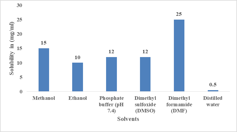
Fig. 2 Solubility studies of Gallic acid.
Plotting calibration curve and Estimation of
Gallic acid showed a peak wavelength at 260nm, consistent with standard values. This wavelength was used for further studies with dilutions ranging from 10–60 µg/ml. The linearity curve was an R² value of 0.9992. (Fig. 3).
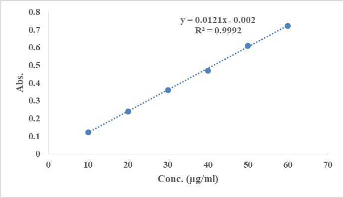
Fig. 3: Linearity curve of Gallic acid.
FT-IR spectrum of Gallic acid
FTIR study of gallic acid has stretching of vibrations of hydroxyl (–OH) and carbonyl (C=O) groups are shown by a wide band between 3600 and 2500 cm⁻¹ and a sharp peak at 1702 cm⁻¹. Additionally, the stretching vibrations of C–C bonds inside the aromatic ring of gallic acid are responsible for the distinctive bands at 1617, 1540, and 1451 cm⁻¹. These frequencies match values that have been previously documented in the literature
Evaluation of Gallic acid loaded ethosomes
Particle size analysis and polydispersity index
The particle size of gallic acid ethosomes formulation (GE1-GE9) was measured with a Litesizer 500 as 267.5-288.9 nm, confirming its nano-scale suitability for skin penetration (Fig. 4). The gallic acid ethosomes formulation GE1-GE9 showed a PDI of 0.308 to 0.62, indicating uniform size distribution and narrow dispersion (Fig. 5).
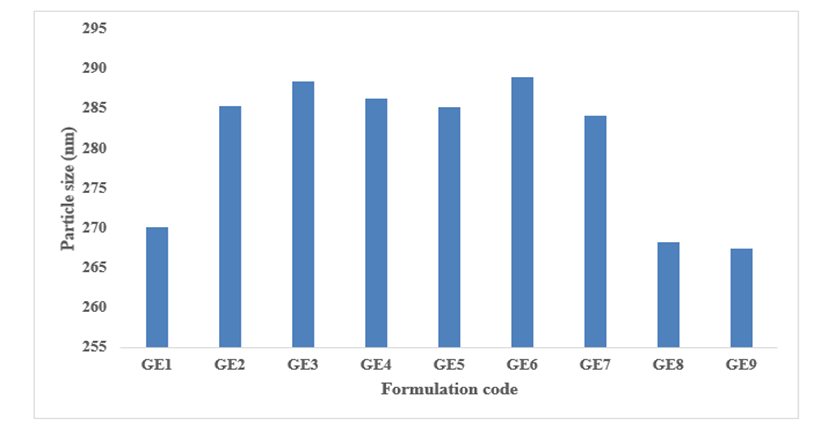
Fig. 4 Particle size of gallic acid loaded ethosomes (GE1-GE6).
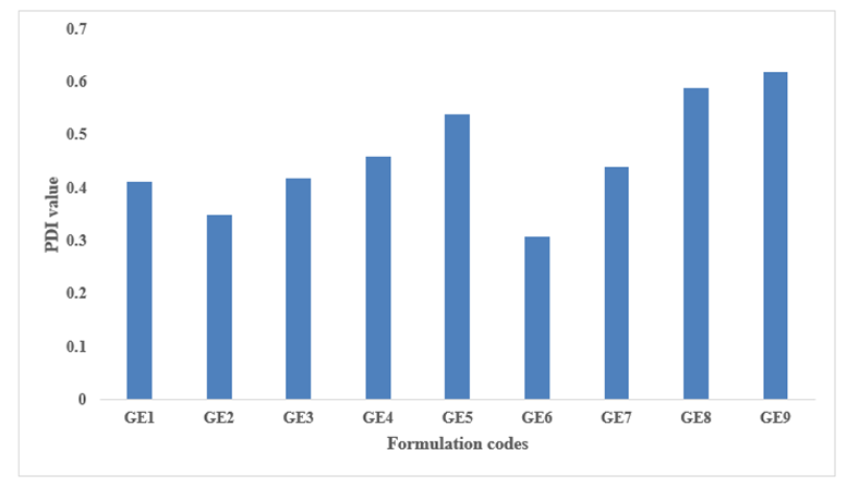
Fig. 5 PDI of gallic acid loaded ethosomes (GE1-GE6).
Zeta potential
Higher zeta potential increases particle repulsion, preventing aggregation and improving stability. The ethosomes showed a zeta potential of gallic acid loaded ethosomes G1 -GE9 was -21.4 to –38.3 mV, confirming good stability. (Fig. 6).
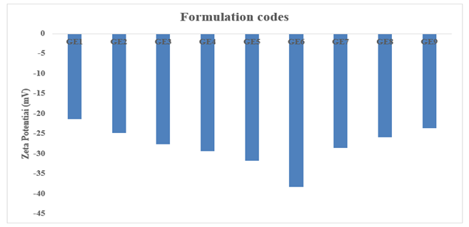
Fig. 6: Zeta potential of gallic acid loaded ethosomes (GE1-GE6).
Entrapment efficiency (%EE)
%EE of the composition ranged from 56 ± 0.70% to 88.5 ± 0.26%. Formulation GE6 showed the highest %EE at 88.5 ± 0.26% (Fig. 7). The results suggest that %EE of ethosomes increases with soya lecithin ratio.
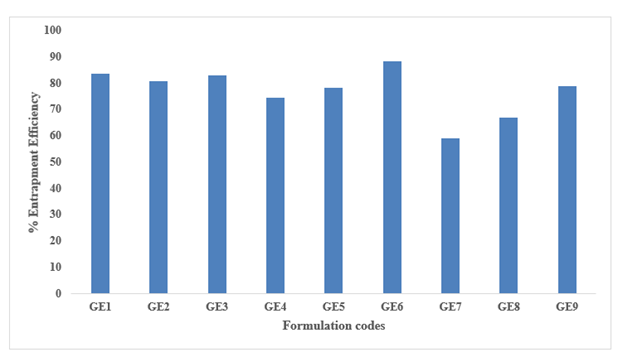
Fig. 7: EE (%) of various compositions of Gallic acid loaded ethosomes.
Characterization of Gallic acid-loaded ethosomes gel
Clarity
The colour of the ethosomes-loaded gel formulations was found to be whitish (refer to Table 2).
pH
The results of the pH prepared formulation were determined to be 6.9±0.02, indicating that the formulation was suitable and better (refer to Table 2).
Gelling Time
Gelling time of ethosomes loaded gel was found to be 65±0.123 sec (refer in Table 2).
Viscosity
The results of viscosity of the prepared formulation were 19237±67.8 cps (refer to Table 2).
Gelation Temperature
The results of the gelation temp of the prepared formulation were 31.34±0.23 (℃). The optimised formulations showed an acceptable gelling temperature (refer to Table 2).
Drug content
The amount of the drug preparation was determined to be 79 ±0.12% (refer to Table 2).
Table 2: Analysis of Gallic acid-loaded ethosomes-loaded gel.
|
Parameters |
Formulation (n=3) |
|
Physical appearance |
Whitish |
|
Drug content (%) |
98 ±0.12 |
|
pH |
5.8±0.02 |
|
Gelling Time (sec.) |
65±0.123 |
|
Viscosity (cps) |
19237±67.8 |
|
Gelation Temperature (℃) |
31.34±0.23 |
(mean ± S.D)
In-vitro diffusion study
The in vitro delivery of drug of Gallic acid from different formulations was carried out over a 24-hour period to compare the release profiles of the Gallic acid solution, plain gel, and ethosomal gel. The Gallic acid solution exhibited a rapid initial release, with 23.12% released within 0.5 h, reaching 54.52% at 2 h and gradually increasing to 78.43% at 24 h. The plain gel showed a slower initial release (12.45% at 0.5 h) compared to the solution, followed by a sustained release pattern, achieving 38.42% at 2 h, 60.12% at 4 h, and 84.32% at 24 h. In contrast, the ethosomal gel demonstrated the most controlled and prolonged release, with only 8.76% drug release at 0.5 h, 28.12% at 2 h, and 45.61% at 4 h, followed by a gradual increase to 82.23% at 12 h and the highest overall release of 98.82% at 24 h (Fig. 8). These results indicate that ethosomal incorporation within the gel matrix significantly retarded the initial drug release and provided sustained delivery, making it a promising approach for prolonged therapeutic action of Gallic acid.
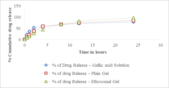
Fig. 8: % Cumulative drug release of drug solution, plain gel, ethosomal gel formulation.
In-vitro drug release kinetics studies
The rate of release was calculated from graph slopes, and R² values were assessed. Release kinetics data [Fig. 9 (A, B, C, D)] showed that the first‑order model had the maximum R², shows the model fit and it is analyzed by making chart of time vs % of drug release giving an R² of 0.9998.
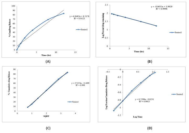
Fig. 9: (A) Zero order plot of ethosomal loaded gel. (B) First order plot of ethosomal loaded gel. (C) Higuchi plot of Ethosomal loaded gel. (D)Peppa's plot of ethosomal loaded gel.
Stability studies
The otimized ethosomal gel formulation remained stable throughout a three-month storage period, showing no detectable alterations in physical characteristics, pH values, or gelation time (Table 3).
Table 3 Stability data of ethosomal loaded gel at 0, 2 and 3 months.
|
Parameters |
For 0 month |
For 2 months |
For 3 months |
|
Physical Appearance |
Whitish |
Whitish |
Whitish |
|
pH |
6.9±0.43 |
6.8±0.28 |
6.9±0.11 |
|
Gelling Time (sec) |
64±0.181 |
65±0.111 |
65±0.165 |
CONCLUSION :
The present investigation successfully developed and characterized a Gallic acid-loaded ethosomal gel intended for topical anti-inflammatory therapy. The optimized ethosomal formulation demonstrated nano-sized vesicles, high entrapment efficiency, good stability, and uniform particle distribution, which are essential for effective skin penetration and drug delivery. Incorporation of ethosomes into the gel system provided suitable physicochemical characteristics and enhanced sustained drug release behavior compared to conventional formulations. The in-vitro diffusion and release kinetic studies confirmed prolonged drug release with improved delivery performance. Furthermore, the formulation remained stable during the storage period, indicating its suitability for pharmaceutical application. Therefore, the developed Gallic acid-loaded ethosomal gel represents a promising and effective carrier system for topical anti-inflammatory treatment with potential for enhanced therapeutic efficacy and patient compliance.
BIBLIOGRAPHY:
1. Agarwal, S, and G Gautam. 2020. Formulation, Development and Evaluation of Atorvastatin Ethosomal Gel. International Journal of Pharmaceutical Investigation 10(4).
2. Fernandes, Felipe Hugo Alencar, and Hérida Regina Nunes Salgado. 2016. Gallic Acid: Review of the Methods of Determination and Quantification. Critical reviews in analytical chemistry 46(3): 257–65.
3. Hadidi, Milad, Rafael Liñán-Atero, Mohammad Tarahi, Marios C Christodoulou, and Fatemeh Aghababaei. 2024. The Potential Health Benefits of Gallic Acid: Therapeutic and Food Applications. Antioxidants 13(8): 1001.
4. Hamdi, Nurul Ain Mohammad, Nurul Atifah Azmi, Nurul Hanis Mohd Sabari, Ahmad Fahmi Harun, and Muhammad Salahuddin Haris. 2023. An Insight into the Use and Advantages of Carbopol in Topical Mucoadhesive Drug Delivery System: A Systematic Review. Journal of Pharmacy 3(1): 53–65.
5. Huanbutta, Kampanart, Napapat Rattanachitthawat, Kunlathida Luangpraditkun, Pornsak Sriamornsak, Vivek Puri, Inderbir Singh, and Tanikan Sangnim. 2022. Development and Evaluation of Ethosomes Loaded with Zingiber Zerumbet Linn Rhizome Extract for Antifungal Skin Infection in Deep Layer Skin. Pharmaceutics 14(12): 2765.
6. Medzhitov, Ruslan. 2021. The Spectrum of Inflammatory Responses. Science 374(6571): 1070–75.
7. Munir, Minahal, Muhammad Zaman, Muhammad Ahsan Waqar, Huma Hameed, and Tehseen Riaz. 2024. A Comprehensive Review on Transethosomes as a Novel Vesicular Approach for Drug Delivery through Transdermal Route. Journal of Liposome Research 34(1): 203–18.
8. Syed, Shoaeb Mohammad, Deepak Jadhav, Somnath Kalegaonkar, Ajinkya Holkar, Satyapal Gaikwad, and Priyanka Thore. 2025. From Lab to Shelf: A Reliable UV Spectrophotometric Technique for Gallic Acid Analysis in Medicines and Nutraceuticals. Asian Journal of Pharmaceutical Research and Development 13(5): 107–11.
9. Tsioptsias, C, and Ioannis Tsivintzelis. 2022. Insights on Thermodynamic Thermal Properties and Infrared Spectroscopic Band Assignments of Gallic Acid. Journal of Pharmaceutical and Biomedical Analysis 221: 115065.
10. Ujiie, Hideyuki, David Rosmarin, Michael P Schön, Sonja Ständer, Katharina Boch, Martin Metz, Marcus Maurer, et al. 2022. Unmet Medical Needs in Chronic, Non-Communicable Inflammatory Skin Diseases. Frontiers in medicine 9: 875492.
11. Wianowska, Dorota, and Małgorzata Olszowy-Tomczyk. 2023. A Concise Profile of Gallic Acid—From Its Natural Sources through Biological Properties and Chemical Methods of Determination. Molecules 28(3): 1186.
12. Yang, Li, Lifang Wu, Dongze Wu, Deshun Shi, Tai Wang, and Xiaoliang Zhu. 2017. Mechanism of Transdermal Permeation Promotion of Lipophilic Drugs by Ethosomes. International journal of nanomedicine: 3357–64.