FORMULATION AND CHARACTERIZATION OF LEFLUNOMIDE LOADED TRANSETHOSOMES LOADED HYDROGEL FOR RHEUMATOID ARTHRITIS
- Sandeep kumar , NKBR College of Pharmacy and Research Centre, Meerut-Hapur Road, Phaphunda, Meerut, Uttar Pradesh, 245206, India
- Sachin Kumar , NKBR College of Pharmacy and Research Centre, Meerut-Hapur Road, Phaphunda, Meerut, Uttar Pradesh, 245206, India
- Ravi Jayant , NKBR College of Pharmacy and Research Centre, Meerut-Hapur Road, Phaphunda, Meerut, Uttar Pradesh, 245206, India
Article Information:
Abstract:
The present study aimed to develop and evaluate a leflunomide-loaded transethosomal hydrogel for enhanced topical delivery and sustained drug release. Leflunomide-loaded transethosomes were prepared using the thin-film hydration method employing Phospholipon® 90 G as phospholipid and sodium cholate as edge activator. The prepared formulations were evaluated for particle size, polydispersity index (PDI), zeta potential, entrapment efficiency, and in-vitro drug release behaviour. Preformulation studies confirmed the identity and purity of leflunomide through melting point determination, FTIR analysis, solubility studies, and UV spectrophotometric analysis. Among the developed formulations, formulation A5 exhibited the most desirable characteristics with particle size of 146 nm, PDI of 0.142, zeta potential of −27.04 mV, and entrapment efficiency of 82%, indicating excellent stability and drug encapsulation efficiency. The optimized transethosomal formulation was incorporated into Carbopol 934-based hydrogel systems (TH1–TH3) and evaluated for pH, viscosity, spreadability, extrudability, washability, and drug content. The developed hydrogels showed satisfactory physicochemical characteristics suitable for topical administration. In-vitro drug release studies demonstrated sustained drug release over 12 h, with formulation TH2 showing the highest cumulative drug release of 94.31%. Release kinetic analysis revealed that the formulation predominantly followed zero-order release kinetics, indicating controlled drug release behaviour. The findings suggest that the developed leflunomide-loaded transethosomal hydrogel may serve as a promising topical drug delivery system with enhanced stability, improved skin penetration, and prolonged therapeutic action.
Keywords:
Article :
INTRODUCTION:
Leflunomide is a widely prescribed disease-modifying anti-rheumatic drug (DMARD) used for the management of rheumatoid arthritis and other chronic inflammatory disorders [1]. Rheumatoid arthritis is a progressive autoimmune disease characterized by persistent synovial inflammation, cartilage degradation, joint stiffness, and gradual destruction of bone tissue, which ultimately leads to functional disability and reduced quality of life [2]. Conventional oral administration of leflunomide has demonstrated significant therapeutic efficacy by inhibiting pyrimidine synthesis and suppressing activated T-lymphocyte proliferation [3]. However, long-term oral therapy is frequently associated with systemic adverse effects such as hepatotoxicity, gastrointestinal irritation, hypertension, and poor patient compliance due to repeated dosing requirements [4]. These limitations necessitate the development of alternative drug delivery systems capable of improving therapeutic effectiveness while minimizing systemic toxicity. The present study was therefore designed to formulate and evaluate leflunomide-loaded transethosomal hydrogel for enhanced topical delivery. The transethosomes were prepared using the thin-film hydration technique employing Phospholipon 90G and sodium cholate as key formulation components. The prepared vesicles were characterized for particle size, polydispersity index, zeta potential, and entrapment efficiency. The optimized formulation was further incorporated into Carbopol 934 hydrogel and evaluated for physicochemical characteristics, spreadability, viscosity, drug content, extrudability, washability, and in-vitro drug release behaviour. The developed transethosomal hydrogel system is expected to provide sustained drug release, improved skin permeation, and enhanced therapeutic performance for the effective management of inflammatory disorders.
Materials and methods:
Materials
Leflunomide was obtained as a gift sample from Genome Pharmaceuticals Pvt. Ltd. Phospholipon 90 G and sodium cholate were procured from Molychem Pvt. Ltd. and were used as the phospholipid and edge activator, respectively. Ethanol and chloroform were purchased from SRL Chem Pvt. Ltd., whereas methanol was obtained from Molychem Pvt. Ltd.. All the chemicals and solvents used in the study were of analytical reagent grade and utilized as received without any further purification.
Pre-formulation studies
Organoleptic properties
The organoleptic properties of the drug, including its appearance and other sensory characteristics, were examined and recorded.
Melting point
Using a digital melting point device and the capillary tube technique, the melting point of LF was determined.
FTIR spectroscopy
The identification of characteristic functional groups and confirmation of the chemical nature of pure drug sample was done by the help of FTIR spectroscopy. The drug was finely ground with IR grade potassium bromide and prepared as a pellet with a hydraulic press at 5.5 metric-ton pressure for analysis. The prepared pellet was then loaded into the sample chamber of the FTIR and the spectrum was obtained in the range of 4000–450 cm⁻¹. The characteristic absorbance peaks of the functional groups obtained in the experiment were compared and analyzed with the reported reference values (B.P., 2009).
Solubility study
The drug's solubility was evaluated using the pharmacopeial method. The selection of suitable diffusible and dispersible media for drug release and pharmaceutical studies was based on the solubility profile of LF in different solvents. Briefly, one part of the drug was mixed separately with varying proportions of different organic solvents, including ethanol, dimethyl sulfoxide (DMSO), and dimethylformamide (DMF). The mixtures were then subjected to reciprocal shaking at 37°C for 5 minutes
Determination of Absorption Maxima (λmax) of LF
The UV-Visible spectrophotometer (Labindia UV 3000+) was used to determine the maximal absorption and purity of LF. The drug was quantitatively estimated using UV-visible spectroscopy. Phosphate-buffered saline (PBS, pH 7.4) was used to generate the medication LF's main stock at a concentration of 1 mg/mL, which was subsequently diluted to a concentration of 10µg/mL. The produced sample's maximum absorption wavelength (λmax) was determined by scanning it in a standard 1 cm quartz cuvette using the UV–Visible spectrophotometer UV–1110, which has a wavelength range of 200 to 800 nm.
Preparation of Standard Calibration Curve of LF in PBS (pH 7.4)
LF stock solution was prepared by dissolving 10 mg of LF in PBS (1000 µg/mL). From this solution, 1 mL was further diluted up to 10 mL with PBS (pH 7.4) to get a secondary stock solution with concentration 100 µg/mL Appropriate aliquots (1 - 10 mL) were then diluted with PBS (pH 7.4) to prepare working concentrations between 10 and 100 µg/mL in separate 10 mL volumetric flasks. The absorbance of each solution was measured at 260 nm that corresponds to the value of the maximum λ for the drug using PBS (pH 7.4) as blank. An absorbance-concentration graph was created. [8,9].
Formulation of LF-loaded transethosomes
Transethosomes loaded with LF were prepared by the thin-film hydration technique. Accurately weighed quantities of LF, Phospholipon® 90 G (phospholipid), and sodium cholate (edge activator) were dissolved in a mixture of methanol and chloroform (3:1, v/v) in a round-bottom flask. The obtained organic solution was subjected to rotary evaporation under reduced pressure to remove the solvents and form a thin, homogeneous lipid film on the inner wall of the flask. After obtaining the film it was then stored for 24 h in a desiccator to ensure that all solvents were removed. After that, the dried lipid film was hydrated with the hydroalcoholic mixture (7:3 v/v) of water and ethanol with continuous stirring for 1 h to form vesicles. The dispersion was then allowed to cool and it was allowed to swell and to become stabilized. The obtained vesicular suspension was further subjected to probe sonication on a titanium probe probe ultrasonicator for 4 min for reducing the size of vesicles and getting homogeneous formulation (Table 1). Lastly, the vesicle size, polydispersity index, zeta potential and entrapment efficiency of the developed LF loaded transethosomes were determined [10,11].
Table 1: Composition of LF-loaded transethosomes
|
Excipients |
Batch Code |
|||||
|
A1 |
A2 |
A3 |
A4 |
A5 |
A6 |
|
|
Leflunomide (mg) |
10 |
10 |
10 |
10 |
10 |
10 |
|
Phospholipon 90 G (mg) |
100 |
100 |
100 |
150 |
150 |
150 |
|
Sodium Cholate (mg) |
10 |
20 |
30 |
10 |
20 |
30 |
|
Ethanol (%) |
30 |
30 |
30 |
30 |
30 |
30 |
|
Water (%) |
70 |
70 |
70 |
70 |
70 |
70 |
|
Methanol: Chloroform (3:1) (mL) |
10 |
10 |
10 |
10 |
10 |
10 |
Characterisation of LF-loaded transethsomoal
Particle size and PDI
The particle size and PDI of the LF loaded transethosomes were measured using a dynamic light scattering (DLS) technique with a particle size analyzer (Litesizer 500) [12].
Zeta potentials
A dynamic light scattering-based zeta potential analyser (Litesizer 500) was used to measure the zeta potential of LF-loaded transethosomes in order to assess the vesicular system's stability and surface charge. To prevent multiple scattering and guarantee precise measurement, the transethosomal dispersion was suitably diluted with distilled water. Measurements were performed at 25°C with an applied electric field after the diluted sample was put into a customised zeta potential cell (folded capillary cell). The average value was provided after each sample was examined three times. The zeta potential values were expressed in mV, where higher absolute values typically range from ±20 mV or above. This indicated good physical stability of the vesicular system, which may be attributed to adequate electrostatic repulsion among the vesicles, thereby preventing particle aggregation. [13].
3 % Entrapment efficiency
The entrapment efficiency of LF-loaded transethosomes was determined using the centrifugation method. Briefly, 1 mL of the formulation was centrifuged at 15,000 rpm for 30 minutes at 4 °C. After centrifugation, the supernatant containing the free drug was collected and analysed at 260 nm using a UV spectrophotometer. The amount of unentrapped drug was measured, and the percentage entrapment efficiency was calculated using the following formula [14].
Preparation of hydrogel base
To prepare a 0.5% Carbopol 934 gel, 0.5 g of Carbopol 934 was accurately weighed and dispersed in 100 mL of distilled water with continuous gentle stirring. The mixture was allowed to hydrate for 24 hours (Table 2). To maintain consistency and improve the gel's texture, 2 mL of glycerine was subsequently added. Methylparaben and propylparaben were incorporated as preservatives. Carbopol gels at 1% and 2% were made [15].
Table 2: Composition of different hydrogel bases.
|
Formulation |
Carbopol (%) |
|
TH1 |
0.5 |
|
TH2 |
1 |
|
TH3 |
2 |
Preparation of transethosomes-loaded hydrogel
To remove the unencapsulated drug, a specified quantity of the optimized L-loaded transethosomal formulation (F5) (equivalent to 10 mg of drug) was centrifuged at 6000 rpm for 20 min. While the sedimented vesicles were collected and redispersed in an appropriate medium, the clear supernatant containing free medication was carefully separated. To create a homogeneous transethosomal gel, the purified transethosomal dispersion was added to the pre-made gel foundation while continuously stirring for 10 minutes at 25 rpm using a mechanical stirrer. Transethosomes equal to 0.1% w/w of LF were added to create the optimal LF-loaded transethosomal formulation (F5). Additionally, gels with different polymer concentrations (0.5%, 1%, and 2% w/w) were made in order to assess how they affected spreadability, viscosity, and drug-release behaviour [16].
Evaluation of LF-Loaded Transethosomal Hydrogel
Physical Evaluation
The prepared LF-loaded transethosomal hydrogel was subjected to visual inspection to evaluate its physical characteristics, including appearance, clarity, consistency, occlusiveness, washability, and other organoleptic properties [17].
Determination of pH
The pH of developed LF loaded transethosomal hydrogel was determined by using a calibrated digital pH meter. Triplicates were measured for all measurements and the mean value recorded [18].
Spreadability
The spreadability of the prepared LF loaded transethosomal hydrogel was calculated by introducing 1 g of the prepared formulation between two square glass plates (20 × 20 cm²). Precise (500 g) was carefully placed on top of the upper plate and the diameter of the spread gel was measured after 5 minutes [19]. The spreadability was calculated using the following equation:
Where:
- S = Spreadability
- M = Weight placed on the upper plate
- L = Length of the glass plate (cm)
- T = Time required for separation (seconds)
Viscosity
A Brookfield viscometer was used to measure the viscosity of the LF-loaded transethosomal hydrogel. Spindle number six was used for the measurement, and it was rotating at 100 rpm [19].
Extrudability study
The extrudability of the formulated hydrogel was evaluated by filling the gel into collapsible tubes. The force required to extrude a ribbon of hydrogel measuring 0.8 cm from the tube was determined in terms of applied weight (g), and the obtained value was used to assess the extrudability of the formulation [20].
Washability
The washability of the prepared hydrogel formulation was assessed by applying a small quantity of gel onto the skin surface followed by rinsing with warm water. The formulation was considered acceptable if it could be removed easily without leaving any visible residue on the skin [21].
Drug content
By precisely transferring 1 g of the formulation into the volumetric flask (10 mL), the drug content of the produced LF loaded in transethosomal hydrogel was ascertained. After adding a little quantity of methanol, the liquid was constantly agitated until the hydrogel completely dispersed, resulting in a clear solution. Methanol was then added to get the final amount up to 10 mL. Spectrophotometric analysis was performed at 260 nm on the residual solution following filtering, and the analytical technique was verified [22].
In-vitro drug release study
In vitro drug release research was conducted to evaluate and improve the formulation of LF loaded transethosomal hydrogel. For the investigation, a Franz diffusion cell assembly with a dialysis membrane pore size of 0.4 μm was utilised. Prior to the experiment, a phosphate buffer solution (pH 7.4) was used to hydrate the dialysis membrane for a whole day. The donor compartment was filled with about 1 g of the transethosomal hydrogel loaded with LF, while the receptor compartment was filled with 13.3 mL of phosphate buffer solution (pH 7.4). To maintain consistent conditions throughout the investigation, the whole assembly was maintained at 37 ± 1°C, and a magnetic stirrer operating at 400 rpm was used to assure agitation. The release experiment lasted for twelve hours. Samples were taken from the receptor compartment and replaced with a fresh phosphate buffer solution of the same volume at different time intervals to maintain the receptor chamber in "sink" condition. The release of the drug from the samples was then quantified by a validated UV spectrophotometric method at 260nm [22].
In-vitro drug release kinetics studies
The in-vitro release data of the optimized leflunomide-loaded transethosomal formulation were subjected to various kinetic models in order to evaluate the mechanism and pattern of drug release. The cumulative percentage drug release obtained from the diffusion study was fitted into different mathematical models including zero-order, first-order, Higuchi, and Korsmeyer–Peppas models. Zero-order kinetics was evaluated by plotting cumulative percentage drug release versus time, whereas first-order kinetics was determined by plotting log cumulative percentage drug remaining versus time. Higuchi’s model was analyzed by plotting cumulative percentage drug release against the square root of time to determine diffusion-controlled release behavior. The Korsmeyer–Peppas model was applied by plotting log cumulative percentage drug release versus log time to identify the mechanism of drug transport from the vesicular system. The regression coefficient (R²) values obtained from each kinetic model were compared, and the model showing the highest R² value was considered as the best-fit model for describing the drug release kinetics of the formulation.
RESULT AND DISCUSSION:
Preformulation Studies
Organoleptic Properties
The organoleptic assessment revealed that leflunomide was obtained as a white-to-off-white crystalline powder with a characteristic bitter taste and an almost odourless nature.
Melting point analysis
The melting point of Leflunomide was determined using a digital melting point apparatus. The analysis was performed in triplicate, and the average melting point was 166.3 °C. The observed value was in close agreement with the reported pharmacopeial range of 164–168 °C (USP), indicating the drug sample's purity and identity. 3.1.3
Solubility study
Distilled water, methanol, ethanol (95%), dimethyl sulfoxide (DMSO), dimethylformamide (DMF), and dichloromethane were among the solvents in which the solubility of LF was assessed (Fig. 2). LF's poor water solubility was confirmed by the findings, which showed very low solubility in distilled water (3.25 µg/mL). On the other hand, far greater solubility was found in organic solvents; ethanol (95%) had the highest solubility (24.1 mg/mL), followed by methanol (23.4 mg/mL), DMSO (20.4 mg/mL), and DMF (15.7 mg/mL). Dichloromethane similarly showed moderate solubility (3.6 mg/mL).
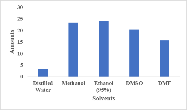
Fig. 2: Solubilities studies of LF.
Preparation of Calibration Curve and Determination of λmax
The UV absorption spectrum of LF was analysed to determine its wavelength of maximum absorption (λmax). The drug exhibited maximum absorbance at 260 nm, consistent with the observed experimental data [Fig. 3 (A)]. Therefore, 260 nm was selected for all subsequent analytical estimations. A UV-visible spectrophotometer was used to measure the absorbance values of standard solutions of LF with concentrations of 5, 10, 15, 20, 25, and 30 µg/mL at 260 nm. The calibration curve demonstrated a direct linear relationship between drug concentration and absorbance. The regression coefficient (R²) value of 0.999 indicated excellent linearity of the developed analytical method [Fig. 3(B)].
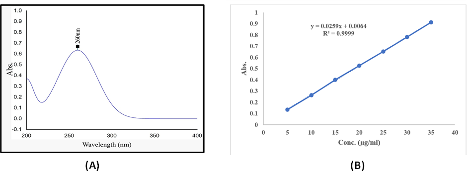
Fig. 3: (A) UV-spectrum of LF in PBS (pH 7.4), (B) Linearity curve of LF.
FT-IR spectrum of chlorhexidine
The FTIR spectrum of LF demonstrated the presence of characteristic functional groups and confirmed the compatibility of the drug with formulation excipients. A broad band detected between 3413–3450 cm⁻¹ was related to O–H stretching vibrations. The peak observed near 2924 cm⁻¹ represented aliphatic C–H stretching. A distinct absorption peak around 1632 cm⁻¹ indicated C=O stretching of carbonyl groups. Additionally, peaks at approximately 1543 cm⁻¹ and 1388 cm⁻¹ were attributed to bending vibrations of the molecular structure. Strong absorption bands within 1233–1020 cm⁻¹ corresponded to C–O stretching vibrations. The overall FTIR findings confirmed that LF retained its chemical structure and showed no noticeable incompatibility with the excipients used in the formulation (Fig. 4).
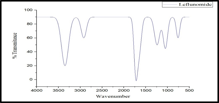
Fig. 4: FTIR Spectrum of LF.
Evaluation of LF-loaded transdermal
Particle size analysis and polydispersity index
An instrument, the Litesizer 500, used to analyse the particle size of the prepared formulations (A1-A6), showed a range of 134-268 nm [Fig. 5 (A)]. The transethosomes' Polydispersity Index (PDI) ranged from 0.18 to 0.24 [Fig. 5 (B)].
Zeta potential
Zeta potential is a widely utilized method for determining the stability of colloidal dispersion. A1 through A6 were discovered to have zeta potentials between -16.08 and -27.04 mV [Fig. 5 (C)].
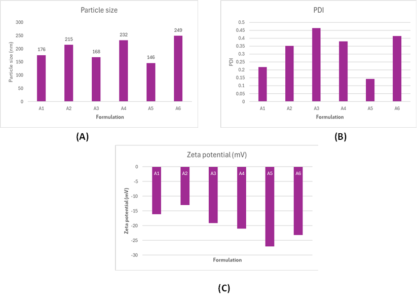
Fig. 5: (A) Particle size of different formulations of LF-loaded transethosomal. (B) PDI of different formulations of LF-loaded transethosomal. (C) Zeta potentail of different formulations of LF-loaded transethosomal.
4 % Entrapment efficiency (%EE)
The entrapment efficiency of Leflunomide-loaded transethosomal formulations (A1–A6) was evaluated, and the results are presented in Fig. 6. Effective drug encapsulation inside the vesicular system was demonstrated by the entrapment efficiency of all formulations, which ranged from 66% to 82%. Due to the optimal amounts of phospholipid, ethanol, and edge activator, which enhance vesicle formation and drug retention, F5 showed the highest entrapment efficiency (82%) among all formulations. Additionally, formulations F3 and F4 had relatively high entrapment efficiencies of 74% and 76%, respectively. F1, on the other hand, had the lowest entrapment effectiveness (66%), presumably as a result of decreased vesicle stability and lower lipid content. The results indicate that increasing lipid concentration and the optimal surfactant level enhance drug entrapment, whereas excessive or insufficient surfactant levels can lead to drug leakage or poor encapsulation. Therefore, F5 was considered the optimized formulation based on its superior entrapment efficiency.
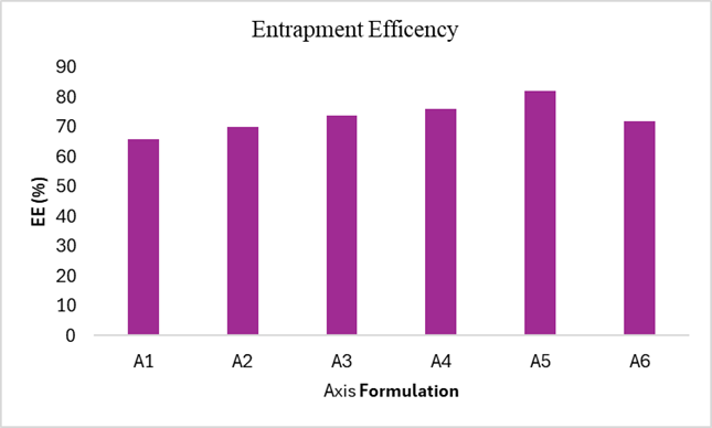
Fig. 6: EE (%) of different formulations of LF-loaded transethosomal.
Characterisation of LF-loaded transethosomal hydrogel
Among the eight prepared transethosomal batches, formulation F5 demonstrated the most desirable characteristics, including a suitable particle size, high entrapment efficiency (%EE), an optimum polydispersity index (PDI), and a favourable zeta potential (ZP). Therefore, batch F5 was selected for incorporation into hydrogel formulations prepared using Carbopol 934P as the gelling agent. Three different hydrogel batches were subsequently developed and evaluated.
Physical Evaluation
The results obtained from the physical evaluation of the prepared hydrogel formulations are presented in Table 3.
Determination of pH
The pH of a topical hydrogel formulation plays a crucial role in maintaining formulation stability, ensuring skin compatibility, and improving patient acceptability. The pH of the prepared LF-loaded transethosomal hydrogels was measured using a digital pH meter fitted with a calibrated electrode probe. The observed pH values were found to be within the acceptable physiological range for topical administration, suggesting a lower possibility of skin irritation upon application. The recorded pH values are presented in Table 3.
Drug Content
The drug content of the developed transethosomal hydrogel formulations was determined by a UV–visible spectrophotometric method in order to quantify the amount of LF uniformly incorporated within the hydrogel system. The results indicated satisfactory and uniform distribution of the drug throughout the hydrogel matrix. The obtained drug content values are summarised in Table 3
Spreadability
The spreadability of the prepared hydrogel formulations was evaluated by the glass slide method to determine the ease with which the formulation could be applied over the skin surface. The diameter of the gel before and after spreading was measured, and the results reflected satisfactory spreading characteristics of the developed hydrogels. The spreadability values are presented in Table 3.
Washability
The formulated hydrogel exhibited satisfactory washability characteristics. The applied formulation could be easily removed from the skin surface using lukewarm water without leaving any visible residue, stain, or film. These findings indicate the suitability of the hydrogel for convenient topical administration and routine use. The washability results are presented in Table 3.
Extrudability
The prepared hydrogel formulation demonstrated good extrudability, showing smooth and continuous extrusion under moderate pressure without blockage or deformation of the gel structure. The extrudability results are provided in Table 3.
Viscosity of Hydrogel
The viscosity of the prepared LF-loaded transethosomal hydrogel was determined using a Brookfield viscometer employing spindle number 7 at a rotational speed of 100 rpm. The obtained viscosity values demonstrated appropriate rheological characteristics suitable for topical administration and ensured proper consistency of the hydrogel formulation. The viscosity data are presented in Table 3.
Table 3: Results of LF-loaded transethosomes hydrogel.
|
Parameters |
TH1 |
TH2 |
TH3 |
|
Physical appearance |
Turbid White |
Turbid White |
Turbid White |
|
pH |
6.6±0.123 |
6.5±0.153 |
6.7±0.159 |
|
Viscosity (cps) |
8167 |
8795 |
8563 |
|
Drug content (%) |
91 |
93 |
92 |
|
Extrudibility |
++ |
+++ |
++ |
|
Spreadability |
++ |
+++ |
++ |
|
Washability |
++ + |
+++ |
++ |
Where: Excellent: +++, Good: ++, Average: +, Poor: -
In-vitro Drug Release of LF-Loaded Transethosomal Hydrogel
The in vitro drug release study of LF-loaded transethosomal formulations (TH1, TH2, and TH3) showed a gradual and sustained release pattern over 12 hours. Among all formulations, TH2 exhibited the highest drug release of 94.31%, followed by TH1 (92.87%) and TH3 (92.16%) at the end of 12 hours. The results indicated controlled and prolonged drug release behaviour of the developed transethosomal formulations (Fig. 7).
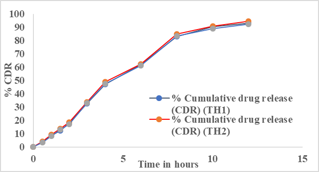
Fig. 7: In-vitro cumulative drug release profile of LF-loaded transethosomal hydrogel.
In-vitro Drug Release Kinetics Studies
The drug release kinetics of the LF-loaded transethosomal hydrogel were evaluated by analysing the release data using different kinetic models. The release rate was determined from the slope of the respective plots, and the coefficient of determination (R²) was calculated to identify the best-fitting model. The kinetic model-fitting data are presented in[Figs. 8(A,B,C,D)]. Among the evaluated models, the zero-order kinetic model showed the highest R² value, indicating that the drug release pattern best followed zero-order kinetics. This finding was further supported by the linear relationship observed in the plot of cumulative percentage drug release versus time (h), which exhibited an R² value of 0.9911.
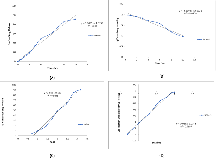
Fig. 8: (A) Zero-order plot for release kinetics. (B) First order plot for release kinetics. (C) Higuchi plot for release kinetics. (D) Peppa's plot for release kinetics.
CONCLUSION :
The present investigation successfully developed and evaluated a leflunomide-loaded transethosomal hydrogel for topical drug delivery. The prepared transethosomal formulations demonstrated favourable vesicular characteristics, including nanosized particle distribution, good stability, and high drug entrapment efficiency. Among all formulations, A5 was identified as the optimized formulation based on its optimum particle size, low PDI, high zeta potential, and maximum entrapment efficiency. Incorporation of the optimized transethosomal dispersion into Carbopol-based hydrogel resulted in formulations with acceptable pH, viscosity, spreadability, washability, and extrudability, confirming their suitability for dermal application. The in-vitro drug release study revealed prolonged and controlled release of leflunomide over 12 h, with TH2 exhibiting the highest cumulative drug release. Furthermore, kinetic modelling suggested that the drug release followed predominantly zero-order kinetics, indicating sustained and controlled release behaviour. Overall, the developed leflunomide-loaded transethosomal hydrogel represents a promising nanocarrier-based topical delivery system that may improve drug permeation, enhance therapeutic efficacy, and minimize systemic side effects associated with conventional therapy.
BIBLIOGRAPHY:
1. Herrmann ML, Schleyerbach R, Kirschbaum BJ. Leflunomide: an immunomodulatory drug for the treatment of rheumatoid arthritis and other autoimmune diseases. Immunopharmacology. 2000;47(2–3):273–89.
2. Jahid M, Khan KU, Ahmed RS. Overview of rheumatoid arthritis and scientific understanding of the disease. Mediterranean journal of rheumatology. 2023;34(3):284–91.
3. Breedveld FC, Dayer JM. Leflunomide: mode of action in the treatment of rheumatoid arthritis. Annals of the rheumatic diseases. 2000;59(11):841–9.
4. Boussios S, Pentheroudakis G, Katsanos K, Pavlidis N. Systemic treatment-induced gastrointestinal toxicity: incidence, clinical presentation and management. Annals of gastroenterology. 2012;25(2):106.
5. Mahanur V, Rajge R, Tawar M. A review on emerging oral dosage forms which helps to bypass the hepatic first pass metabolism. Asian journal of pharmacy and technology. 2022;12(1):47–52.
6. Labie H, Blanzat M. Hydrogels for dermal and transdermal drug delivery. Biomaterials science. 2023;11(12):4073–93.
7. Elkhoury K, Koçak P, Kang A, Arab-Tehrany E, Ellis Ward J, Shin SR. Engineering smart targeting nanovesicles and their combination with hydrogels for controlled drug delivery. Pharmaceutics. 2020;12(9):849.
8. Lakshmana PS, Prakash STNK, Shanmugarathinam A. Development of difference spectrophotometric method for the estimation of leflunomide in tablet dosage form. Chemical Industry and Chemical Engineering Quarterly. 2012;18(3):407–10.
9. Gothi S, Patani P, Rajan V, Tiwari N. A Comprehensive Review of Leflunomide Analytical Analysis: Spectroscopic Technique, Chromatographic Condition, Detection Limits and Validation Parameters. African Journal of Biomedical Research. 2025;28.
10. El-Sonbaty MM, Akl MA, Khalid M, Kassem AA. Does the technical methodology influence the quality attributes and the potential of skin permeation of Luliconazole loaded transethosomes? Journal of Drug Delivery Science and Technology. 2022;68:103096.
11. Sreedharan Nair R, Billa N, Morris AP. In vitro skin permeation and rheological evaluation of a transethosomal formulation containing curcumin-tocotrienol combinations for enhanced topical applications. Journal of Dispersion Science and Technology. 2025;1–11.
12. Yurtsever AG, Ekmekcioglu A, Muftuoglu M, Güngör S, Erdal MS. Formulation development and evaluation of fluvastatin loaded transethosomes: Characterization, stability, in vitro dermal penetration, cytotoxicity and antipsoriatic activity studies. Journal of Drug Delivery Science and Technology. 2024;91:105234.
13. Alam P, Imran M, Jahan S, Akhtar A, Hasan Z. Formulation and characterization of hesperidin-loaded transethosomal gel for dermal delivery to enhance antibacterial activity: comprehension of in vitro, ex vivo, and dermatokinetic analysis. Gels. 2023;9(10):791.
14. Adin SN, Gupta I, Aqil M, Mujeeb M. Baicalin loaded transethosomes for rheumatoid arthritis: Development, characterization, pharmacokinetic and pharmacodynamic evaluation. Journal of drug delivery science and technology. 2023;81:104209.
15. Sarfraz M, Iqbal R, Khan KU, Minhas MU. Carbopol based hydrogels for itopride hydrochloride delivery; synthesis, characterization and comparative assessment with various monomers. Journal of Functional Biomaterials. 2022;13(4):295.
16. Aodah AH, Hashmi S, Akhtar N, Ullah Z, Zafar A, Zaki RM, et al. Formulation development, optimization by box–behnken design, and in vitro and ex vivo characterization of hexatriacontane-loaded transethosomal gel for antimicrobial treatment for skin infections. Gels. 2023;9(4):322.
17. Garg V, Singh H, Bhatia A, Raza K, Singh SK, Singh B, et al. Systematic development of transethosomal gel system of piroxicam: formulation optimization, in vitro evaluation, and ex vivo assessment. AAPS pharmscitech. 2017;18(1):58–71.
18. Nousheen K, Din FU, Jamshaid H, Afza R, Khan SU, Malik M, et al. Metformin HCl-loaded transethosomal gel; development, characterization, and antidiabetic potential evaluation in the diabetes-induced rat model. Drug delivery. 2023;30(1):2251720.
19. Zaki RM, Seshadri VD, Mutayran AS, Elsawaf LA, Hamad AM, Almurshedi AS, et al. Wound healing efficacy of rosuvastatin transethosomal gel, I optimal optimization, histological and in vivo evaluation. Pharmaceutics. 2022;14(11):2521.
20. Majumdar S, Dave R. Formulation study of gel containing pterocarpus santalinus extract for its antiinflammatory activity. World J of Phar and Pharmaceu Sci. 2013;2(6):4951–64.
21. Shrikhande BK, Goupale DC. Development and evaluation of anti-inflammatory oleogels of Bosewellia serrata (gugul) and Curcuma longa (turmeric). Indian Drugs. 2001;38(12):613–6.
22. Sahu A, Rathee S, Saraf S, Raikwar S, Das Bidla P, Pawar RS, et al. Development and characterization of leflunomide and resveratrol loaded nanostructured lipid carrier based in-situ hydrogel system for effective management of rheumatoid arthritis. Journal of Pharmaceutical Innovation. 2025;20(3):107.