Study on the branching pattern of Middle Cerebral Artery – A Cadaveric Study
- Neeraja M , Assistant professor, Department of Anatomy, Chalmeda Anand Rao Institute of Medical Sciences, Karimnagar, Telangana
- Raja Sekhar Katiki Reddi , Associate professor, Department of Anatomy, Government Medical College Quthbullapur, Medchal, Malkajgiri district, Telangana
- Takkallapally Anitha , Professor & Head, Department of Anatomy, Chalmeda Anand Rao Institute of Medical Sciences, Karimnagar, Telangana
Article Information:
Abstract:
Abstract: Background: The middle cerebral artery (MCA) is the largest terminal branch of the internal carotid artery and supplies a major part of the cerebral hemisphere. Its branching pattern is clinically important during cerebral angiography, aneurysm surgery and other neurovascular procedures. Aim: To study the branching pattern of the middle cerebral artery in adult human cadaveric cerebral hemispheres. Materials and Methods: The present descriptive cadaveric observational study was conducted on 30 adult human cerebral hemispheres, including 15 right and 15 left hemispheres. Formalin-fixed brain specimens were dissected carefully to expose the circle of Willis and the MCA. The artery was traced from its origin to the Sylvian fissure, and its branching pattern was classified as bifurcation, trifurcation, quadrifurcation, ramification or single trunk. Data were recorded and analyzed using frequencies and percentages. Results: Bifurcation was the most common MCA branching pattern, observed in 20 specimens (66.67%), followed by trifurcation in 6 specimens (20.00%), ramification in 3 specimens (10.00%), and quadrifurcation in 1 specimen (3.33%). No single trunk pattern was observed. Bifurcation and trifurcation were equally distributed on both right and left sides. Variant branching patterns were observed in 33.33% of specimens. Conclusion: The study showed that bifurcation is the predominant branching pattern of the MCA, while trifurcation, ramification and quadrifurcation are important anatomical variations. Knowledge of these variations is useful for anatomists, radiologists and neurosurgeons in cerebral imaging and operative procedures.
Keywords:
Article :
INTRODUCTION:
The middle cerebral artery (MCA) is the largest and one of the most clinically important terminal branches of the internal carotid artery. It runs laterally through the Sylvian fissure and supplies a wide cortical and deep territory, including major parts of the frontal, parietal and temporal lobes, insula, basal ganglia and internal capsule. Because of this large vascular territory, anatomical knowledge of the MCA is important in understanding cerebrovascular accidents, intracranial aneurysms and neurosurgical approaches involving the lateral cerebral surface [1,2].
The branching pattern of the MCA is known to show considerable anatomical variation. Classically, the MCA is divided into M1, M2, M3 and M4 segments, and its main trunk may terminate as bifurcation, trifurcation, quadrifurcation, ramification or, rarely, as a single trunk. Gibo et al. described the microsurgical anatomy of the MCA and reported that bifurcation was the most common pattern, followed by trifurcation and multiple trunks [2]. Recent cadaveric studies have also shown variable branching patterns; for example, Mohan reported bifurcation in 69.2%, trifurcation in 20% and ramification in 10.8% of specimens [3].
A precise understanding of MCA branching is essential for anatomists, radiologists and neurosurgeons, especially during interpretation of angiographic images, clipping of MCA aneurysms, resection of vascular malformations and planning of microneurosurgical procedures. Variations in early cortical branches and lenticulostriate arteries may increase the risk of intraoperative vascular injury and postoperative neurological deficit. Therefore, a cadaveric study on the branching pattern of the middle cerebral artery is useful for documenting anatomical variations in the studied population and for improving the safety of diagnostic and surgical interventions [3,4].
Materials and Method:
The present study was designed as a descriptive cadaveric observational study to assess the branching pattern of the middle cerebral artery in adult human cadaveric brains.
The study was carried out in the Department of Anatomy of a medical college. Formalin-fixed adult human cadaveric brains available in the department were used for the study.
A total of 30 cerebral hemispheres from adult human cadaveric brains were included in the study. Both right and left cerebral hemispheres were examined for the origin, course and branching pattern of the middle cerebral artery.
Inclusion criteria
Adult human cadaveric brains with intact cerebral hemispheres and clearly identifiable middle cerebral arteries were included in the study. Specimens in which the circle of Willis and MCA region were well preserved were selected.
Exclusion criteria
Cadaveric brains with damaged cerebral hemispheres, poorly preserved vessels, previous dissection injury, vascular distortion, gross pathological lesions, or incomplete visualization of the middle cerebral artery were excluded from the study.
Method of dissection
The formalin-fixed brain specimens were carefully removed from the cranial cavity during routine dissection. The meninges were gently removed to expose the base of the brain and the circle of Willis. The internal carotid artery was identified, and its terminal branches were traced. The middle cerebral artery was then identified as the lateral terminal branch of the internal carotid artery.
The MCA was followed laterally into the lateral sulcus/Sylvian fissure. Fine dissection was carried out using forceps, scissors and blunt probes to expose the main trunk and its branches without damaging the vessels. The M1 segment and the point of division of the MCA were carefully observed.
Parameters studied
The following anatomical parameters were recorded for each cerebral hemisphere:
1. Side of the cerebral hemisphere: right or left.
2. Origin of the middle cerebral artery.
3. Course of the MCA from its origin to the Sylvian fissure.
4. Type of branching pattern of the MCA.
5. Presence of bifurcation, trifurcation, quadrifurcation, ramification or single trunk pattern.
6. Any anatomical variation in the branching pattern.
7. Comparison of MCA branching pattern between right and left cerebral hemispheres.
Classification of branching pattern
The branching pattern of the middle cerebral artery was classified as follows:
|
Branching pattern |
Description |
|
Bifurcation |
Division of MCA into two main trunks |
|
Trifurcation |
Division of MCA into three main trunks |
|
Quadrifurcation |
Division of MCA into four main trunks |
|
Ramification |
Division into multiple small branches without a clear main trunk pattern |
|
Single trunk |
MCA continuing as a single trunk without major division at the observed point |
Data collection
All findings were recorded in a predesigned proforma. Each specimen was numbered, and observations were entered separately for the right and left sides. Photographs were taken wherever required for documentation of normal and variant branching patterns.
Statistical analysis
The collected data were entered into Microsoft Excel and analyzed using descriptive statistics. The frequency and percentage of each type of MCA branching pattern were calculated. Right-sided and left-sided patterns were compared using simple percentage distribution. Results were presented in the form of tables and figures.
Ethical consideration
As the study was conducted on formalin-fixed cadaveric brain specimens available in the Department of Anatomy, no direct human participation was involved. Permission from the concerned department/institutional authority was obtained before conducting the study.
Observation and Results:
Table 1 : Distribution of Cerebral Hemispheres Studied
|
Side of cerebral hemisphere |
Number of specimens |
Percentage |
|
Right side |
15 |
50.00% |
|
Left side |
15 |
50.00% |
|
Total |
30 |
100.00% |
In the present study, equal numbers of right and left cerebral hemispheres were examined. This allowed comparison of the branching pattern of the middle cerebral artery on both sides.
Table 2 : Overall Branching Pattern of Middle Cerebral Artery
|
Branching pattern of MCA |
Number of specimens |
Percentage |
|
Bifurcation |
20 |
66.67% |
|
Trifurcation |
6 |
20.00% |
|
Quadrifurcation |
1 |
3.33% |
|
Ramification |
3 |
10.00% |
|
Single trunk |
0 |
0.00% |
|
Total |
30 |
100.00% |
The most common branching pattern of the MCA was bifurcation, seen in 20 specimens (66.67%). Trifurcation was observed in 6 specimens (20.00%), while ramification was noted in 3 specimens (10.00%). Quadrifurcation was rare and was seen in only 1 specimen (3.33%). No single trunk pattern was observed.
Table 3 : Side-wise Distribution of Branching Pattern of Middle Cerebral Artery
|
Branching pattern |
Right side,( n=15) |
Left side,( n=15) |
Total |
|
Bifurcation |
10(66.67%) |
10(66.67%) |
20 |
|
Trifurcation |
3(20.0%) |
3(20.0%) |
6 |
|
Quadrifurcation |
1(6.67%) |
0(0%) |
1 |
|
Ramification |
1(6.67%) |
2(13.33%) |
3 |
|
Single trunk |
0(0%) |
0(0%) |
0 |
|
Total |
15(100%) |
15(100%) |
30 |
On the right side, bifurcation was observed in 10 specimens (66.67%), trifurcation in 3 specimens (20.00%), quadrifurcation in 1 specimen (6.67%), and ramification in 1 specimen (6.67%). On the left side, bifurcation was also observed in 10 specimens (66.67%), trifurcation in 3 specimens (20.00%), and ramification in 2 specimens (13.33%). Quadrifurcation was not observed on the left side.
Table 4 : Frequency of Major and Variant Branching Patterns of MCA
|
Type of pattern |
Branching pattern included |
Number of specimens |
Percentage |
|
Common pattern |
Bifurcation |
20 |
66.67% |
|
Variant patterns |
Trifurcation, quadrifurcation and ramification |
10 |
33.33% |
|
Total |
|
30 |
100.00% |
Above table 4. showed, the common pattern of MCA branching, that is bifurcation, was observed in 66.67% of specimens. Variant branching patterns were observed in 33.33% of specimens, indicating that anatomical variation in the branching pattern of the MCA is relatively frequent.
DISCUSSION :
The present cadaveric study was conducted on 30 cerebral hemispheres, including 15 right and 15 left hemispheres, to observe the branching pattern of the middle cerebral artery (MCA). In the present study, bifurcation was the most common branching pattern, observed in 20 specimens (66.67%), followed by trifurcation in 6 specimens (20.00%), ramification in 3 specimens (10.00%), and quadrifurcation in 1 specimen (3.33%). No single trunk pattern was observed. Side-wise analysis showed equal bifurcation and trifurcation patterns on both right and left sides, while quadrifurcation was seen only on the right side and ramification was slightly more common on the left side. Thus, the present findings indicate that although bifurcation is the usual pattern, variant branching patterns of MCA are not uncommon.
The findings of the present study are almost similar to the Indian cadaveric study by Mohan, who studied anatomical variations in the MCA and reported bifurcation in 69.2%, trifurcation in 20%, and ramification in 10.8% of specimens [3]. In the present study, bifurcation was observed in 66.67%, trifurcation in 20.00%, and ramification in 10.00%, which closely corresponds with Mohan’s observations. This similarity suggests that bifurcation remains the predominant pattern of MCA division in Indian cadaveric material, while trifurcation and ramification represent important anatomical variations that should be carefully noted during dissection, radiological interpretation and neurosurgical planning. Mohan also emphasized that knowledge of MCA branching pattern is important for minimizing neurovascular morbidity during surgical procedures [3].
The present findings are also comparable with the study by Jeyakumar and Veerapandian, who studied the microsurgical anatomy of MCA in 15 fresh adult cadavers at Madurai Medical College and observed bifurcation in 73% and trifurcation in 27% of cases [5]. In comparison, the present study showed a slightly lower frequency of bifurcation (66.67%) and trifurcation (20.00%), but additionally observed ramification and quadrifurcation patterns. The difference may be due to variation in sample size, method of dissection, population differences and criteria used for classification of branching patterns. However, both studies clearly indicate that bifurcation is the commonest branching pattern of MCA in Indian cadaveric specimens [5].
In another Indian study, Gunnal et al. studied the middle cerebral artery in human cadaveric brains and described important morphological features and branching variations of MCA [3]. Their study highlighted that the MCA has considerable anatomical variation and that detailed knowledge of its branching pattern is essential because the MCA supplies a large functional area of the cerebral hemisphere. The predominance of bifurcation in the present study supports the general observation made by previous anatomical studies that bifurcation is the most frequent terminal division pattern of the MCA, while other patterns such as trifurcation, ramification and multiple division occur less frequently [1].
When compared with a recent Indian microsurgical study by Rakshith et al., the present study also supports the observation that the MCA shows variable branching anatomy in Indian cadavers [6]. Rakshith et al. studied microsurgical anatomy and variations of MCA in Northwest Indian cadavers and emphasized the relevance of MCA variations for aneurysm surgery, stroke evaluation and other neurovascular procedures [6]. The present observation of 33.33% variant branching patterns also supports the view that a considerable proportion of cases may show non-bifurcating patterns, which should be identified before or during operative procedures.
From a clinical point of view, the MCA is the largest terminal branch of the internal carotid artery and supplies important motor, sensory and language-related cortical areas. Variations in branching pattern may influence the site of vascular occlusion, clinical presentation of stroke, interpretation of angiographic images and surgical management of aneurysms or vascular malformations. Jeyakumar and Veerapandian also stated that a detailed angiographic evaluation is mandatory before surgery and that the branching pattern of MCA should be understood clearly because it is a common site for aneurysm formation [5].
|
Study |
Present study |
Mohan et al |
Jeyakumar and Veerapandian et al |
Gunnal et al. |
|
Sample |
30 hemispheres |
60 cadavers / specimens |
15 fresh adult cadavers |
Human cadaveric brains |
|
Bifurcation |
66.67% |
69.20% |
73.00% |
Bifurcation common |
|
Trifurcation |
20.00% |
20.00% |
27.00% |
Trifurcation present |
|
Ramification |
10.00% |
10.80% |
Not specified |
Variations present |
|
Other pattern |
Quadrifurcation 3.33% |
Not reported |
Multiple branches mentioned |
Multiple variations described |
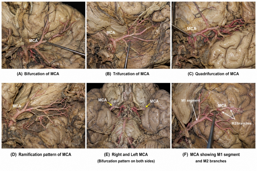
Image 1 : Middle cerebral artery dissection patterns
CONCLUSION :
The present cadaveric study showed that bifurcation was the most common branching pattern of the middle cerebral artery, observed in 66.67% of specimens, followed by trifurcation (20.00%), ramification (10.00%), and quadrifurcation (3.33%). No single trunk pattern was observed. The branching pattern was almost similar on both right and left sides, although minor variations were noted. These findings are comparable with previous Indian studies and confirm that, while bifurcation is the usual MCA pattern, anatomical variations are relatively frequent. Therefore, detailed knowledge of MCA branching is important for anatomists, radiologists and neurosurgeons during cerebral angiography, aneurysm surgery and other neurovascular procedures.
Conflict of Interest : None
BIBLIOGRAPHY:
1. Gunnal SA, Farooqui MS, Wabale RN. Study of middle cerebral artery in human cadaveric brain. Ann Indian Acad Neurol. 2019;22(2):187-194. doi:10.4103/aian.AIAN_315_18.
2. Gibo H, Carver CC, Rhoton AL Jr, Lenkey C, Mitchell RJ. Microsurgical anatomy of the middle cerebral artery. J Neurosurg. 1981;54(2):151-169. doi:10.3171/jns.1981.54.2.0151.
3. Mohan K. A study of the anatomical variations in branching pattern of middle cerebral artery. Indian J Clin Anat Physiol. 2021;8(2):105-109. doi:10.18231/j.ijcap.2021.023.
4. Umansky F, Dujovny M, Ausman JI, Diaz FG, Mirchandani HG. Anomalies and variations of the middle cerebral artery: a microanatomical study. Neurosurgery. 1988;22(6 Pt 1):1023-1027.
5. Jeyakumar R, Veerapandian R. Study of anatomical variations in middle cerebral artery. Int J Sci Stud. 2018;5(12):6-11. doi:10.17354/ijss/2018/72.
6. Rakshith S, Gupta V, Pathak A, et al. Microsurgical anatomy of middle cerebral artery in Northwest Indian cadavers and its neurosurgical implications. J Cerebrovasc Sci. 2024;12(2):91-98.