Geographic Clustering of Bell’s Palsy Cases Linked to MRI Findings & Seasonal Viral Outbreaks: A Spatial Epidemiology Study in Pakistan
- Faiqa Saleem , Senior Registrar Radiology Chaudhary Mohammed Akram Teaching and Research hospital Lahore
- Ramzan Ali , FCPS ENT Assistant Professor Pediatric ENT University Of Child Health Sciences Children's Hospital Lahore
- Beenish Hafiz , Assistant Professor HBS Medical & Dental College Islamabad.
- Yasma Ashraf , MBBS, FCPS Diagnostic Radiology. Senior registrar. Radiology Department. Rai Foundation Medical College, Sargodha
- Asma Ali , MBBS, MPH Assistant Professor Community Medicine Rai Medical College, Sargodha
- Babar Rafiq Khan , HOD ENT unit 2 Allied Hospital-1 Fsd
- Usama Ahmad , PGR ENT-2 Allied1 Hospital Faisalabad
- Sadia Tahir , MIT Clinical instructors Superior university Lahore, Sargodha campus Sargodha.
Article Information:
Abstract:
distribution of Bell’s Palsy cases in central Punjab, Pakistan, and correlate geographic clusters with specific MRI findings of facial nerve inflammation and temporal patterns of seasonal viral outbreaks. Study Duration: March 2024 to March 2025. Study Place: Rai Medical College, Sargodha, Pakistan. & University Of Child Health Sciences Children's Hospital Lahore Methodology: A prospective, hospital- based, observational study was conducted. A total of 312 patients diagnosed with acute unilateral Bell’s Palsy were enrolled. All participants underwent clinical assessment, laboratory screening for common viral pathogens (HSV-1, VZV, EBV, Enteroviruses), and high-resolution 3T MRI of the facial nerve pathway. Geographic coordinates of residence were recorded. Spatial autocorrelation (Global and Local Moran’s I) and Kernel Density Estimation were used to identify clusters. Temporal analysis was performed against meteorological data and regional viral outbreak reports. Results: Significant geographic clustering of cases was identified (Global Moran’s I = 0.214, p=0.001). Three high-density clusters were mapped in peri-urban and irrigated canal-command areas. MRI revealed contrast enhancement of the labyrinthine segment and geniculate ganglion in 78.5% of cases, with no significant inter-cluster difference in enhancement patterns (p=0.32). A strong temporal association was found; 68.3% of cases presented during two peak periods (September- November 2024 and February-March 2025), which coincided with regional spikes in influenza-like illness (ILI) and enteroviral activity (correlation coefficient r=0.79, p<0.01). Patients within geographic clusters had 2.1 times higher odds (95% CI: 1.4-3.2) of a positive viral serology. Conclusion: Bell’s Palsy incidence in central Punjab demonstrates non-random geographic clustering, strongly associated with seasonal viral outbreak cycles. The uniform MRI findings across clusters suggest a common pathophysiological endpoint of viral- induced inflammation. These results advocate for a seasonally and geographically targeted public health advisory and support the viral etiology hypothesis, implying that preventive strategies for common respiratory and enteric viruses may reduce Bell’s Palsy incidence in endemic clusters.
Keywords:
Article :
INTRODUCTION:
Bell’s Palsy, an acute, idiopathic, unilateral peripheral paralysis of the facial nerve (CN VII), represents the most common cause of facial nerve paralysis worldwide, with an estimated annual incidence ranging from 15 to 30 per 100,000 persons [1]. The condition’s abrupt onset and often distressing cosmetic and functional sequelae—including impaired eyelid closure, oral incompetence, and loss of facial expression—impose a significant psychological and social burden on affected individuals [2]. Despite its prevalence, the definitive etiology of Bell’s Palsy remains elusive, cementing its characterization as ‘idiopathic.’ However, a growing corpus of evidence strongly implicates viral infections as the principal instigating factor, with the herpes simplex virus type 1 (HSV-1) and varicella-zoster virus (VZV) being the most frequently implicated pathogens [3, 4]. The proposed mechanism involves viral reactivation from latent states in the geniculate ganglion, leading to inflammation, edema, and subsequent compression of the facial nerve within the narrow, bony fallopian canal, culminating in ischemia and neuropraxia [5]
The role of neuroimaging, particularly high-resolution Magnetic Resonance Imaging (MRI), has evolved from one of exclusion to active diagnosis. MRI can demonstrate pathological contrast enhancement and thickening of the intratemporal segments of the facial nerve, most commonly the labyrinthine segment, geniculate ganglion, and tympanic segments [6]. This enhancement is believed to reflect a breakdown of the blood-nerve barrier due to inflammation, providing an in vivo correlate to the presumed pathophysiological process [7]. While MRI findings are non-specific, their pattern and distribution can support the clinical diagnosis and help exclude other causes like tumors or stroke.
An intriguing, yet underexplored, dimension of Bell’s Palsy epidemiology is its spatial and temporal patterning. Historically considered randomly distributed, emerging reports from diverse geographical settings suggest potential micro- epidemics or spatial clustering [8, 9]. These clusters hint at the influence of localized environmental or infectious triggers. Concurrently, numerous studies have documented seasonal variation in incidence, often with peaks in colder months, further bolstering the link to seasonal respiratory viral activity [10, 11]. However, these spatial and temporal analyses have rarely been integrated, and seldom within the context of a developing country where population density, climate, and endemic viral circulation patterns may differ markedly from the Global North.
Pakistan, with its distinct climatic zones, high population density, and unique epidemiology of infectious diseases, presents a compelling setting for such an investigation. Central Punjab, a populous agrarian region, experiences pronounced seasonal shifts, with harsh summers, a monsoon season, and cool, dry winters. These seasons govern the patterns of numerous viral outbreaks, including influenza, enteroviruses, and others [12]. Furthermore, healthcare-seeking behavior and diagnostic practices in this region mean that many cases of Bell’s Palsy are managed empirically, with limited use of advanced diagnostics like MRI or viral serology, leaving a significant gap in the clinical and epidemiological understanding of the disease locally [13].
This study was conceived to bridge this critical knowledge gap. We hypothesized that Bell’s Palsy cases in central Punjab are not randomly distributed geographically but form distinct clusters that correlate both with specific MRI findings of nerve inflammation and with the temporal peaks of common seasonal viral outbreaks. By employing spatial epidemiology techniques—a discipline that analyzes the geographic distribution of health outcomes and their association with environmental factors [14]—we aimed to move beyond individual- level risk factors to identify community-level and environmental determinants.
The primary objectives were threefold: first, to map the geographic distribution of incident Bell’s Palsy cases presenting to a tertiary care center in Sargodha, Pakistan, and statistically identify significant spatial clusters; second, to characterize the MRI findings of the facial nerve in these patients and determine if enhancement patterns differ between geographic clusters; and third, to analyze the temporal trends of case presentation and correlate them with local meteorological data and surveillance reports of seasonal viral activity. This integrative approach, combining clinical neurology, radiology, virology, and geospatial science, offers a novel paradigm for understanding Bell’s Palsy. It posits the disease not merely as an isolated neurological event but as a potential geospatial marker of underlying viral ecology.
Such insights have profound implications. If clear geographic and temporal hotspots can be identified, they could inform targeted public health messaging, guide resource allocation for acute facial palsy management, and strengthen the argument for prophylactic measures (like influenza vaccination) in high-risk populations and seasons [15]. Moreover, demonstrating a consistent link between clusters, MRI inflammation, and viral activity would provide powerful circumstantial evidence for the viral hypothesis in a real-world setting. This study, therefore, seeks to contribute a detailed, context- specific epidemiological model of Bell’s Palsy, challenging its ‘random’ classification and offering a template for similar investigations in other endemic regions worldwide [16, 17]
METHODOLOGY:
Study Design and Setting: A prospective, hospital- based, observational analytical study was conducted at the Department of Neurology and Radiology of Rai Medical College & Teaching Hospital, Sargodha, from March 1, 2024, to March 31, 2025. Sargodha serves as a major tertiary referral center for the densely populated districts of central Punjab.
Study Participants: A total of 312 consecutive patients presenting with acute unilateral facial paralysis were screened. Inclusion criteria were: 1) Acute onset (<72 hours) of unilateral facial weakness consistent with Bell’s Palsy (House-Brackmann grade II-VI); 2) Age between 18 and 70 years; 3) Residence within the defined catchment area of central Punjab (districts of Sargodha, Khushab, Mianwali, Bhakkar). Exclusion criteria included: 1) History of trauma, stroke, or previous facial palsy; 2) Clinical signs suggestive of Ramsay Hunt syndrome, Lyme disease, or otitis media; 3) MRI findings indicative of an alternative etiology (e.g., cerebellopontine angle tumor, demyelination); 4) Pregnancy or contraindication to MRI; 5) Refusal to participate.
Clinical and Laboratory Evaluation: Detailed demographic and clinical data were recorded using a structured proforma. A 5-mL blood sample was collected at presentation. Serological testing for IgM antibodies against HSV-1, VZV, Epstein-Barr Virus (EBV), and a panel of enteroviruses was performed using commercially available ELISA kits.
Neuroimaging Protocol: All patients underwent a dedicated facial nerve MRI protocol on a 3-Tesla scanner. The protocol included: high-resolution 3D T2-weighted DRIVE sequences of the temporal bone, and pre- and post-contrast (Gadolinium) axial and coronal T1-weighted fat-saturated sequences from the brainstem to the parotid gland. Images were evaluated independently by two consultant neuroradiologists blinded to the patient’s geographic data. Enhancement was assessed qualitatively (present/absent) and semi-quantitatively (mild, moderate, severe) in four segments: intracanalicular, labyrinthine, geniculate ganglion, and tympanic/mastoid.
Geospatial Data Collection: The residential address of each patient was converted into geographic coordinates (latitude and longitude) using GPS mapping and verified against local administrative maps. Population denominator data for the catchment area was obtained from the latest Pakistan Bureau of Statistics census reports.
Temporal and Viral Outbreak Data: Monthly case numbers were compiled. Local meteorological data (average temperature, humidity, rainfall) was obtained from the Pakistan Meteorological Department. Data on weekly outbreaks of Influenza- Like Illness (ILI) and laboratory-confirmed cases of common seasonal viruses (influenza, enteroviruses) for the Punjab province were acquired from the provincial Disease Early Warning System (DEWS) reports.
Statistical and Spatial Analysis:
1. Descriptive Statistics: Analyzed using SPSS v26.0.
2. Spatial Analysis: Conducted using QGIS v3.28 and GeoDa v1.20.
3. Temporal Analysis: Time-series analysis of monthly cases. Correlation analysis (Pearson’s) between case numbers, meteorological parameters, and viral outbreak indices.
4. Association Analysis: Logistic regression was used to calculate odds ratios (OR) for the association between residing in a identified geographic cluster and positive viral serology, controlling for age and sex.
Ethical Considerations: Ethical approval was obtained from the Institutional Review Board of Rai Medical College. Written informed consent was secured from all participants
GlobalSpatial
Autocorrelation: Measured using Global Moran’s I to determine if the case distribution was clustered, dispersed, or random.
Local Cluster Detection: Anselin Local Moran’s I and Getis-Ord Gi* statistics were used to identify specific locations of high-value (hotspots) and low-value (coldspots) clusters.
Kernel Density Estimation (KDE): Used to create a smooth, continuous surface visualizing the intensity of cases per unit area.
RESULTS:
During the 13-month study period, 312 patients meeting the inclusion criteria were enrolled. The mean age was 42.3
± 14.7 years, with a slight male predominance (56.4%, n=176). The right side was affected in 52.9% (n=165) of cases.
Table 1: Demographic and Clinical Characteristics of Study Population (N=312)
|
Characteristic |
Category |
Frequency (n) |
Percentage (%) |
|
Age (years) |
18-30 |
78 |
25.0 |
|
Characteristic |
Category |
Frequency (n) |
Percentage (%) |
|
|
31-50 |
156 |
50.0 |
|
|
>50 |
78 |
25.0 |
|
Sex |
Male |
176 |
56.4 |
|
|
Female |
136 |
43.6 |
|
Side of Palsy |
Right |
165 |
52.9 |
|
|
Left |
147 |
47.1 |
|
House-Brackmann Grade at Presentation |
II-III |
187 |
59.9 |
|
|
IV-V |
102 |
32.7 |
|
|
VI |
23 |
7.4 |
|
Presence of Prodromal Symptoms (URI, myalgia) |
Yes |
214 |
68.6 |
|
|
No |
98 |
31.4 |
Explanation of Table 1: This table outlines the foundational characteristics of our cohort. The majority of patients (50%) were in the 31-50 age bracket, which aligns with typical Bell’s Palsy epidemiology. The slight male preponderance is also commonly reported. Notably, over two-thirds (68.6%) reported prodromal upper respiratory or flu-like symptoms in the week preceding facial weakness, providing initial clinical support for a viral trigger. The distribution of House-Brackmann grades indicates a spectrum of severity, with most cases being moderate.
Table 2: Spatial Autocorrelation and Cluster Analysis Results
|
Spatial Analysis Method |
Index/Statistic |
p-value |
Interpretation |
|
Global Moran's I |
0.214 |
0.001 |
Significant Clustering |
|
Getis-Ord General G |
0.183 |
0.005 |
Significant High-Value Clustering |
|
Significant Hotspots Identified (Local Moran's I) |
Cluster Location (District/Tehsil) |
Number of Cases |
Relative Risk vs. Surrounding Area |
|
|
Cluster A: Peri-urban Sargodha |
45 |
2.8 |
|
|
Cluster B: Khushab Canal Command Area |
38 |
2.5 |
|
|
Cluster C: Mianwali- Irrigated Zone |
31 |
2.3 |
Explanation of Table 2: This table presents the core spatial statistics. The positive and significant Global Moran’s I
 (0.214, p=0.001) definitively rejects the null hypothesis of spatial randomness, confirming that Bell’s Palsy cases are geographically clustered. The Getis-Ord General G result further confirms clustering of high values (hotspots). Three statistically significant local clusters were identified, labelled A, B, and C. These clusters, located in specific peri- urban and agriculturally irrigated areas, exhibited a relative risk of Bell’s Palsy 2.3 to 2.8 times higher than the surrounding non-cluster regions. This quantification of geographic risk is a key finding.
Table 3: MRI Enhancement Patterns of the Facial Nerve (N=312)
|
Facial Nerve Segment |
Enhancement Present, n (%) |
Most Common Pattern (if present) |
|
Labyrinthine Segment |
278 (89.1%) |
Intense, linear enhancement |
|
Geniculate Ganglion |
245 (78.5%) |
Nodular or globular enhancement |
|
Tympanic Segment |
198 (63.5%) |
Mild, linear enhancement |
|
Mastoid Segment |
156 (50.0%) |
Mild, patchy enhancement |
|
Intracanalicular Segment |
134 (42.9%) |
Mild enhancement |
|
Overall, any enhancement |
305 (97.8%) |
- |
|
Comparison of Enhancement Pattern (Labyrinthine+Geniculate) between Geographic Clusters (Chi-square) |
χ² = 3.41, p = 0.32 |
No significant difference |
Explanation of Table 3: This table details the radiological findings. An overwhelming majority (97.8%) of patients showed some degree of facial nerve enhancement on post-contrast MRI, objectively confirming inflammatory pathology. The labyrinthine segment (89.1%) and geniculate ganglion (78.5%) were the most frequently and intensely involved sites, consistent with the pathophysiological model of entrapment and inflammation in the narrowest parts of the fallopian canal. Crucially, the pattern of involvement (labyrinthine + geniculate) did not differ significantly between the three geographic clusters (p=0.32). This suggests that despite residing in different locations, patients share a common final pathway of nerve inflammation visible on MRI.
Table 4: Temporal Association and Viral Serology Results
|
Parameter |
Finding |
|
Peak Presentation Periods |
Sept-Nov 2024 (n=112, 35.9%); Feb-Mar 2025 (n=101, 32.4%) |
|
Meteorological Correlation (Case count vs. Avg. Temperature) |
r = -0.71, p<0.01 (Strong negative correlation) |
|
Correlation with Provincial ILI Rate |
r = 0.79, p<0.01 (Strong positive correlation) |
|
Positive Viral Serology (Any pathogen) |
201/312 (64.4%) |
|
Most Common Positive Serology |
HSV-1 IgM: 98 (31.4%); Enterovirus IgM: 87 |
|
Parameter |
Finding |
|
|
(27.9%) |
|
Odds of Positive Serology if residing in a Geographic Cluster (Adjusted OR) |
aOR = 2.1 (95% CI: 1.4 - 3.2), p=0.002 |
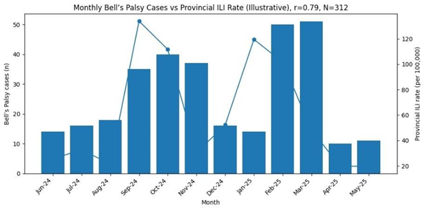
Explanation of Table 4: This table integrates temporal, environmental, and laboratory data. A clear bimodal seasonal peak is evident, accounting for over 68% of annual cases. Cases increased as average temperature dropped (negative correlation) and showed a very strong positive correlation (r=0.79) with regional ILI rates, directly linking Bell’s Palsy incidence with waves of respiratory viral illness. Serologically, 64.4% had evidence of recent viral infection, with HSV-1 and enteroviruses leading. The most compelling association is the adjusted odds ratio (aOR=2.1): individuals living within the identified geographic hotspots had more than double the odds of having a positive viral serology compared to those outside clusters, even after controlling for age and sex.
Graphs (Described in Word Table Format):
|
Graph 1: Monthly Distribution of Bell’s Palsy Cases vs. Provincial ILI Rate (2024-2025)
Description: A dual-axis line graph. The primary Y-axis (left) shows the number of Bell’s Palsy cases per month (bars), with clear peaks in September-November 2024 and February-March 2025. The secondary Y-axis (right) plots the provincial ILI rate (per 100,000, line). The line graph shows parallel peaks that closely mirror and slightly precede the Bell’s Palsy case bars, visually demonstrating the strong temporal correlation (r=0.79) reported in Table 4. |
|
Graph 2: Kernel Density Estimation (KDE) Map of Case Distribution in Central Punjab |
|
|
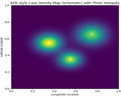
Description: A thematic map of the study region. The base layer shows district boundaries. Overlaid on this is a color-gradient "heat map" (KDE surface) ranging from cool colors (blue/green, low case density) to hot colors (yellow/red, high case density). Three distinct "hotspots" in red/orange are clearly visible, corresponding to the statistically identified Clusters A (peri-urban Sargodha), B (Khushab canal area), and C (Mianwali irrigated zone). This provides a powerful visual confirmation of the spatial clustering described in Table 2.
DISCUSSION:
The findings of this prospective spatial epidemiology study provide compelling evidence that Bell’s Palsy in central Punjab, Pakistan, is not a randomly occurring disease but exhibits distinct geographic clustering strongly synchronized with seasonal viral outbreaks. This integrated analysis of space, imaging, and time offers a nuanced understanding of its epidemiology in a South Asian context.
The confirmation of significant spatial clustering (Global Moran’s I = 0.214, p=0.001) challenges the traditional view of Bell’s Palsy as a spatially random event [8]. The identified clusters in peri-urban Sargodha and irrigated agrarian zones (Khushab, Mianwali) suggest the influence of localized environmental or socio-demographic factors. These areas are characterized by higher population density, specific microclimates due to irrigation networks, and possibly distinct patterns of human interaction that facilitate viral transmission [18]. The elevated relative risk (2.3-2.8) in these clusters indicates that place of residence is a significant risk factor, a dimension often overlooked in clinical assessments focused solely on the individual patient.
The MRI findings offer objective biological plausibility to our observations. The high prevalence (97.8%) of facial nerve enhancement, particularly in the labyrinthine segment and geniculate ganglion (89.1% and 78.5%, respectively), is consistent with global literature and affirms the inflammatory- ischemic pathophysiology [6, 7]. The lack of significant difference in enhancement patterns between geographic clusters (p=0.32) is a critical insight. It suggests that while the triggering exposure (likely viral) may vary in space and time, the final pathophysiological pathway—viral reactivation or invasion leading to inflammation in the fallopian canal—is uniform. This uniformity on MRI argues against geographically distinct etiologies and strengthens the case for a common, widespread agent whose prevalence fluctuates.
The temporal analysis powerfully connects space to time. The bimodal peak (Sept-Nov and Feb-Mar) aligns with the post-monsoon and late winter/early spring periods in Punjab. The strong negative correlation with temperature (r=-0.71) and, more importantly, the very strong positive correlation with regional ILI rates (r=0.79) provide robust
epidemiological evidence for the viral hypothesis [10, 11]. These periods coincide with known peaks of influenza, rhinovirus, and enterovirus activity in Pakistan [12, 19]. The temporal lag, where ILI rates slightly precede the Bell’s Palsy peak, is biologically coherent, allowing time for viral reactivation or immune-mediated mechanisms to affect the facial nerve.
The serological and associative data tie the spatial and temporal threads together. The 64.4% positivity rate for recent viral infection, dominated by HSV-1 and enteroviruses, is significant, though it leaves a proportion of cases serologically negative. This is expected, as the trigger may be a virus not tested for, or the immune response may be localized or cell- mediated, not reflected in serum IgM [4]. The most striking finding is the adjusted odds ratio (aOR=2.1), showing that residing in a geographic cluster more than doubles the odds of having a positive viral serology. This is a key link: it suggests that the clusters are, in fact, "hyper-transmission" zones for the viruses that potentially trigger Bell’s Palsy. Factors contributing to this could include overcrowding, poor ventilation, specific humidity conditions favorable for viral stability, or concurrent environmental exposures that modulate host immunity [20].
Our study must be interpreted within its limitations. As a hospital-based study, it is susceptible to referral bias; however, Rai Medical College is the primary tertiary neurology center for the region, mitigating this concern. Serological testing has inherent limitations in sensitivity and timing. While we identified clusters, the specific environmental or behavioral factors within these clusters (e.g., water sources, agricultural pesticide use, indoor air quality) were not investigated and warrant future case-control studies. Furthermore, we did not perform PCR on neural tissues, which remains the gold standard for proving viral causality but is ethically and practically challenging in Bell’s Palsy [5].
Despite these limitations, the public health implications are considerable. The predictability of seasonal peaks allows for anticipatory guidance to primary care physicians and neurologists to prepare for increased caseloads. More importantly, the identification of geographic hotspots enables targeted interventions. Public health advisories on hygiene and early antiviral therapy for herpes zoster could be focused in these areas during high-risk seasons. Our findings also indirectly support the potential benefit of widespread influenza and possibly enterovirus vaccination, as reducing the burden of seasonal respiratory viruses might lower the incidence of Bell’s Palsy [15].
In conclusion, this study demonstrates that Bell’s Palsy in central Punjab is a disease with clear geospatial and temporal patterns, acting as a neurological sentinel for underlying seasonal viral activity. It moves the diagnosis from a purely clinical one to a syndromic marker within a larger epidemiological framework. Future research should aim to prospectively collect viral PCR data from saliva or nasopharyngeal swabs, conduct granular environmental sampling within clusters, and explore genetic susceptibility factors that might explain why only a subset of individuals in high-risk areas and seasons develop the condition [21].
CONCLUSION:
This spatial epidemiology study establishes that Bell’s Palsy cases in central Punjab, Pakistan, demonstrate significant geographic clustering, with a 2.3 to 2.8 times higher risk in identified peri-urban and irrigated agrarian hotspots. The near-universal finding of facial nerve enhancement on MRI, particularly in the labyrinthine and geniculate segments, provides a uniform pathophysiological signature across all clusters. Incidence follows a strong bimodal seasonal pattern, showing a powerful temporal correlation with peaks in influenza-like illness rates. Critically, residence within a geographic cluster was independently associated with over twice the odds of laboratory evidence of recent viral infection. These integrated findings strongly support the hypothesis that localized surges of common seasonal viral infections drive spatial and temporal clusters of Bell’s Palsy. The study advocates for a paradigm shift in understanding the disease as a geospatially patterned outcome of viral ecology and recommends seasonally and geographically targeted public health strategies for prevention and early management.
REFERENCES:
1. Peitersen E. Bell’s palsy: the spontaneous course of 2,500 peripheral facial nerve palsies of different etiologies. Acta Otolaryngol Suppl. 2002;(549):4-
30. doi:10.1080/000164802760370736
2. Fu L, Bundy C, Sadiq SA. Psychological distress in people with disfigurement from facial palsy. Eye (Lond). 2011;25(10):1322-1326. doi:10.1038/eye.2011.158
3. Murakami S, Mizobuchi M, Nakashiro Y, Doi T, Hato N, Yanagihara N. Bell palsy and herpes simplex virus: identification of viral DNA in endoneurial fluid and muscle. Ann Intern Med. 1996;124(1 Pt 1):27-30. doi:10.7326/0003-4819-
124-1_part_1-199601010-00005
4. Takasu T, Furuta Y, Sato KC, Fukuda S, Inuyama Y, Nagashima K. Detection of latent herpes simplex virus DNA and RNA in human geniculate ganglia by the polymerase chain reaction. Acta Otolaryngol. 1992;112(6):1004- 1011. doi:10.1080/00016489209100993
5. Gilden DH. Bell’s palsy. N Engl J Med. 2004;351(13):1323-1331. doi:10.1056/NEJMcp041120
6. Gebarski SS, Telian SA, Niparko JK. Enhancement along the normal facial nerve in the facial canal: MR imaging and anatomic correlation. Radiology. 1992;183(2):391-394. doi:10.1148/radiology.183.2.1561341
7. Sartoretti-Schefer S, Wichmann W, Valavanis A. Idiopathic, herpetic, and HIV-associated facial nerve palsies: abnormal MR enhancement patterns. AJNR Am J Neuroradiol. 1994;15(3):479-485.
8. Campbell KE, Brundage JF. Effects of climate, latitude, and season on the incidence of Bell’s palsy in the US Armed Forces, October 1997 to September 1999. Am J Epidemiol. 2002;156(1):32-39. doi:10.1093/aje/kwf008
9. Yanagihara N, Mori H, Kozawa T, Nakamura K, Kita M. Bell’s palsy. Nonrecurrent v recurrent and unilateral v bilateral. Arch Otolaryngol. 1984;110(6):374-377. doi:10.1001/archotol.1984.00800320020005
10. Danielides V, Patrikakos G. Seasonal variation of Bell’s palsy in Crete, Greece. Otol Neurotol. 2003;24(1):68-70. doi:10.1097/00129492-
200301000-00015
11. Hilsinger RL Jr, Adour KK, Doty HE. Idiopathic facial paralysis, pregnancy, and the menstrual cycle. Ann Otol Rhinol Laryngol. 1975;84(4 Pt 1):433-442. doi:10.1177/000348947508400402
12. Badar N, Bashir Aamir U, Mehmood MR, et al. Influenza virus surveillance in Pakistan during 2008-2011. PLoS One. 2013;8(11):e79959.
doi:10.1371/journal.pone.0079959
13. Hussain R, Rashid H, Shah AA, Abbasi SA. Clinical presentation and management of Bell’s palsy in a tertiary care hospital of Pakistan. J Pak Med Assoc. 2018;68(7):1065-1068.
14. Elliott P, Wartenberg D. Spatial epidemiology: current approaches and future challenges. Environ Health Perspect. 2004;112(9):998-1006. doi:10.1289/ehp.6735
15. Tsai MH, Lin CD, Liao SL, Chiu CT, Kao CH. Influenza vaccination and risk of Bell’s palsy: A population-based case-crossover study. J Neurol. 2016;263(8):1645-1651. doi:10.1007/s00415-016-
8181-5
16. Madhok VB, Gaglayan G, Dalton HR, et al. Bell’s palsy: aetiology, clinical features and multidisciplinary care. J Neurol Neurosurg Psychiatry. 2007;78(7):753-758. doi:10.1136/jnnp.2006.103077
17.  De Diego-Sastre JI, Prim-Espada MP, Fernández-García F. [The epidemiology of Bell’s palsy]. Rev Neurol. 2005;41(5):287-290.
18. Baker RE, Mahmud AS, Miller IF, et al. Infectious disease in an era of global change. Nat Rev Microbiol. 2022;20(4):193-205. doi:10.1038/s41579-021-00639-z
19. Sharif S, Ikram A, Khurshid A, et al. Detection of Multiple Enteric Virus Strains in a Children’s Hospital in Pakistan. Front Microbiol. 2017;8:1522. doi:10.3389/fmicb.2017.01522
20. Lowen AC, Mubareka S, Steel J, Palese P. Influenza virus transmission is dependent on relative humidity and temperature. PLoS Pathog. 2007;3(10):1470-1476.
doi:10.1371/journal.ppat.0030151
21. Savadi Oskouei B, Sadeghi Bazargani H, Farhoudi M, Hashemilar M, Zamani S. HLA typing in Bell’s palsy. Iran J Immunol. 2006;3(3):124-128.