Anatomical Challenges and Operative Strategies in Cholecystoduodenal Fistula
- Muhammad Rashed , Surgical Specialist, Type D Hospital, Garhi Habibullah, Mansehra, Pakistan,
- Muhammad Salman , Assistant Professor, Jinnah Medical College, Peshawar, Pakistan
- Salman Ahmad Khan , MPhil Scholar, Khyber Medical University, Peshawar, Pakistan
- Muhammad Ishaq , DHQ Hospital, Charsadda, Pakistan
- Muhammad Haroon , MPhil Scholar, Khyber Medical University, Peshawar, Pakistan
- Amir Hamza , MPhil Scholar, Khyber Medical University, Peshawar, Pakistan
Article Information:
Abstract:
The aim of this retrospective observational study was to assess anatomical considerations and surgical approaches in the management of cholecystoduodenal fistula. Patients were treated at a Type D Hospital, Garhi Habibullah, Mansehra, where surgery was an integral part of patient care.Data regarding clinical, operative and postoperative details were obtained from patient records. Data were stratified according to operative method and operative findings. A limited survey of surgeons was also performed to determine surgeon opinion of difficulty of surgery and factors affecting surgical decision-making.Cholecystoduodenal fistula was often seen in the context of chronic gallbladder stones and significant adhesions. Fistula was frequently diagnosed intraoperatively. Open surgery was more likely to be used in complex cases and laparoscopic surgery was linked with a shorter hospital length of stay but higher conversion rate.his indicates that cholecystoduodenal fistula is a complex entity with variable surgical strategies and individual considerations
Keywords:
Article :
INTRODUCTION:
Cholecystoduodenal fistula is a form of internal biliary fistula that develops as a complication of long-standing gallstone disease. It occurs when chronic inflammation of the gallbladder leads to adhesion formation with adjacent structures, most commonly the duodenum. Over time, persistent inflammation and pressure from gallstones result in erosion of the gallbladder wall and eventual formation of a fistulous tract between the gallbladder and the duodenum (1,2).
Although the condition is relatively uncommon, it represents the most frequent type of biliary-enteric fistula, accounting for approximately 70–80% of cases (3). It occurs most commonly in the elderly and patients with a long history of gallbladder stones. The importance of cholecystoduodenal fistula extends from the disease process itself to the diagnostic challenge and surgical complexities.
The clinical presentation can be vague. This may include right upper abdominal pain, vomiting, nausea or dyspepsia, which are also seen in benign gallbladder diseases (4). Severe complications like gallstone ileus, gastrointestinal bleeding or recurrent cholangitis may be present, but these are rare and many are only diagnosed intraoperatively.
Cholecystoduodenal fistula can be challenging to diagnose pre-operatively. Imaging techniques such as ultrasound are used frequently but have poor sensitivity to detect fistulous tracts. The use of computed tomography (CT) has increased the diagnostic sensitivity, particularly when looking for indirect signs like pneumobilia, contracted gallbladder or misplaced gallbladder stones (5). Magnetic resonance cholangiopancreatography (MRCP) can demonstrate biliary anatomy but may not be accessible. So, many patients are still diagnosed at surgery (6).
Cholecystoduodenal fistula is difficult for surgeons. Infection results in scarring and distortion of anatomy. Landmarks like Calot's triangle can become obscured, which poses a risk of bile duct injury during surgery (7). Further, the fistula demands consideration of the duodenal wall, adding complexity.
Traditionally, open cholecystectomy with fistula repair is preferred, especially in the presence of severe inflammation and anatomical distortion (8). Open procedures offer improved visibility and safer dissection techniques. But as experience with minimally invasive surgery grows, more surgeons are considering laparoscopic surgery.
Laparoscopy has benefits including less pain, shorter hospital stay and quicker recovery (9). But its effectiveness in the treatment of cholecystoduodenal fistula is debatable. Adhesions and anatomical distortion can pose technical challenges and a greater risk of conversion to open surgery (10). Thus, patient selection and surgical skill are crucial.
Methods of repairing the fistula vary, such as closure of the duodenal defect, omental patch, or more complex procedures for complicated cases (11). No one technique is strictly followed and treatment is sometimes determined intraoperatively and by the surgeon's discretion.
While there is literature on cholecystoduodenal fistula, the majority of the research has been performed in well-equipped hospitals with advanced diagnostic and surgical equipment. There is paucity of data from low-income countries such as Pakistan where the healthcare system may have limited resources, patients may have lower socioeconomic status and may not have access to advanced imaging and laparoscopic equipment. This may affect the diagnosis and surgical techniques used. So, there is a need for research on this condition in resource-limited countries.
This study sought to evaluate anatomical complicating factors, surgical options and outcomes of cases of cholecystoduodenal fistula managed at Type D Hospital, Garhi Habibullah, Mansehra. It also sought to compare the results with previous studies and to assess surgeons' views on surgical decision-making and challenges.
By combining clinical evidence and surgeons' insights, this research offers practical guidance on the management of this complex condition, and identifies factors that impact surgical outcomes in clinical practice.
MATERIALS AND METHODS:
Study Design and Setting
This is a retrospective observational study that was done at Type D Hospital, Garhi Habibullah, Mansehra with the help of a previously recorded patient data. The patients were included in the study based on having cholecystoduodenal fistula and undergoing surgical management as a part of routine clinical practice between January 2018 and December 2024.
The aim of the study was to assess the anatomical difficulties, operative techniques, and postoperative results of this condition. No intervention of any kind was done experimentally and all the treatment decisions were taken by the treating surgical team according to the normal clinical judgment.
Study Population
Inclusions and exclusions were applied to come with a total of 30 patients included in the study. Hospital medical records and operative logs were used to identify patients.
Inclusion Criteria
· Patients with cholecystoduodenal fistula.
· Radiologically or intraoperatively diagnosed.
· Surgically treated patients.
Exclusion Criteria
· Incomplete medical records
· Patients that are treated conservatively without surgery.
· Lost to follow-up patients.
A small sample of 30 patients was obtained after eliminating a few of the initially identified cases because of incomplete documentation.
Data Collection
The retrospective method was used to collect data based on hospital records. Data were documented in a systematic manner without affecting care of the patients.
· The collected variables were:
· Demographic data (age, gender)
· Clinical presentation
· Comorbid conditions
· Imaging findings
· Mode of diagnosis (preoperative vs intraoperative)
· Open vs Laparoscopic Procedure
· Intraoperative findings
· Postoperative outcomes
· Complications
Grouping of Patients
The two groups of patients were identified according to the type of surgery performed:
1. Open surgery group
2. Laparoscopic surgery group
The decision of the surgical method to be adopted was made by the operating surgeon depending on the condition of the patient, imaging and intraoperative evaluation.
Survey Component
addition to the clinical data, a questionnaire survey was conducted of 22 surgeons involved in hepatobiliary surgery or general surgery.
The survey included questions related to:
· Perceived intraoperative challenges
· Preferred surgical approach
· Factors influencing decision-making
· Perception of the risk of bile duct injury
A structured format was used to record responses and a descriptive analysis was performed.
Outcome Measures
Primary Outcomes
· Operative difficulty
· Length of hospital stay
Secondary Outcomes
· Postoperative complications
· Conversion (open to laparoscopic)
· Intraoperative findings
· Surgeon perception
Definitions
· Preoperative diagnosis: Diagnosis of fistula by imaging prior to surgery
Intraoperative diagnosis: Diagnosis during
RESULTS:
Study Population
The study used a total of 30 patients after the inclusion criteria were applied. All patients were confirmed to have cholecystoduodenal fistula diagnosed either preoperative by imaging or during the surgery.
Most of the patients had a history of chronic gallstone disease with most of the patients reporting with non-specific symptoms in the upper abdomen. There were similar demographic and clinical baseline factors in the study population.
Baseline Characteristics
Table 1 gives a summary of the demographic and clinical profile of the patients who were included in the study
Table 1: Baseline Demographic and Clinical Characteristics
|
Variable |
Value |
|
Mean age (years) |
59.2 ± 11.4 |
|
Female (%) |
18 (60%) |
|
History of gallstones (%) |
27 (90%) |
|
Diabetes mellitus (%) |
9 (30%) |
|
Hypertension (%) |
11 (36.7%) |
The population used in the study was mostly female, with an average age of nearly 60 years old. A significant percentage of the patients reported having a history of gallstone disease in the past, which justifies its importance in the development of fistulas. There were moderate numbers of comorbid conditions like diabetes and hypertension but they did not play a significant role in the type of surgery selected.
Diagnostic Findings and Surgical Approach
The 13 patients (43 percent) were preoperatively diagnosed with cholecystoduodenal fistula and the rest 17 patients (57 percent) were intraoperatively diagnosed.
The use of both open and laparoscopic surgery was according to intraoperative findings and the preference of the surgeon. Table 2 shows the distribution of surgical approaches.
Table 2: Diagnostic Modality and Surgical Approach
|
Parameter |
Frequency |
Percentage |
|
Preoperative diagnosis |
13 |
43% |
|
Intraoperative diagnosis |
17 |
57% |
|
Open surgery |
24 |
80% |
|
Laparoscopic surgery |
6 |
20% |
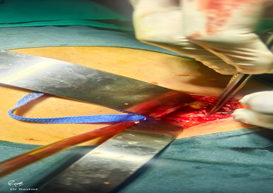
Figure 1 depicts intraoperative observations of thick adhesions
and fistulous communication
Figure 1: Intraoperative image of cholecystoduodenal fistula with dense adhesions between the gallbladder and duodenum, which leads to distorted anatomy.
Open surgery was the dominant approach, performed in the vast majority of cases (80%), particularly in patients with dense adhesions or unclear anatomy. Laparoscopic surgery was attempted in a few cases, but in 2 cases (33.3%) conversion to open surgery was required due to difficulty in dissection and poor visualization.
Intraoperative Findings
Significant anatomical distortion was found in most patients intraoperative assessment. The most regular finding was dense adhesions between the duodenum and the gallbladder that would frequently render normal anatomical landmarks hard to locate.
Table 3: Intraoperative Findings
|
Finding |
Frequency |
Percentage |
|
Dense adhesions |
24 |
80% |
|
Distorted Calot’s triangle |
21 |
70% |
|
Large impacted gallstones |
18 |
60% |
|
Inflammatory tissue changes |
20 |
66.7% |
These results were associated with the augmented operative challenge. The triangle of Calot was not always distinctly defined and in most instances, the conventional method of surgery had to be changed. Dissection was also complicated by the presence of large impacted gallstones.
Complexity during the operations and anatomical distortion in Figure 1, dense adhesions as well as fistulous communication can be seen.
Postoperative Outcomes
The outcomes of the two surgical modalities differed postoperative, in terms of recovery and recurrence of complications. Table 4 summarizes these results.
Table 4: Postoperative Outcomes
|
Outcome |
Open Surgery (n=24) |
Laparoscopic (n=6) |
|
Mean hospital stay (days) |
8.5 ± 2.1 |
5.4 ± 1.6 |
|
Overall complications |
6 (33%) |
2 (16%) |
|
Bile leak |
2 (11%) |
1 (8%) |
|
Wound infection |
3 (17%) |
1 (8%) |
Laparoscopic surgery patients stayed in the hospital a shorter period and experienced fewer complications than those who had an open surgery. Nevertheless, technically challenging cases were more likely to be operated open, possibly explaining the increased rate of complications recorded in this population.
None of the mortality was reported in this study. All complications were treated conservatively or by performing minor procedures and none of the patients had to be re-operated.
Surgeon Survey Findings
The survey was structured and carried out among surgeons, revealing that the most important intraoperative challenges were the anatomical distortions (73) and the risk of bile duct injury (64). Preoperative planning did not have a major impact on the decision of the surgical approach to be used, with intraoperative findings and surgeon experience playing the major role
DISCUSSION:
This study examined the anatomy issues and the surgical approach in the case of a patient with cholecystoduodenal fistula based on retrospective clinical outcomes of the Type D Hospital, Garhi Habibullah, Mansehra. The results indicate that despite the fact that this condition is not very common, it still poses serious challenges to diagnosis and surgery in regular clinical settings.
The low rate of preoperative diagnosis was one of the most significant findings of this study. A significant percentage of patients were intraoperatively diagnosed as opposed to imaging. This observation is in line with the prior literature, in which preoperative detection rates are rather fluctuating and usually not optimal (5,6). This is primarily because of the nonspecific clinical presentation. The patients most of them present with the symptoms which overlap with the uncomplicated forms of gallstone disease e.g. the abdominal pain, dyspepsia or nausea. Thus, the existence of a fistula can be overlooked at first.
Imaging is very essential in diagnosis although its ability to be effective depends on the type of modality being employed. The first-line investigation, ultrasound, has a low sensitivity in the identification of fistulous tracts. CT scans are more visualized and can depict indirect complications like pneumobilia, a shrunken gallbladder or ectopic gallstones. Nonetheless, the fistula can be not clearly shown on the CT even in some instances. MRCP provides better definition of biliary structure but is not a standard procedure and can not be regularly done especially where resources are constrained. That is the reason why a considerable number of cases are still diagnosed during operation.
In this study, we found a large number of dense adhesions and distortion. These changes are due to chronic inflammation and recurrent cholecystitis. The gallbladder gets adherent to surrounding structures, particularly the duodenum, over time and obliteration of tissue planes occurs. The Calot's triangle was in most cases difficult or impossible to define.
This has important implications for surgery. Not being able to identify landmarks increases the risk of bile duct injury, one of the major complications and potentially life-threatening. When this happens, surgeons must change tack. This may involve using a fundus-first (top-down) approach or subtotal cholecystectomy. These approaches allow for the potential of safer dissection when the usual identification of structures is not feasible (7).
Another key conclusion of this study was the strong bias in favour of open surgery as the operative approach. The majority of the surgeries (80%) were open surgery, especially in cases with severe inflammation and altered anatomy. This is probably due to a pragmatic approach where surgeons opted for open surgery to gain better visibility and safe dissection in complicated cases. The open surgery provides a better view of the structures and direct manipulation, which can be helpful in difficult cases.
However, laparoscopic surgery was also successfully performed in some cases, and had superior outcomes, with shorter hospital stay and fewer complications.These results are in line with earlier research which indicates the advantage of the minimally invasive surgery (9). Minimized tissue damage, decreased incisions, and accelerated healing are well-proven benefits of laparoscopy.
Although such advantages existed, the laparoscopic to open surgery conversion rate was quite high. This is a note to pay and a resemblance to the technical challenge of treating cholecystoduodenal fistula laparoscopically. The common causes of conversion are dense adhesions, vague anatomy and inability to locate the fistulous tract. It is necessary to underline that conversion should not be regarded as a failure. Rather, it is a safe and reasonable choice that is taken to avoid complications and guarantee improved results.
The fistula per se treatment was different based on findings during intraoperative. Primary closure of the duodenal defect was done in most instances. Some patients needed reinforcement with an omental patch to have secure closure and minimize the chances of leakage. This study did not normally need more extensive procedures like resection. This implies that, in most instances, even the relatively simple surgical procedures would be adequate, when done in a conscientious manner.
The overall postoperative outcome of this study was good. The rate of complications was more in open surgery group than in laparoscopic. The finding must be interpreted carefully since patients undergoing open surgery were more prone to having severe disease and complicated anatomy. Thus, the increased complication rate might be an indicator of underlying disease severity, but not the surgical approach itself.
Infection of the wound and leakage of bile were the most frequent complications detected. These are complications that are known in hepatobiliary surgery and can be usually treated with proper treatment. Significantly, there was no mortality in this study which means that even using complex cases, good results may be obtained with appropriate surgical control.
The other significant feature of this research was the incorporation of the survey data of the surgeons. Survey outcomes have revealed that the major dilemma that majority of the surgeons face when dealing with cholecystoduodenal fistula is the issue of anatomical distortion. This is in line with the intraoperative results of this study. The danger of bile duct injury was also found out as a significant issue.
This study highlights the importance of surgical experience and intra-operative judgment. The treatment of cholecystoduodenal fistula is not a very rigid process, but requires flexibility and adjustment to different circumstances during surgery. Surgeons should also be prepared to change their approach based on the intraoperative findings and must not be reluctant to convert to open surgery if necessary.
The study also reflects the clinical practice in a low-resource setting, such as Pakistan. In these environments, factors such as limited access to advanced imaging techniques, lack of laparoscopic equipment and low socioeconomic status of patients are important factors in clinical practice. This may result in delays in diagnosis and more open surgeries. Additionally, variations in health care delivery and lack of opportunities for training can also impact surgical outcomes. This suggests a need for individualisation of management strategies based on the available resources and calls for more region-specific data from low- and middle-income settings. The most advanced imaging and laparoscopy equipment may not always be available. Open surgery still remains crucial in such settings. This study's findings suggest that open and laparoscopic surgery are valuable and can be selected based on patient factors, disease characteristics and resource availability.
The findings of the current research are generally consistent in comparison with the existing literature. Low rates of preoperative diagnosis, high rates of adhesions, and the necessity of a personal approach to surgical treatment have also been reported previously (8,10). The advantages of laparoscopic surgery in the selected patients are well known, as well as its disadvantages in the complex cases.
There are a number of limitations to this study. The retrospective design implies that the existing records were used in gathering data, and the decisions on treatments were not standardized. This presents the risk of selection bias, with more complicated cases possibly preferentially treated with open surgery. The sample was also rather small and it does not allow to conduct the detailed statistical analysis. Moreover, there was no evaluation of long-term outcomes and the emphasis was more on perioperative outcomes.
While this study has limitations, it can be regarded as an insight into the clinical management of cholecystoduodenal fistula. It highlights the importance of early diagnosis, adequate planning for surgery and adaptability in the theatre. The combination of clinical data and surgeons' opinion will improve the quality of the results and will reflect the real life management rather than the controlled environment of the experiment.
Studies need to be done in bigger and in multiple centres to provide more reliable data. Future studies will allow better control of confounding factors and a better comparison of surgical techniques. Long-term data including recurrence and quality of life after surgery should be evaluated.
Innovations in imaging and minimally invasive procedures can enhance treatment of this condition in future. More use of CT and MRCP would help in the preoperative diagnosis and hence, improved planning of the surgery. Likewise, conversion rates could be minimized and the contribution of minimally invasive surgery could be extended by the enhancement of laparoscopic equipment and training.
To conclude, cholecystoduodenal fistula is a difficult condition that needs a unique approach in order to be managed successfully. The results of this work underline that there is no particular surgical method that fits any situation. Rather, the decision of the approach is to be informed by the intraoperative findings, patient status, and the experience of the surgeon. Open and laparoscopic procedures are equally essential and patient safety and best outcomes should always be the first priority.
Cholecystoduodenal fistula is a rare yet clinically important complication of chronic gallstone disease. As it is not regularly met in the everyday surgical practice, it is both challenging to diagnose and to operatively address it. The results of this research point to the complexity of this condition and the need to have a close surgical planning and an individual approach to the treatment.
Among the main observations of this research was that preoperative diagnosis was challenging to make. A large percentage of the cases were diagnosed only at the time of surgery even with the availability of the imaging modalities. This is indicative of the nonspecific clinical presentation of the disease, in which the symptoms frequently resemble uncomplicated gallstone disease. This causes the diagnosis to often be missed during primary assessment. The results indicate that a high suspicion index is required especially in patients whose history of cholelithiasis is long or who experience frequent biliary symptoms.
Imaging is a significant aspect of diagnosis, but it has its shortcomings. Although ultrasound is the most widely utilized first-line test, it is not a very effective test in the detection of fistulous communication. Computed tomography offers superior imaging and can show indirect findings like pneumobilia or inflammatory alterations, yet this too is not a conclusive modality. Consequently, the surgeons must be ready to face unforeseen intraoperative discoveries and respond to them.
The results of the intraoperative observation of the present study indicate that anatomical distortion is one of the significant issues in the treatment of cholecystoduodenal fistula. Obliteration of normal tissue planes, fibrosis, and dense adhesions were frequent. In most instances, key landmarks like Calot triangle in anatomy were not evident. This drastically raises the chances of bile duct damage and necessitates alteration of conventional surgical methods.
Surgical method is another factor that should be taken into account. Often, open surgery was used in this study and especially in those patients whose inflammation was severe or they had a complex anatomy. The open procedures were more exposed and could do the dissection safely in problematic cases. This proves the applicability of open surgery in the treatment of complex conditions of the biliary.
Meanwhile, laparoscopic surgery had been successfully done in a few patients and was linked with better postoperative recovery. Patients receiving laparoscopic surgery experienced less hospitalization and complication. The results are in line with the current evidence that advocates the advantages of minimally invasive surgery. Nevertheless, the conversion rate in this study is relatively high and it demonstrates the shortcomings of laparoscopy when there are a lot of adhesions and distorted anatomy.
It should be stressed that conversion to open surgery should not be considered a complication or a failure. Rather, it is a safe and suitable choice that is undertaken in reaction to intraoperative challenge. Patient safety should always come first in such situations and not following a certain surgical method.
The fistula was treated differently depending on the findings made during the operation. Primary closure of the duodenal defect was adequate in the majority of situations. Additional reinforcement with an omental patch was needed in others. The fact that there is no standard way of doing things shows that the presentation is very diverse and that this necessitates personalized management. Surgeons are left to use their judgment and experience to decide on the best technique in each situation.
The overall outcome of the postoperative outcomes was positive in this study. Even though the incidence of complications was more in the open surgery group, this could be attributed to the complications that come with cases that are handled using this method. The usual complications encountered, including the wound infection and bile leak were common but were dealt with successfully. Notably, none of the mortality was reported, meaning that good outcomes are possible even in complicated cases with proper management.
The fact that data on survey of the surgeons has been included adds more information on real-life practice. The majority of surgeons named the anatomy distortion and bile duct injury risk as the main obstacles. These results support the role of experience in surgery and intraoperative decision-making in addressing this condition. Cholecystoduodenal fistula unlike the more routine ones needs flexibility and the ability to adapt to the changing circumstances intraoperative conditions on findings.
There are some limitations in this study. It is retrospective and has a relatively small sample. The decisions about the treatment were not standardized and were based on clinical judgment and this could lead to selection bias. Also, the long-term outcomes were not evaluated. In spite of these shortcomings, the study is useful in its contribution towards understanding how this condition is managed in a clinical practice.
CONCLUSION :
In conclusion, cholecystoduodenal fistula is a surgical challenge which requires a tailor-made approach for its successful treatment. Early diagnosis, proper preparation for surgery and adaptability in the surgical procedure are important to achieve good results. Traditional open surgery is important as laparoscopic surgery and the choice of the technique should be based on the patients, anatomical findings and the surgeon's experience.
This study shows that a middle way in management is the right approach and safety should be the priority. Even the complicated cases can be managed successfully. Further studies with large number of patients and longer follow up are needed to clarify the optimal management of this condition.
REFERENCES:
1. Glenn F, Reed C. Biliary enteric fistula. Ann Surg. 1981;193(3):308–312.
2. Costi R, Randone B, Violi V. Cholecystoduodenal fistula: review. World J Gastroenterol. 2015;21(26):7879-7885.
3. Lassandro F, et al. Gallstone ileus analysis. AJR Am J Roentgenol. 2016;186:115–120.
4. Chowbey PK, et al. Laparoscopic management of biliary fistulas. Surg Endosc. 2018;32:178–185.
5. Li XY, et al. Laparoscopic vs open management. Surg Today. 2019;49:103–110.
6. Malik AA, et al. Internal biliary fistula outcomes. Int J Surg. 2021;85:12–18.
7. Chen Y, et al. CT diagnosis of biliary fistulas. Radiology. 2023;307:210–218.
8. Ahmed M, et al. Preoperative imaging challenges. J Hepatobiliary Surg. 2020;27:55–62.
9. Gupta V, et al. Surgical anatomy distortion in CDF. HPB Surg. 2019;2019:1–7.
10. Singh K, et al. Conversion rates in laparoscopy. Surg Laparosc Endosc Percutan Tech. 2022;32:88–94.
11. Kumar S, et al. Surgical repair techniques. Ann Hepatobiliary Pancreat Surg. 2021;25:45–52.
12. Rahman S, et al. Outcomes in elderly patients. World J Surg. 2020;44:221–228.
13. Zhang L, et al. Endoscopic approaches. Gastrointest Endosc. 2024;99:345–352.
14. Verma S, et al. Surgical complication analysis. J Surg Res. 2018;230:150–156.
15. Ali R, et al. Clinical outcomes in biliary fistula. BMC Surg. 2022;22:114