Assessing Dental Panoramic Radiographs for Third Molar Eruption as a Tool for Forensic Age Estimation in Living Populations: An Observational Study
- K Dhruv Kiran , Saveetha Dental College and Hospitals, Saveetha Institute of Medical and Technical Sciences (SIMATS),Saveetha University,Chennai – 600077
- Dinesh Kumar , 2Assistant Professor Department of Pedodontics, Saveetha Dental College and Hospitals,Saveetha Institute of Medical and Technical sciences (SIMATS),Saveetha University, Chennai – 600077,
Article Information:
Abstract:
Forensic age estimation in living individuals plays a crucial role in legal and medico-social contexts, including criminal responsibility, immigration, and sports eligibility. Dental age estimation using panoramic radiographs is considered reliable due to the relative stability of dental development compared to other biological indicators. Aim: To assess the accuracy and reliability of dental panoramic radiographs (OPGs) in estimating age based on the eruption status of third molars. Materials and Methods: This observational study was conducted in the Department of Oral Medicine and Radiology, Saveetha Dental College, Chennai. A total of 250 orthopantomograms (OPGs) of individuals aged 15–25 years were analyzed. The eruption status of all four third molars was categorized into three stages and further grouped into phases (A–D). Statistical analysis was performed using **SPSS version 23.0. Results: A progressive increase in the eruption stage of third molars was observed with advancing age. Phase C and Phase D were predominantly associated with individuals aged above 18 years. However, overlap between age groups was noted in earlier stages.Conclusion: Third molar eruption alone is not sufficient for precise age determination. However, the presence of all four third molars at the occlusal plane strongly correlates with individuals being above 18 years of age, making it a useful adjunct tool in forensic age estimation.
Keywords:
Article :
INTRODUCTION :
Dental panoramic radiographs (DPRs), also known as orthopantomograms, have emerged as a highly valuable diagnostic modality in forensic age estimation due to their ability to provide a comprehensive and simultaneous view of the maxillofacial region, including both jaws and all developing teeth. Unlike intraoral radiographs, DPRs allow for the assessment of multiple teeth in a single image, making them particularly useful in large-scale forensic and medico-legal investigations. Their relatively low radiation dose, cost-effectiveness, and widespread availability in dental practice further enhance their applicability in routine forensic assessments. [1-5]
Among all teeth, third molars—commonly referred to as wisdom teeth—are considered the most reliable indicators for age estimation during late adolescence and early adulthood. This is primarily because they are the last teeth to develop and erupt, typically between the ages of 15 and 25 years, a critical period where other skeletal and dental maturity indicators may have already completed development. The stages of third molar formation, mineralization, and eruption thus provide crucial information for distinguishing between minors and adults, especially in legal contexts such as criminal responsibility, asylum cases, and age verification in sports. [6-12]
Several standardized methods have been developed to evaluate dental maturity using DPRs. Among these, Demirjian’s method and the Modified Gleiser and Hunt method are widely recognized and extensively utilized. These techniques categorize tooth development into distinct stages based on morphological and radiographic features such as crown formation, root elongation, and apical closure. The use of such standardized staging systems enhances reproducibility and allows for comparative analysis across different studies and populations.
One of the key advantages of DPR-based age estimation is its non-invasive nature, which makes it ethically acceptable and clinically feasible for use in living individuals. Additionally, dental development is considered to be less influenced by external environmental factors such as nutrition and socioeconomic status when compared to skeletal maturation, thereby offering relatively greater consistency and reliability. [13-18]
However, despite these advantages, third molar development is subject to considerable variability influenced by genetic, environmental, ethnic, and hormonal factors. Differences in eruption timing and developmental stages have been reported across populations, which can limit the universal applicability of existing age estimation methods. This variability underscores the need for population-specific studies and the development of localized reference standards to improve accuracy and reliability in forensic applications. [19-25]
Furthermore, recent advancements in imaging technologies and digital analysis, including cone-beam computed tomography (CBCT) and artificial intelligence-based predictive models, hold promise for enhancing the precision of dental age estimation. These technologies enable more detailed visualization and objective assessment of dental structures, potentially reducing observer bias and improving diagnostic accuracy. [26]
In conclusion, the assessment of third molar eruption using dental panoramic radiographs represents a practical, non-invasive, and widely accepted approach for forensic age estimation in living populations. Although variability in developmental patterns presents certain limitations, ongoing research incorporating diverse population data and advanced imaging techniques is expected to refine and standardize this method. Such developments will contribute significantly to the establishment of reliable, evidence-based protocols, ultimately supporting accurate decision-making in legal, medical, and social contexts. [27]
AIM
To assess the reliability of dental panoramic radiographs in estimating age based on third molar eruption status.
OBJECTIVES
1. To evaluate eruption stages of third molars in individuals aged 15–25 years
2. To categorize eruption patterns into defined phases
3. To analyze the correlation between eruption stages and chronological age
4. To assess the applicability of third molar eruption in forensic age estimation
MATERIALS AND METHODS: :
Study Design and Setting
This observational study was conducted in the Department of Oral Medicine and Radiology, Saveetha Dental College, Chennai.
Sample Size
A total of 250 OPGs of individuals aged between 15–25 years were included in the study.
Inclusion Criteria
● Presence or developmental stages of all four third molars
Exclusion Criteria
● Impacted third molars
● Missing third molars
● Developmental anomalies affecting tooth eruption
Assessment of Third Molar Stages
Each third molar was categorized into the following stages:
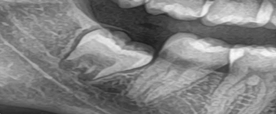
● Stage 1: Developmental stage with encircled follicle
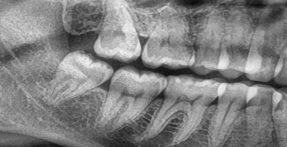
● Stage 2: Eruption within bone
● Stage 3: Fully erupted and aligned with occlusal plane
Phase Classification
Based on the stages of all four third molars:
● Phase A: All four molars in Stage 1
● Phase B: At least one molar in Stage 2
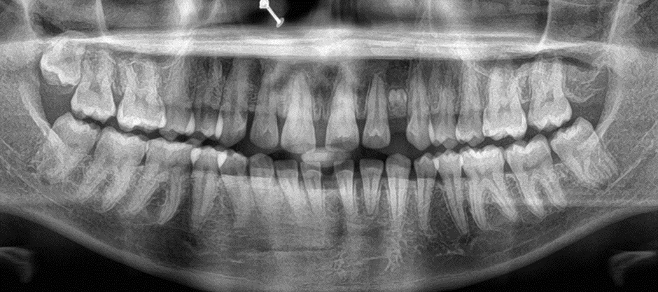
● Phase C: At least one molar in Stage 3
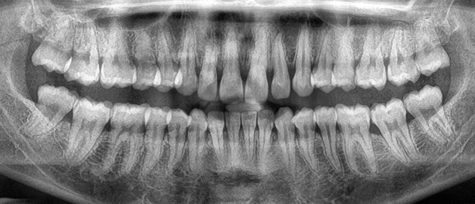
● Phase D: All four molars in Stage 3
Statistical Analysis
Data were analyzed using SPSS (Version 23.0). Descriptive statistics and correlation analysis were performed to determine the association between age and eruption stages.
Statistical analysis was conducted post analysis of the data with the help of SPSS v23.0.
Stage 1
Stage 2
Stage 3
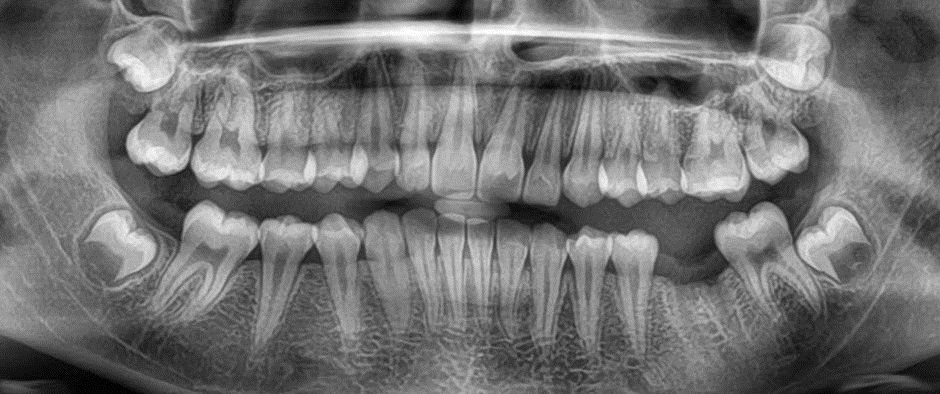
Phase A
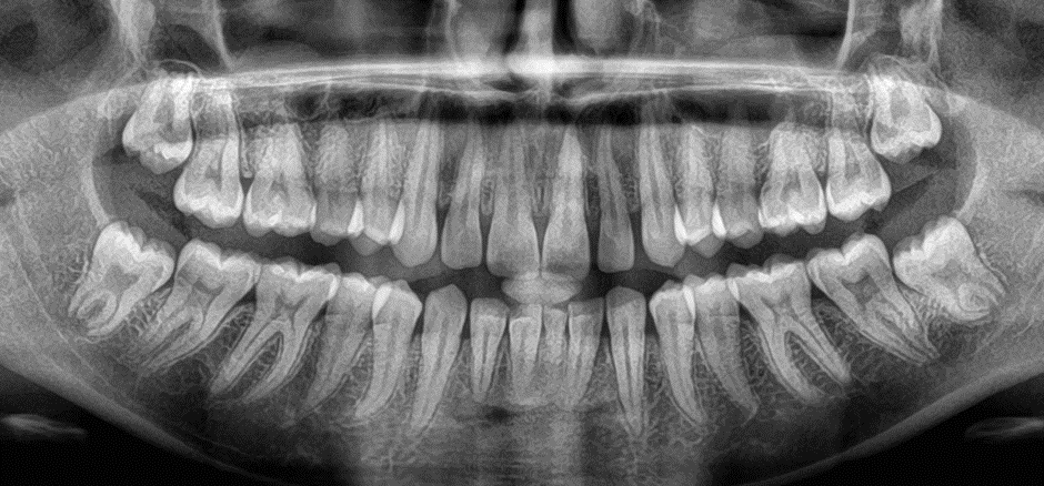
Phase B
Phase C
Phase D
RESULTS: :
The present study demonstrated a clear and progressive relationship between third molar eruption stages and chronological age among the study population. A distinct pattern of dental maturation was observed, supporting the utility of third molars as indicators in forensic age estimation.
· Stage 1 was predominantly observed in individuals aged 15–17 years, representing the early developmental phase characterized by follicular presence and incomplete eruption. This stage reflects the initial phases of mineralization and is indicative of individuals in mid-adolescence.
· Stage 2 was more frequently identified in the 16–20 years age group, corresponding to the transitional eruption phase where the tooth is actively emerging but remains partially embedded in bone. This stage showed considerable overlap with both earlier and later stages, indicating variability in eruption timing.
· Stage 3 was primarily observed in individuals aged 18–25 years, signifying complete eruption with the third molar reaching the occlusal planeThis stage is considered a strong indicator of late adolescence or early adulthood.
Phase-wise Observations
· Phase A (All molars in Stage 1): Predominantly associated with younger individuals below 17 years, confirming early developmental status.
· Phase B (At least one molar in Stage 2): Displayed significant overlap across mid-adolescent age groups, reflecting variability in eruption progression.
· Phase C (At least one molar in Stage 3): Strongly associated with individuals approaching or exceeding 18 years of age, indicating transition toward dental maturity.
· Phase D (All molars in Stage 3): Demonstrated a strong correlation with individuals above 18 years, suggesting completion of third molar eruption.
Statistical analysis revealed a significant association (p < 0.05) between eruption phases and chronological age, confirming the reliability of third molar assessment as an age indicator. However, overlapping age ranges across different stages limited the ability to determine exact chronological age, emphasizing that this method is more suitable for age estimation ranges rather than precise age determination.
DISCUSSION
The present study aimed to evaluate the reliability of third molar eruption status as a tool for forensic age estimation using dental panoramic radiographs. The findings revealed a consistent and progressive pattern of third molar development with increasing chronological age, aligning with observations reported in previous studies.
The strong association between Stage 3 eruption and Phase D classification with individuals above 18 years is of particular forensic significance. This finding supports the application of third molar eruption status in legal contexts, especially in determining whether an individual has attained the age of majority. The presence of all four third molars in the occlusal plane serves as a practical and clinically observable marker suggesting adulthood.
However, the overlap observed in Stage 1 and Stage 2 across multiple age groups highlights a key limitation of this method. Such variability may arise due to differences in genetic predisposition, nutritional status, hormonal influences, and environmental conditions. These factors can affect the timing and rate of tooth development, thereby reducing the precision of age estimation when relying solely on third molars.
Compared to skeletal age estimation methods, dental development is generally considered more stable and less influenced by external environmental factors. This enhances its reliability in forensic applications. Nevertheless, population-specific variations necessitate the establishment of localized reference standards to improve diagnostic accuracy.
The use of dental panoramic radiographs offers several practical advantages, including non-invasiveness, ease of access, and cost-effectiveness. Additionally, the method does not require highly specialized training, allowing it to be applied in routine clinical settings as well as forensic investigations. This makes it particularly useful in situations where rapid and preliminary age assessment is required.
Despite these advantages, it is important to emphasize that third molar evaluation should not be used as a standalone method for definitive age determination. Instead, it should be integrated with other assessment techniques, such as skeletal analysis and biochemical markers, to enhance overall accuracy and reliability.
LIMITATIONS: The present study has several limitations that must be considered when interpreting the findings:
● Biological Variability: Third molar eruption is influenced by genetic, environmental, and ethnic factors, leading to significant inter-individual variability.
● Stage Overlap: Considerable overlap exists between different eruption stages across age groups, making precise age determination challenging.
● Population Specificity: Findings are based on a specific population and may not be directly applicable to other ethnic or geographic groups.
● Age Range Limitation: The method is most reliable within the 17–25 year age range; accuracy decreases significantly outside this period.
● Radiographic Interpretation Bias: Variations in interpretation of radiographs may introduce observer bias.
SCOPE OF FUTURE RESEARCH: Future research should focus on enhancing the accuracy and applicability of third molar-based age estimation through the following approaches:
● Larger and Diverse Sample Populations: Inclusion of multi-ethnic and geographically diverse populations to improve generalizability.
● Multimodal Age Estimation: Integration of dental, skeletal, and biochemical markers to develop comprehensive age estimation models.
● Standardized Reference Databases: Development of global and population-specific reference standards for third molar development.
● Advanced Imaging Techniques: Utilization of cone-beam computed tomography (CBCT) and other high-resolution imaging modalities for detailed analysis.
● Artificial Intelligence and Machine Learning: Implementation of AI-based predictive models to reduce observer bias and improve accuracy.
Understanding these correlations in greater detail will significantly enhance the reliability of forensic age estimation and support more accurate legal decision-making.
CONCLUSION :
Assessment of third molar eruption using dental panoramic radiographs is a valuable and practical adjunct tool for forensic age estimation in living individuals. The study demonstrates a significant correlation between eruption stages and chronological age, particularly highlighting that the presence of all four third molars in the occlusal plane strongly suggests that an individual is above 18 years of age.
However, due to variability in eruption patterns and overlap between developmental stages, this method cannot be relied upon for precise age determination when used in isolation. It is best utilized as part of a combined approach alongside other diagnostic methods to improve overall accuracy.
The simplicity, non-invasive nature, and accessibility of this technique make it highly suitable for routine clinical and forensic applications. With further research and integration of advanced technologies, third molar assessment has the potential to become an even more reliable tool in forensic odontology.
BIBLIOGRAPHY:
1. Timme M, Viktorov J, Steffens L, Streeter A, Karch A, Schmeling A. Third molar eruption in dental panoramic radiographs as a feature for forensic age assessment–new reference data from a German population. Head & Face Medicine. 2024 May 10;20(1):29.
2. Timme M, Bender J, Steffens L, Shay D, Schmeling A. Third Molar Eruption in Dental Panoramic Radiographs as a Feature for Forensic Age Assessment—Presentation of a New Non-Staging Method Based on Measurements. Biology. 2023 Nov 5;12(11):1403.
3. Gambier A, Rérolle C, Faisant M, Lemarchand J, Paré A, Saint-Martin P. Contribution of third molar eruption to the estimation of the forensic age of living individuals. International Journal of legal medicine. 2019 Mar 5;133(2):625-32.
4. Timme M, Viktorov J, Steffens L, Streeter A, Karch A, Schmeling A. Third molar eruption in orthopantomograms as a feature for forensic age assessment—a comparison study of different classification systems. International Journal of Legal Medicine. 2023 May;137(3):765-72.
5. Kutesa AM, Rwenyonyi CM, Mwesigwa CL, Muhammad M, Nabaggala GS, Kalyango J. Dental age estimation using radiographic assessment of third molar eruption among 10–20-year-old Ugandan population. Journal of Forensic Dental Sciences. 2019 Jan 1;11(1):16-21.
6. Timme M, Viktorov J, Steffens L, Streeter A, Karch A, Schmeling A. Third molar eruption in orthopantomograms as a feature for forensic age assessment—a comparison study of different classification systems. International Journal of Legal Medicine. 2023 May;137(3):765-72.
7. Kutesa AM, Rwenyonyi CM, Mwesigwa CL, Muhammad M, Nabaggala GS, Kalyango J. Dental age estimation using radiographic assessment of third molar eruption among 10–20-year-old Ugandan population. Journal of Forensic Dental Sciences. 2019 Jan 1;11(1):16-21.
8. Pinto R, Cordeiro R, Galego Z, Turkina A, Boedi R, Franco A. Radio-morphologic assessment of dental formation leading to permanent teeth and third molar age estimation models. Morphologie. 2025 Sep 1;109(366):100966.
9. Timme M, Viktorov J, Steffens L, Streeter A, Karch A, Guo YC, Schmeling A. Age-related patterns in mandibular third molar eruption: presenting new forensic age assessment reference data from a Chinese population. Forensic Science, Medicine and Pathology. 2025 Mar;21(1):173-82.
10. Velandia Palacio LA. Development of third molar and Third molar index: I3M Global observations.
11. Gunacar DN, Bayrak S, Sinanoglu EA. Three-dimensional verification of the radiographic visibility of the root pulp used for forensic age estimation in mandibular third molars. Dentomaxillofacial Radiology. 2022 Mar 1;51(3):20210368.
12. Sharma S. Forensic Science, Age Estimation, Open Apex, Panoramic Radiographs (Master's thesis, Rajiv Gandhi University of Health Sciences (India)).
13. Chulamanee P, Panyarak W. Dental radiography in age determination: contemporary methods and trends. Oral Sci Rep. 2023 Nov;44:54-76.
14. Dalessandri D, Tonni I, Laffranchi L, Migliorati M, Isola G, Visconti L, Bonetti S, Paganelli C. 2D vs. 3D radiological methods for dental age determination around 18 years: a systematic review. Applied Sciences. 2020 Apr 29;10(9):3094.
15. De Luca S, Paladugu S, Balla SB, Moukarzel M, Angelakopoulos N. Accuracy of third molar eruption for legal age estimation using the Gambier method in Lebanese subadults. Odontology. 2025 Oct 23:1-0.
16. Balla SB, Ali SH, Mula AP, Tadakamadla SK, Rizky MB, Franco A, Angelakopoulos N, Tadakamadla J. The effect of impaction on the mineralisation of third molars and forensic age estimation: A systematic review and meta-analysis. Forensic Science International. 2025 Sep 6:112650.
17. Kihara EN. Utilization and Accuracy of Dental Age Estimation Methods and Development of Tooth Maturity Reference Values in a Kenyan Population (Doctoral dissertation, COHES-JKUAT).
18. Ashifa N, Parakh MK, Ulaganambi S. Estimation of age using third molar development: A radiological cross-sectional study. The American journal of forensic medicine and pathology. 2020 Jun 1;41(2):115-8.
19. Abdullah NA, Al-Aghbari BA, Mokbel AH. Radiographic evaluation of third molars development in relation to chronological age among children and youth in Aden city. University of Aden Journal of Natural and Applied Sciences. 2020 Apr 30;24(1):251-61.
20. Palmela Pereira C. Dental Age Assessment Methods in Children and Adolescents. InDental Age Assessment: A Global Perspective 2026 Jan 14 (pp. 63-96). Cham: Springer Nature Switzerland.
21. Helal SO. Age Estimation Through Radiographic Evaluation of Third Molar Mineralization (Doctoral dissertation, University of Illinois Chicago).
22. Sartori V, Franco A, Linden MS, Cardoso M, de Castro D, Sartori A, Silva C, Trentin M, De Carli JP. Testing international techniques for the radiographic assessment of third molar maturation. Journal of Clinical and Experimental Dentistry. 2021 Dec 1;13(12):e1182.
23. Abuabara A, Antunes LA, Antunes LS, Rodrigues da Cruz K, do Nascimento TV, França PH, Küchler EC, Miranda de Araujo C, Baratto-Filho F. Age estimation using dental radiography in the Brazilian population: a systematic review and meta-analysis. Egyptian Journal of Forensic Sciences. 2025 Jul 30;15(1):56.
24. Holm Kvist L, Sabo S. How old are you? Age assessments based on dental radiographs of third molars around the 18-year-threshold in different populations, a systematic review.
25. Cantekin K, Sekerci AE, Buyuk SK. Dental computed tomographic imaging as age estimation: morphological analysis of the third molar of a group of Turkish population. The American Journal of Forensic Medicine and Pathology. 2013 Dec 1;34(4):357-62.
26. Ergin MO, Erciyas K, Gunes SS, Isır AB. Assessment of periodontal ligament visibility in third molars for threshold-based forensic age estimation: a cross-sectional study in a Turkish population. International Journal of Legal Medicine. 2026 Feb 24:1-8.
27. Fonseca GM, Capitaneanu C. Historical Perspective of Dental Age Assessment. InDental Age Assessment: A Global Perspective 2026 Jan 14 (pp. 9-32). Cham: Springer Nature Switzerland.