Single Stage Reconstruction of Atrophic Tibial Non-Union with Critical-Sized Bone Defect, Limb Length Discrepancy, And Equinus Deformity Using A Single Ilizarov Construct: A Case Report
- Jeffry Andrianus , Department of Orthopaedic and Traumatology, Faculty of Medicine, Airlangga University, Surabaya, 60286, Indonesia
- Maulana Makdum Ibrahim , Faculty of Medicine, Airlangga University, Surabaya, Indonesia
- Alfin Bachtiar Ilhami , Department of Orthopaedic and Traumatology, Faculty of Medicine, Airlangga University, Surabaya, 60286, Indonesia
Article Information:
Abstract:
Atrophic tibial non-union with critical-sized bone defect, limb length discrepancy (LLD), and equinus deformity presents a complex reconstructive challenge. We report a 28-year-old female with a 6 cm tibial defect, 7 cm LLD, and severe equinus (talotibial angle 77°) following trauma. Reconstruction was performed using a single Ilizarov system, combining trifocal bone transport, limb lengthening, and gradual deformity correction within one construct. Early outcomes demonstrate progressive regenerate formation, impro ved alignment, and correction of deformity. Preliminary ASAMI assessment suggests good functional progress, pending final evaluation after frame removal. This case highlights the effectiveness of a single-stage Ilizarov strategy for simultaneous management of the combined pathology, potentially reducing the need for staged procedures.
Keywords:
Article :
INTRODUCTION :
High-energy trauma or infection commonly cause critical-sized bone defects, which require reconstruction strategies to restore both bone continuity and alignment. Ilizarov external fixation technique may be used as distraction osteogenesis as it enables biological reconstruction while allowing deformity correction.
The trifocal bone transport technique, which involves double-level distraction osteogenesis, allows for the simultaneous transport of two bone segments, potentially speeding up docking and reducing external fixation time compared to bifocal methods1–4. Liu et al. reported improved efficiency in managing large tibial defects with a trifocal approach1. Most studies have focused primarily on defect reconstruction, with few reports addressing the simultaneous management of limb length discrepancy and joint deformity. A case of atrophic tibial non-union with critical bone loss, significant limb length discrepancy, and equinus deformity was managed using a single Ilizarov external fixation construct.
CASE PRESENTATION:
A 28-year-old female presented with a post-traumatic left tibial non-union following failed external fixation (2023). The patient was non–weight bearing at presentation.
Clinical examination demonstrated a malaligned limb with restricted ankle motion (plantarflexion 10–13°), fixed equinus deformity (tibiotalar angle 77°), and a limb length discrepancy of 7 cm. The soft tissue envelope showed post-operative scarring with areas of prior bone exposure, without active sinus formation (Fig. 1).
Figure 1
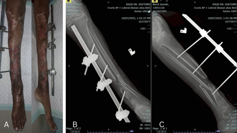
Laboratory investigations revealed no evidence of active infection. Radiographs confirmed an atrophic tibial non-union with an approximately 6 cm segmental bone defect (Fig. 1). No neurovascular deficit was identified, and there was no history of flap coverage or degloving injury.
On January 20, 2026, the patient underwent removal of the previous fixation followed by application of Ilizarov frame. A longitudinal incision was made along the tibial axis over the defect site. Fibrous tissue was excised, and the non-union site was debrided with removal of non-viable bone until healthy bleeding bone was achieved. Trifocal osteotomy was then performed at the proximal tibia, with two corticotomy sites created approximately 5 cm distal to the tibial tuberosity and 5 cm apart. A four-ring Ilizarov construct was applied in a trifocal configuration: one ring at the proximal segment, two rings at the transport segments, and one ring at the distal segment. This configuration enabled independent segmental transport without mechanical interference. An additional foot ring was incorporated to allow gradual correction of the equinus deformity through controlled distraction, avoiding the need for Achilles tendon lengthening (Fig. 2).
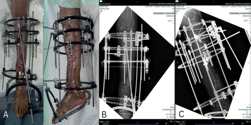
Figure 2
Distraction commenced after 7 days of latency phase at a rate of 1 mm/day, divided into 6 increments, applied across the transport segments (rings 2–4). Gradual ankle correction was achieved simultaneously at a rate of 1 mm/day until a plantigrade position was obtained. The patient then allowed toe-touch weight bearing.
At 2-month follow-up, alignment had improved, with correction of the tibiotalar angle to 88° and reduction of limb length discrepancy to 2.3 cm (Fig. 3). The patient progressed to partial weight bearing with crutches. There were no clinical features suggestive of complex regional pain syndrome observed. Motor assessment demonstrated ankle stiffness with a range of motion of 0–20° plantarflexion. The docking site had not yet consolidated, and no signs of infection were identified.
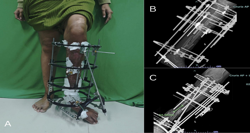
Figure 3
DISCUSSION :
This case highlights the combined challenge of biological insufficiency and structural deformity in atrophic tibial non-union. The coexistence of a 6 cm bone defect, 7cm limb length discrepancy, and equinus contracture requires a strategy that restores both bone continuity and functional alignment, which is often difficult to achieve with conventional methods. The Ilizarov technique enables biological reconstruction through distraction osteogenesis. Traditional bifocal transport, as described by Paley, is effective but often limited by prolonged external fixation time in large defects. Trifocal transport, supported by more recent studies such as Liu et al.1 and Yang et al.5, improves efficiency by accelerating docking and reducing treatment duration. Similarly, contemporary series (e.g., Maimaiti et al.6) have explored strategies to optimize fixation time and regenerate quality in complex tibial defects.
These approaches primarily focus on defect reconstruction, but may still necessitate additional or staged procedures to address limb length discrepancy and joint deformity. In contrast, the single-construct strategy enabled simultaneous bone transport, limb lengthening, and equinus deformity correction within one mechanical system. A unified approach minimizes the need for staged interventions, simplifies construct design, and facilitates the coordinated correction of both biological and mechanical pathologies. The addition of a hinged foot ring allowed for gradual correction of deformity without the requirement for extra soft tissue procedures.
Early outcomes were favorable, with progressive callus formation, improved alignment, and no evidence of infection. According to the Association for the Study and Application of the Methods of Ilizarov (ASAMI) criteria, the current bone result is consistent with a good outcome during the transport phase, with final results pending docking and full weight bearing. A limitation of this report is the short follow-up period, with definitive bone and functional outcomes yet to be established.
In conclusion, a single Ilizarov construct may provide an effective single-stage solution for complex tibial non-union by simultaneously addressing bone defect, limb length discrepancy, and deformity, potentially reducing the need for staged reconstruction.
CONCLUSION :
The Ilizarov technique enabled early closure of a 6 cm tibial defect, correction of a significant ankle deformity, and progressive restoration of a 7 cm limb length discrepancy. Preliminary ASAMI assessment indicates favorable reconstructive progress, although continued follow-up is required to confirm complete union and full functional recovery.
BIBLIOGRAPHY:
1. lizarov GA. The tension-stress effect on the genesis and growth of tissues. Part I. The influence of stability of fixation and soft-tissue preservation. Clin Orthop Relat Res. 1989 Jan;(238):249–81. PubMed PMID: 2910611.
2. Paley D. Problems, obstacles, and complications of limb lengthening by the Ilizarov technique. Clin Orthop Relat Res. 1990 Jan;(250):81–104. PubMed PMID: 2403498.
3. Catagni MA, Bolano L, Cattaneo R. Management of fibular hemimelia using the Ilizarov method. Orthop Clin North Am. 1991 Oct;22(4):715–22. PubMed PMID: 1945347.
4. Liu K, Zhang H, Maimaiti X, Yusufu A. Bifocal versus trifocal bone transport for the management of tibial bone defects caused by fracture-related infection: a meta-analysis. J Orthop Surg Res. 2023 Feb 25;18(1):140. doi:10.1186/s13018-023-03636-5 PubMed PMID: 36841800.
5. Yang X, Hamiti Y, Liu K, Wang S, Kadier X, Xiong D, et al. Optimizing bone transport strategies: a pixel value ratio-based evaluation of regeneration rates in bifocal and trifocal techniques. Front Surg. 2024 Dec 10;11. doi:10.3389/fsurg.2024.1494658
6. Maimaiti X, Liu K, Yusufu A, Xie Z. Treatment of tibial bone defects caused by infection: a retrospective comparative study of bone transport using a combined technique of unilateral external fixation over an intramedullary nail versus circular external fixation over an intramedullary nail. BMC Musculoskelet Disord. 2024 Apr 12;25(1):284. doi:10.1186/s12891-024-07377-2
- Figure 1. Clinical Picture of soft tissue scar (left) and radiograph of the left lower leg (right) preoperatively to Ilizarov
- Figure 2. Immidiate Postoperative clinical picture and radiograph
- Figure 3. 2 months postoperative clinical picture and radiograph