Diagnostic Accuracy of Magnetic Resonance Imaging in Detecting Optic Nerve Invasion in Retinoblastoma Patients Taking Histopathology as Gold Standard
- Safa Khalil , MBBS, PGR in Radiology Department of Radiology, SKMCH and RC, Lahore
- Anis ur Rehman , MBBS, FCPS, FRCR, Consultant, Department of Radiology, SKMCH and RC, Lahore
- Ammar Zahid Sheikh , MBBS, PGR in Radiology Department of Radiology, SKMCH and RC, Lahore
- Hooria Shumail , MBBS, PGR in Radiology Department of Radiology, SKMCH and RC, Lahore
Article Information:
Abstract:
Retinoblastoma is the most common intraocular malignancy in children, and early detection of optic nerve invasion is crucial as it is associated with poor prognosis and increased risk of metastasis. Histopathology is considered the gold standard for detecting optic nerve invasion; however, it is only available after enucleation. Magnetic resonance imaging (MRI), being a non-invasive modality with excellent soft tissue resolution, plays an important role in preoperative assessment, but its diagnostic accuracy needs further evaluation.Objectives: To determine the diagnostic accuracy of magnetic resonance imaging in detecting optic nerve invasion in retinoblastoma patients, taking histopathology as the gold standard. Study design & Setting: A cross-sectional validation study was conducted in the Department of Radiology at Shaukat Khanum Hospital, Lahore from 2 March 2025 to 2 June 2025.Methodology: A total of 139 patients aged 6 months to 10 years with suspected retinoblastoma were included using non-probability consecutive sampling. MRI of the brain and orbits was performed using a 3 Tesla scanner. Findings were compared with histopathology. Sensitivity, specificity, positive predictive value, negative predictive value, and diagnostic accuracy were calculated. ROC curve and likelihood ratios were also determined.Results: MRI showed a sensitivity of 83.3% and specificity of 77.2%. The positive and negative predictive values were 73.5% and 85.9%, respectively, with an overall diagnostic accuracy of 79.9%. ROC analysis demonstrated an AUC of 0.803. Stratified analysis showed consistent diagnostic performance across age, gender, and duration groups (p < 0.001).Conclusion: MRI is a reliable non-invasive modality with good diagnostic accuracy for detecting optic nerve invasion in retinoblastoma patients and can be effectively used in preoperative assessment.
Keywords:
Article :
INTRODUCTION :
Congenital melanocytic nevi (CMN) are pigmented Retinoblastoma is an oncologic disease of the developing retina, typically presenting in patients younger than 5 years of age. Retinoblastoma occurs in a heritable form (40% of cases, mostly bilateral) and a nonheritable form (60% of cases, always unilateral).1 Mortality ranges from < 5% in high-income countries to 40% to 70% in low-income countries. Survival is poor in the presence of central nervous system metastases.2 Histopathological postlaminar optic nerve invasion (PLONI) of the tumor is a risk factor for developing metastatic disease. Optic nerve invasion can be detected on magnetic resonance imaging (MRI) as contrast enhancement in the optic nerve beyond the lamina cribrosa: postlaminar optic nerve enhancement (PLONE).3,4
MRI plays a crucial role in the preoperative evaluation of retinoblastoma. While it helps in confirming the clinical diagnosis, it also enables assessment of tumor invasion of the choroid, sclera, and optic nerve, all of which are high-risk features for systemic dissemination. Additionally, it helps identify concomitant intracranial metastasis and embryonal tumors.5 The development of retinoblastoma is primarily attributed to mutations in the RB1 tumor suppressor gene, leading to uncontrolled proliferation of retinal cells. The heritable form is often associated with germline mutations and may present earlier with bilateral or multifocal tumors, whereas the nonheritable form typically arises from somatic mutations and is usually unilateral.6 The pathophysiology involves disruption of normal cell cycle regulation, resulting in rapid tumor growth and potential extension beyond the globe.7 Clinically, patients commonly present with leukocoria, strabismus, decreased vision, or ocular inflammation, and in advanced stages, proptosis or orbital cellulitis–like features may be observed. If left untreated, the tumor may invade the optic nerve, choroid, and orbit, eventually leading to intracranial extension and distant metastasis, which significantly worsens prognosis.8
In addition to clinical examination and fundoscopic findings, imaging modalities play a key role in disease evaluation. Magnetic resonance imaging is the preferred modality due to its superior soft tissue contrast and ability to delineate intraocular and extraocular tumor extension without radiation exposure. On MRI, optic nerve invasion is suggested by abnormal signal intensity, contrast enhancement, and thickening of the optic nerve beyond the lamina cribrosa.9 Histopathology remains the definitive method for confirming optic nerve involvement, characterized by the presence of malignant cells infiltrating the optic nerve fibers. The correlation between MRI findings and histopathological confirmation is therefore critical in determining the diagnostic performance of MRI in detecting optic nerve invasion in retinoblastoma patients.10
Therefore in such a scenario, MRI can be of important diagnostic value for detection of optic nerve invasion. Many studies highlight the significant association of MRI findings with optic nerve involvement but no local study has been conducted in Pakistan. Therefore the purpose of this study is to find the diagnostic accuracy of magnetic resonance imaging in detecting optic nerve invasion in retinoblastoma patients. Hence providing information about a diagnostic tool which can be more accurate and less time consuming thus helping in decision of treatment plan and prognosis of the disease. My study will not only provide the local stats but will also be a useful addition in the existing literature. The objective of this study is to determine the diagnostic accuracy of magnetic resonance imaging in detecting optic nerve invasion in retinoblastoma patients, taking histopathology as the gold standard.
MATERIALS AND METHODS:
After approval from the ethical review committee and CPSP this cross-sectional validation study was conducted in the Department of Radiology at Shaukat Khanum Hospital 2 March 2025 to 2 June 2025. A sample size of 139 cases was calculated with a 95% confidence level, taking the expected percentage of optic nerve invasion in retinoblastoma patients as 46.8% and desired precision of 12% for sensitivity of 40.0% and specificity of 93.55% of MRI.11,12 Non-probability consecutive sampling was used. Patients aged 6 months to 10 years of both genders who were referred to the Radiology Department for evaluation of suspected retinoblastoma with duration of symptoms greater than 3 months were included. Patients who had already received treatment, had recurrent disease, had a history of orbital trauma, or had contraindications to magnetic resonance imaging such as MRI-incompatible prosthesis or cardiac pacemakers were excluded. Informed consent was taken from the parents of each patient. Demographic and clinical variables including age, gender, duration of symptoms, and side affected were recorded.
Magnetic resonance imaging of the brain and orbits was performed in every patient using a 3 Tesla MRI scanner with head coils, with and without contrast, and a slice thickness of 1 mm. All MRI findings were interpreted by a consultant radiologist with at least five years of post-fellowship experience in the presence of the researcher, and optic nerve invasion in retinoblastoma patients was assessed as per operational definition. MRI findings were then compared with histopathology reports, and all data were recorded on a specially designed proforma.
Operational definitions were described as follows: Diagnostic accuracy was measured in terms of sensitivity, specificity, positive predictive value, and negative predictive value. Sensitivity was defined as the ability of MRI to correctly detect optic nerve invasion, while specificity was its ability to correctly identify absence of invasion. Positive predictive value was the probability of optic nerve invasion on MRI among positive cases, and negative predictive value was the probability of no invasion among negative cases. True positive and true negative were defined as agreement of MRI findings with histopathology for presence and absence of invasion, respectively, while false positive and false negative indicated disagreement. Suspected retinoblastoma cases were identified by clinical features such as ptosis, leukocoria, and calcifications on CT scan. Posterior laminar optic nerve invasion on MRI was defined by characteristic signal changes with contrast enhancement and optic nerve thickening ≥2 mm, whereas on histopathology it was confirmed by typical tumor cells and involvement of the lamina cribrosa with or without extension beyond it.
The collected data were analyzed using SPSS version 25.0. The Shapiro–Wilk test was applied to assess normality of data. Age and duration of symptoms were presented as mean and standard deviation or median with interquartile range, while gender, side affected, and optic nerve invasion on MRI and histopathology were presented as frequencies and percentages. A 2×2 contingency table was used to calculate sensitivity, specificity, positive predictive value, negative predictive value, and diagnostic accuracy of MRI in detecting optic nerve invasion, taking histopathology as the gold standard. Receiver operating characteristic (ROC) curve and likelihood ratios were also calculated. Effect modifiers including age, gender, duration of symptoms, and side affected were controlled through stratification, and post-stratification diagnostic accuracy was determined. The chi-square test was applied, and a p-value of ≤0.05 was considered statistically significant.
RESULTS:
The mean age of the study participants was 5.41 ± 2.83 years, with a median age of 5.0 years (IQR = 5.0), ranging from 1 to 10 years. The majority of patients were aged ≤5 years (66.2%), while 33.8% were older than 5 years. There was a slight male predominance with 56.8% males and 43.2% females. Most patients had a duration of symptoms ≤3 months (64.7%), whereas 35.3% had symptoms for more than 3 months. Regarding laterality, the right eye was more commonly affected (59.7%) compared to the left eye (40.3%), as given in Table 1
.
Table 1: Demographic and Clinical Characteristics of Study Participants (n = 139)
|
Variable |
Parameters |
Frequency (%) / Mean ± SD |
|
Age |
Mean ± SD |
5.41 ± 2.83 |
|
Median (IQR) |
5.0 (5.0) |
|
|
Min – Max |
1 – 10 |
|
|
≤5 years |
92 (66.2%) |
|
|
>5 years |
47 (33.8%) |
|
|
Gender |
Male |
79 (56.8%) |
|
Female |
60 (43.2%) |
|
|
Duration of symptoms |
≤3 months |
90 (64.7%) |
|
>3 months |
49 (35.3%) |
|
|
Side affected |
Right |
83 (59.7%) |
|
Left |
56 (40.3%) |
Optic nerve invasion was detected on MRI in 48.9% of patients, while 51.1% showed no evidence of invasion. On histopathological examination, optic nerve invasion was present in 43.2% of cases and absent in 56.8% of patients, as given in Table 2.
Table 2: Frequency of Optic Nerve Invasion on MRI and Histopathology (n = 139)
|
Variable |
Parameter |
Frequency (%) |
|
Optic nerve invasion on MRI |
Present |
68 (48.9%) |
|
Absent |
71 (51.1%) |
|
|
Optic nerve invasion o n Histopathology |
Present |
60 (43.2%) |
|
Absent |
79 (56.8%) |
Among patients with absent MRI findings, 85.9% had no optic nerve invasion on histopathology, while 14.1% were found to have invasion. In contrast, among those with positive MRI findings, 73.5% had confirmed optic nerve invasion on histopathology, whereas 26.5% did not. Overall, histopathology showed optic nerve invasion in 43.2% of patients and absence in 56.8% of cases, as given in Table 3.
Table 3: Comparison of MRI Findings with Histopathology (2×2 Contingency Table) (n = 139)
|
MRI |
Histopathology Absent |
Histopathology Present |
Total |
|
MRI Absent |
61 (85.9%) |
10 (14.1%) |
71 (100.0%) |
|
MRI Present |
18 (26.5%) |
50 (73.5%) |
68 (100.0%) |
|
Total |
79 (56.8%) |
60 (43.2%) |
139 (100.0%) |
Magnetic resonance imaging demonstrated a sensitivity of 83.3% and a specificity of 77.2% in detecting optic nerve invasion. The positive predictive value was 73.5%, while the negative predictive value was 85.9%. The overall diagnostic accuracy of MRI was found to be 79.9%, as given in Table 4.
Table 4: Diagnostic Accuracy of MRI in Detecting Optic Nerve Invasion
|
Parameter |
Value (%) |
|
Sensitivity (a / a+c) |
83.3% |
|
Specificity (d / b+d) |
77.2% |
|
Positive Predictive Value (a / a+b) |
73.5% |
|
Negative Predictive Value (d / c+d) |
85.9% |
|
Diagnostic Accuracy ((a+d) / total) |
79.9% |
Stratified analysis showed that in patients aged ≤5 years, MRI demonstrated a sensitivity of 79.5%, specificity of 77.4%, and diagnostic accuracy of 78.3%, while in those aged >5 years, sensitivity increased to 90.5% with a diagnostic accuracy of 83.0%. Among males, MRI showed a sensitivity of 82.2% and accuracy of 78.5%, whereas in females, sensitivity was 86.7% with a higher accuracy of 81.7%. Regarding duration of symptoms, patients with ≤3 months duration had a sensitivity of 84.6% and accuracy of 81.1%, while those with >3 months duration showed slightly lower sensitivity (81.0%) and accuracy (77.6%). All associations were statistically significant (p < 0.001), as given in Table 5.
Table 5: Stratified diagnostic accuracy of MRI by age, gender, and duration using histopathology as gold standard (n = 139)
|
Variable |
Category |
MRI Absent (Histo Present) n(%) |
MRI Present (Histo Present) n(%) |
Sens (%) |
Spec (%) |
PPV (%) |
NPV (%) |
Acc (%) |
p-value |
|
Age Group |
≤5 years |
8 (16.3) |
31 (72.1) |
79.5 |
77.4 |
72.1 |
83.7 |
78.3 |
<0.001 |
|
>5 years |
2 (9.1) |
19 (76.0) |
90.5 |
76.9 |
76.0 |
90.9 |
83.0 |
<0.001 |
|
|
Gender |
Male |
8 (24.2) |
37 (80.4) |
82.2 |
73.5 |
80.4 |
75.8 |
78.5 |
<0.001 |
|
Female |
2 (5.3) |
13 (59.1) |
86.7 |
80.0 |
59.1 |
94.7 |
81.7 |
<0.001 |
|
|
Duration |
≤3 months |
6 (13.0) |
33 (75.0) |
84.6 |
78.4 |
75.0 |
87.0 |
81.1 |
<0.001 |
|
>3 months |
4 (16.0) |
17 (70.8) |
81.0 |
75.0 |
70.8 |
84.0 |
77.6 |
<0.001 |
Sens = Sensitivity; Spec = Specificity; PPV = Positive Predictive Value; NPV = Negative Predictive Value; Acc = Diagnostic Accuracy.
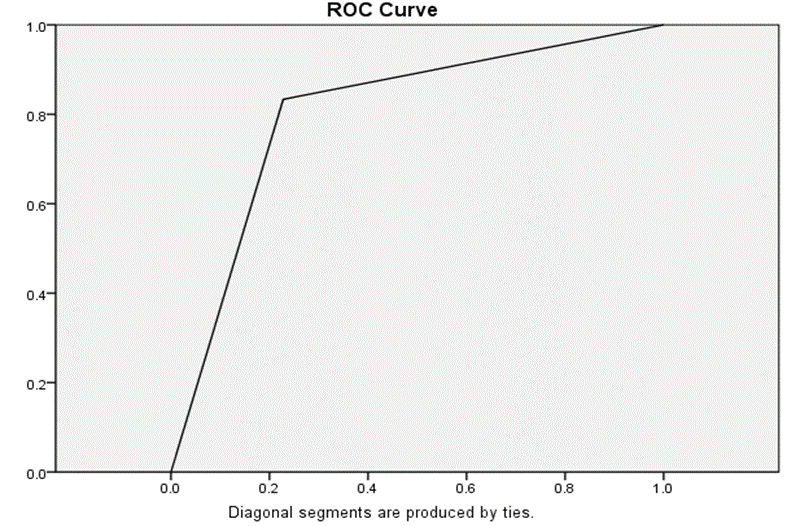
Figure 1: Receiver Operating Characteristic (ROC) Curve Analysis of MRI in Detecting Optic Nerve Invasion
ROC curve analysis demonstrated an area under the curve (AUC) of 0.803, indicating good diagnostic performance of magnetic resonance imaging in detecting optic nerve invasion in retinoblastoma patients. This suggests that MRI has a strong ability to correctly differentiate between patients with and without optic nerve invasion as given in figure 1.
DISCUSSION :
Optic nerve invasion is a critical prognostic factor, associated with increased risk of metastasis and poor survival outcomes. Early and accurate detection of optic nerve involvement is essential for appropriate treatment planning.13 Magnetic resonance imaging (MRI) offers a non-invasive method with excellent soft tissue contrast for preoperative evaluation.14 Therefore, assessing the diagnostic accuracy of MRI in detecting optic nerve invasion is of clinical importance.
In In the present study, magnetic resonance imaging demonstrated a sensitivity of 83.3%, specificity of 77.2%, positive predictive value of 73.5%, negative predictive value of 85.9%, and an overall diagnostic accuracy of 79.9% in detecting optic nerve invasion in retinoblastoma patients. These findings are comparable with previously reported studies, although variations exist across different settings. Usha et al. (2019) reported a sensitivity of 40%, specificity of 93.55%, PPV of 66.67%, and NPV of 82.86% for MRI, indicating significantly lower sensitivity but higher specificity compared to the present study, suggesting that MRI in their cohort was more reliable in confirming absence of disease rather than detecting true positive cases.15
Similarly, Kim et al. (2019) also reported a low sensitivity of 40% and high specificity of 93.55%, with PPV of 66.67% and NPV of 82.86%, findings closely aligned with Usha et al., but in contrast to the present study which demonstrated markedly improved sensitivity with relatively balanced specificity.15,17 Habib et al. (2020) reported sensitivity and specificity of 85.2% and 87%, respectively, along with PPV of 85%, NPV of 87%, and accuracy of 86.2%, which are closely comparable to the present findings, particularly in terms of sensitivity, although their specificity and overall accuracy were slightly higher.20
Abusayf et al. (2020) observed a sensitivity of 88.9%, specificity of 72.4%, PPV of 50%, NPV of 95.5%, and accuracy of 63.3%, showing higher sensitivity but lower specificity and accuracy compared to the present study.16 Wiwatwongwana et al. (2021) reported a sensitivity of 77.8% and specificity of 100%, demonstrating slightly lower sensitivity but higher specificity than the present findings.18 Surukrattanaskul et al. (2022) documented a sensitivity of 75% and specificity of 54%, both of which are lower than those observed in the present study, indicating comparatively better diagnostic performance in our cohort.19 More recently, Kheir et al. (2025) reported a sensitivity of 66.7% and specificity of 71.1%, with a high NPV of 96.9% but a very low PPV of 13.3%; in comparison, the present study showed higher sensitivity, specificity, and PPV, reflecting improved diagnostic balance.21
Overall, the findings of the present study demonstrate that MRI provides good and balanced diagnostic accuracy for detecting optic nerve invasion, with higher sensitivity than several earlier studies and comparable results to more recent literature, supporting its role as a reliable imaging modality in the evaluation of retinoblastoma.
Study Limitations
This was a single-center study, which may limit generalizability of results. The use of a relatively small sample size may affect the robustness of findings. Additionally, MRI interpretation was operator-dependent, which could introduce observer bias.
CONCLUSION :
MRI demonstrated good diagnostic accuracy in detecting optic nerve invasion in retinoblastoma patients. It can serve as a reliable non-invasive tool for preoperative assessment. Its use may aid in early decision-making and improve patient management outcomes.
Acknowledgement: We sincerely acknowledge the support and guidance of our mentors, colleagues, and the staff of the participating hospital for their valuable assistance throughout this study..
Conflict of Interest: No
Funding Disclosure: None
BIBLIOGRAPHY:
1. Tad Moeez Uddin M, Farooque U, Aziz MZ. Different types of clinical presentations and stages of retinoblastoma among children. Cureus. 2020;12(9):e10672.
2. Silvera VM, Guerin JB, Brinjikji W, Dalvin LA. Retinoblastoma: What the neuroradiologist needs to know. AJNR Am J Neuroradiol. 2021;42(4):618–26.
3. Tomar AS, Finger PT, Gallie B. A Multicenter, International Collaborative Study for American Joint Committee on Cancer Staging of Retinoblastoma: Part II: Treatment Success and Globe Salvage. Ophthalmol. 2020;127(12):1733–46.
4. Tomar AS, Finger PT, Gallie B. High-risk Pathologic Features Based on Presenting Findings in Advanced Intraocular Retinoblastoma: A Multicenter, International Data-Sharing American Joint Committee on Cancer Study. Ophthalmol. 2022;129(8):923–32.
5. Tomar AS, Finger PT, Gallie B. Retinoblastoma seeds: impact on American Joint Committee on Cancer clinical staging. Br J Ophthalmol. 2023;107(1):127–32.
6. Cruz-Gálvez CC, Ordaz-Favila JC, Villar-Calvo VM, Cancino-Marentes ME, Bosch-Canto V. Retinoblastoma: Review and new insights. Frontiers in oncology. 2022 Nov 2;12:963780.
7. Welter N, Brzezinski J, Treece A, Chintagumpala M, Young MD, Perotti D, Kieran K, Jongmans MC, Murphy AJ. The pathophysiology of bilateral and multifocal Wilms tumors: what we can learn from the study of predisposition syndromes. Pediatric blood & cancer. 2023 May;70:e29984.
8. Al Akrash LS, Al Semari MA, Al Harithy R. Ocular manifestations of dermatological diseases part I: infectious and inflammatory disorders. International Journal of Dermatology. 2021 Jan;60(1):5-11.
9. Saleh GA, Batouty NM, Haggag S, Elnakib A, Khalifa F, Taher F, Mohamed MA, Farag R, Sandhu H, Sewelam A, El-Baz A. The role of medical image modalities and AI in the early detection, diagnosis and grading of retinal diseases: a survey. Bioengineering. 2022 Aug 4;9(8):366.
10. Pai V, Parra-Fariñas C, Shaikh F, Mallipatna A, Zaarour C, Muthusami P. An updated review of intra-arterial chemotherapy for retinoblastoma. Radiología (English Edition). 2025 Mar 1;67(2):162-75.
11. Hameed M, Siddiqui F, Hussain W, Hanif H, Ram HS, Saleem S, et al. Imaging characteristics of retinoblastoma: a single tertiary care center experience from Pakistan. Pak J Med Health Sci. 2022;16(12):185-7.
12. Wiwatwongwana D, Kulniwatcharoen P, Mahanupab P, Visrutaratna P, Wiwatwongwana A. Accuracy of computed tomography and magnetic resonance imaging for detection of pathologic risk factors in patients diagnosed with retinoblastoma. Curr Eye Res. 2021;46(10):1544-50.
13. Yousef YA, Mohammad M, Khalil H, Khouri T, Alsweiti R, Khzouz J, Abu Laban D, Jaradat I, Ibrahimi AK, Al-Ibraheem A, Masri MA. Ocular and periocular metastasis in breast cancer: clinical characteristics, prognostic factors and treatment outcome. Cancers. 2024 Apr 16;16(8):1518.
14. Al-Haj Husain A, Stadlinger B, Winklhofer S, Piccirelli M, Valdec S. Magnetic resonance imaging for preoperative diagnosis in third molar surgery: a systematic review. Oral Radiology. 2023 Jan;39(1):1-7.
15. Usha K, Gunjan R, Gunja C, Srinavasan KG, Shanthi R, Krishna RS, et al Accuracy of preoperative imaging in predicting optic nerve invasion in retinoblastoma: a retrospective study. Indian J Ophthalmol. 2019;67(12):2019-22.
16. Abusayf MM, Alkatan HM, Elkhamary S, Almesfer SA, Maktabi AM. Histopathological assessment of optic nerve invasion guided by radiological findings in enucleated globes with retinoblastoma. BMC ophthalmology. 2020 Sep 29;20(1):386.
17. Kim U, Rathi G, Chowdhary G, Srinavasan KG, Shanthi R, Krishna RP. Accuracy of preoperative imaging in predicting optic nerve invasion in retinoblastoma: a retrospective study. Indian Journal of Ophthalmology. 2019 Dec 1;67(12).
18. Wiwatwongwana D, Kulniwatcharoen P, Mahanupab P, Visrutaratna P, Wiwatwongwana A. Accuracy of computed tomography and magnetic resonance imaging for detection of pathologic risk factors in patients diagnosed with retinoblastoma. Current Eye Research. 2021 Oct 3;46(10):1544-50.
19. Surukrattanaskul S, Keyurapan B, Wangtiraumnuay N. Correlation between clinical presentations, radiological findings and high risk histopathological features of primary enucleated eyes with advanced retinoblastoma at Queen Sirikit National Institute of Child Health: 5 years result. PloS one. 2022 Jul 20;17(7):e0270362.
20. Habib YS, Youssef AA, AlKiki HA, Ghareeb HT, ElZomor HE. High Resolution MR Imaging guidelines in retinoblastoma: prospective study correlated with histopathological results. Egyptian Journal of Radiology and Nuclear Medicine. 2020 Feb 6;51(1):28.
21. Kheir WJ, Hourani R, Zougheib Y, Slim A, Tamer C, Al-Haddad C. High-risk features in retinoblastoma: the association between histopathology and MRI. BMJ Open Ophthalmology. 2025 Oct 2;10(1).