ADVANCES IN TARGETED DELIVERY OF CRISPR/CAS TECHNOLOGIES FOR COLOREC-TAL CANCER THERAPY
- Nikolay K. Shakhpazyan , Avtsyn Research Institute of Human Morphology, Petrovsky National Research Center of Surgery, 119435, Moscow, Russia
- Liudmila M. Mikhaleva , Avtsyn Research Institute of Human Morphology, Petrovsky National Research Center of Surgery, 119435, Moscow, Russia
- Arcady L. Bedzhanyan , Department of Abdominal Surgery and Oncology II (Coloproctology and Uro-Gynecology), Petrovsky National Research Center of Surgery, 119435, Moscow, Russia
- Alexandra K. Konyukova , Avtsyn Research Institute of Human Morphology, Petrovsky National Research Center of Surgery, 119435, Moscow, Russia
- Andrey S. Kontorschikov , Avtsyn Research Institute of Human Morphology, Petrovsky National Research Center of Surgery, 119435, Moscow, Russia
- Ksenia S. Maslenkina , Avtsyn Research Institute of Human Morphology, Petrovsky National Research Center of Surgery, 119435, Moscow, Russia
- Konstantin Y. Midiber , Avtsyn Research Institute of Human Morphology, Petrovsky National Research Center of Surgery, 119435, Moscow, Russia
- Natalia Vladimirovna Elizova , Laboratory of Angiopathology, Institute of General Pathology and Pathophysiology, Moscow 125315, Russia
- Alexander N. Orekhov , Laboratory of Angiopathology, Institute of General Pathology and Pathophysiology, Moscow 125315, Russia
Article Information:
Abstract:
Colorectal cancer (CRC) remains one of the most prevalent and deadly malignancies, presenting significant treatment challenges due to its complex genetic landscape and resistance to conventional therapies. CRISPR/Cas technology has emerged as a powerful tool for precise genome editing, offering promising opportunities for treating CRC by targeting and modifying cancer-driving genes. However, the effective and safe delivery of CRISPR/Cas components into tumor cells remains a substantial obstacle to the clinical translation of this technology.In this review, we provide a comprehensive overview of the latest advances in targeted delivery systems for CRISPR/Cas technologies in colorectal cancer treatment. We discuss various delivery strategies, including viral vectors, non-viral nanoparticles, cell-penetrating peptides, virus-like particles, exosomes, and other innovative systems, highlighting their potential benefits, challenges, and effectiveness in achieving specific and efficient delivery to CRC cells. Additionally, we explore modifications to the classical CRISPR/Cas systems, such as base editing, prime editing, and epigenetic modulation, which enhance precision and reduce off-target effects, thereby increasing the therapeutic potential of these tools.This review underscores the critical need for developing novel delivery mechanisms that improve the speci-ficity and safety of CRISPR/Cas-based gene therapy for CRC. By critically evaluating existing delivery ap-proaches, their limitations, and potential solutions, this work aims to facilitate the translation of CRISPR/Cas technologies into viable clinical applications for colorectal cancer patients, ultimately enhanc-ing treatment outcomes and quality of life.
Keywords:
Article :
INTRODUCTION:
Colorectal cancer (CRC) is one of the most common and deadly types of cancer, ranking third in prevalence and second in mortality worldwide. Despite advances in diagnosis and treat-ment, the prognosis for CRC patients, especially in the later stages, remains poor [1]. Standard treatment methods, such as surgery, chemotherapy, immunotherapy, and targeted therapy, have major limitations, including severe side effects, drug resistance, and a lack of specificity for tumor cells. This highlights the need for more precise and effective treatment methods aimed at directly modifying cancer cells.
CRISPR/Cas9 technology, which is derived from the adaptive immune system of bacteria, has emerged as a powerful tool for precise genome editing. In recent years, several modifications to the original CRISPR/Cas9 system have been developed, including CRISPR/Cas12a, base edit-ing, prime editing, and dCas9 for epigenetic changes [2]. These new tools expand the possibilities of genome editing and offer more flexible approaches for research and therapy. CRISPR/Cas-based techniques have significant potential for treating CRC due to their ability to precisely target, eliminate specific genetic mutations and regulate gene expression that drive tumor growth and progression. However, one of the biggest challenges in using these technologies in clinical practice is finding effective and safe ways to deliver CRISPR/Cas complexes directly into tumor cells.
Targeted delivery of CRISPR/Cas is particularly challenging in CRC treatment because it is crucial not only to achieve high specificity for cancer cells but also to minimize the impact on healthy tissues. Currently, researchers are exploring a variety of delivery methods, including viral vectors (like adeno-associated viruses and lentiviruses), non-viral nanoparticles (such as lipid na-noparticles and polyethyleneimine complexes), virus-like particles (VLPs), exosomes, and other innovative systems [3]. Additional approaches, such as the use of cell-penetrating peptides and nanoparticles functionalized with aptamers, help improve the specificity and effectiveness of CRISPR/Cas delivery.
The aim of this review is to critically evaluate existing methods for the targeted delivery of CRISPR/Cas systems for in vivo gene therapy of CRC. This review will discuss the latest advanc-es in the field, the main challenges researchers are facing, and the future potential for applying these approaches in clinical settings. Overcoming the challenges in CRISPR/Cas delivery could greatly expand the possibilities for CRC treatment and improve patient outcomes and quality of life.
Principles and Approaches of CRISPR/Cas9 Methods CRISPR/Cas9: The Classical System:
CRISPR/Cas9: The Classical System
The classical CRISPR/Cas9 system is a genome-editing tool that comes from the adaptive immune system of bacteria [4]. It uses the Cas9 protein, which is derived from the bacterial immune system, specifically from Streptococcus pyogenes, guided by a synthetic RNA molecule called guide RNA (gRNA), to introduce double-strand breaks (DSBs) at a specific target site in the DNA. These breaks can be repaired by the cell using either non-homologous end joining (NHEJ), which often results in small insertions or deletions (indels) and leads to gene knockout, or homology-directed repair (HDR), which allows for precise gene modifications [4]. The CRISPR/Cas9 system has become a revolutionary tool for genetic engineering because it is precise and efficient, and it can be used to edit genes involved in many diseases, including cancer (Figure 1).
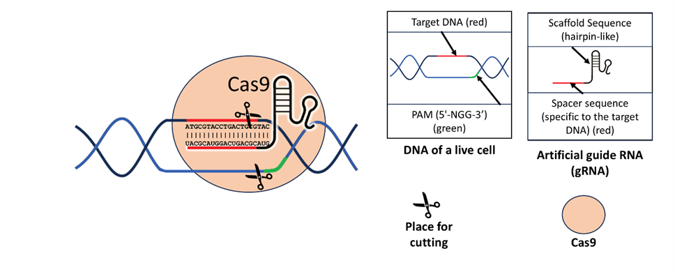
Figure 1: The Classical CRISPR/Cas9 System
This figure illustrates the classical mechanism of the CRISPR/Cas9 system for targeted genome editing. The main panel shows the Cas9 protein bound to the target DNA within a live cell. The artificial guide RNA (gRNA) directs the Cas9 to a specific DNA sequence through complementary base pairing. The gRNA consists of a scaffold sequence, depicted in a hairpin-like structure, and a spacer sequence that is complementary to the target DNA (highlighted in red). The protospacer adjacent motif (PAM), labeled in green, is essential for Cas9 recognition and binding, indicating where Cas9 can effectively operate. The scissors icon represents the site of DNA cleavage, emphasizing Cas9’s role in generating a double-strand break at a specific location within the genome. Insets provide details of the gRNA structure and the interaction with the target DNA, highlighting the precise targeting capabilities of the CRISPR/Cas9 complex. This figure serves as a foundational explanation of the components and function of the CRISPR/Cas9 system used in gene editing.
CRISPR/Cas12a (Cpf1) and Ligase-Assisted Homologous Recombination
CRISPR/Cas12a, also known as Cpf1, is an alternative to the CRISPR/Cas9 system [5]. Unlike Cas9, Cas12a creates staggered cuts in DNA, resulting in sticky ends that can be advantageous for certain gene editing purposes. Cas12a is particularly helpful for restoring the function of mutant genes, using a technique called Ligase-Assisted Homologous Recombination (LAHR) [6]. LAHR, or Ligase-Assisted Homologous Recombination, combines homology-directed repair (HDR) with microhomology-mediated end joining (MMEJ), which enhances the precision and efficiency of the editing process. HDR is a high-fidelity DNA cell repair mechanism that uses a donor DNA template to accurately repair double-strand breaks, allowing precise gene modifications. However, HDR is only efficient during certain phases of the cell cycle, making it inherently limiting in some contexts. MMEJ, on the other hand, is a more flexible repair mechanism that uses short homologous sequences (microhomologies), which are artificial sequences added by researchers to the cells to guide the repair, enabling efficient integration of donor DNA but often resulting in small deletions or insertions. By combining these two mechanisms, LAHR takes advantage of the accuracy of HDR while leveraging the broader applicability of MMEJ [6]. In this process, ligase plays a key role by sealing the nicks and gaps created during DNA repair, which is crucial for joining the DNA ends after the initial cleavage. The ligase enzyme is either naturally present in the cell or supplied exogenously by researchers, ensuring that the donor DNA is properly integrated. This use of ligase helps stabilize the edited DNA and enhances the overall efficiency of the gene repair, allowing for more effective integration of donor DNA into the genome, particularly useful for correcting disease-causing mutations where precision is critical. This combination allows for more efficient integration of donor DNA into the genome, which is especially useful for correcting disease-causing mutations (Figure 2).
Figure 2: CRISPR/Cas12a (Cpf1) and Ligase-Assisted Homologous Recombination
This figure illustrates the mechanism of genome editing using the CRISPR/Cas12a system combined with ligase-assisted homologous recombination to repair targeted deletions. The primary panel depicts the Cas12a protein targeting and cleaving a specific DNA sequence within a live cell, guided by an artificial guide RNA (gRNA). The gRNA consists of a scaffold sequence in a hairpin-like structure and a spacer sequence (in red) that is complementary to the target site. Cas12a generates staggered cuts (sticky ends) in the DNA, resulting in double-strand breaks.
The lower panels illustrate the process of genomic repair. Once Cas12a creates the break, an artificial donor DNA fragment (highlighted in orange) is introduced to facilitate the repair of the deleted region via homologous recombination. This donor fragment carries complementary sequences that align with the sticky ends produced by Cas12a, allowing accurate repair. The final step involves endogenous ligase (denoted by blue "L" symbols) facilitating the rejoining of DNA strands, resulting in the recovery of intact genomic DNA.
Chem-CRISPR/dCas9FCPF: Epigenetic Modification
Chem-CRISPR/dCas9FCPF is a modified version of the CRISPR system that uses a catalytically inactive version of Cas9 (dCas9), combined with chemical compounds, to control gene expression without cutting the DNA [7,8]. The dCas9 protein is guided to specific DNA regions by a gRNA, where it acts as a scaffold to recruit molecules that either activate or suppress gene transcription. In the Chem-CRISPR/dCas9FCPF system, a chemical tag called phenyl-cysteine-proline-phenyl (FCPF) helps recruit co-activators or co-repressors, which modify the chromatin structure or interact with the transcriptional machinery to regulate gene expression. These co-activators and co-repressors are recruited due to their affinity for the FCPF tag, which is engineered to bind specific protein domains. The molecules recruited can include histone acetyltransferases or deacetylases, methyltransferases, or other chromatin modifiers that change the local chromatin environment, thereby either promoting or repressing transcription. The FCPF tag essentially serves as a bridge, allowing dCas9 to bring these regulatory molecules to the target gene site, modulating gene expression dynamically. This mechanism allows researchers to control gene activity dynamically, without creating permanent DNA changes, offering a powerful and reversible tool for gene regulation in research and potential therapeutic applications (Figure 3).
Figure 3: Chem-CRISPR/dCas9FCPF System for Epigenetic Modification
This figure illustrates the mechanism of the Chem-CRISPR/dCas9FCPF system for targeted epigenetic modification aimed at gene repression without causing DNA double-strand breaks. The central component of the system is dCas9FCPF, a catalytically inactive ("dead") version of Cas9 that binds DNA but does not cleave it. The artificial guide RNA (gRNA) directs dCas9 to a specific DNA target sequence, highlighted in red. The FCPF (Phenyl-Cysteine-Proline-Phenyl) tag enables the dCas9 complex to bind with perfluorobiphenyl (denoted as C12F10) - JQ1 complex.
In this system, JQ1 functions as a ligand that binds to bromodomain-containing proteins, such as BRD4, which interact with acetylated histones. By binding to JQ1, dCas9FCPF effectively sequesters the bromodomain protein from its natural binding site on acetylated histones, thereby repressing gene expression in the targeted region. The figure shows how JQ1 - perfluorobiphenyl, in conjunction with dCas9FCPF, lead to the detachment of the
bromodomain protein from the acetylated histone, resulting in chromatin condensation and transcriptional repression.
Prime Editing
Prime editing is a CRISPR-based gene-editing method that allows for precise correction of specific point mutations without causing double-strand breaks [2,9]. It uses a modified version of Cas9 called Cas9 nickase (nCas9), which makes single-strand cuts, along with an artificially added reverse transcriptase enzyme and a special guide RNA (prime editing guide RNA, or pegRNA) that directs the editing. The reverse transcriptase uses the pegRNA as a template to synthesize the corrected DNA sequence at the target site. When nCas9 cuts one strand of the DNA, the reverse transcriptase replaces a short segment of the DNA with the corrected version from the pegRNA, while the original mutated strand is displaced or degraded. The PE3b system, which is a variant of prime editing that involves creating an additional nick on the non-edited strand to promote efficient correction, a component of the prime editing approach, improves the efficiency of editing by using multiple pegRNAs, which helps increase accuracy and reduce unintended changes compared to traditional CRISPR/Cas9 methods. This makes prime editing a powerful tool for treating genetic mutations with minimal risk (Figure 4).
Figure 4: Prime Editing Mechanism for Targeted Gene Modification
This figure illustrates the prime editing approach, which is a CRISPR-based gene-editing method that allows for precise genetic alterations without inducing double-strand breaks. The system relies on a modified Cas9 variant, known as nCas9 (nickase Cas9), which can introduce single-strand nicks rather than creating complete double-strand breaks. This variant is directed to the target DNA sequence by a prime editing guide RNA (pegRNA), depicted in the inset.
The pegRNA consists of three essential components: (1) the spacer sequence, which is complementary to the target DNA region (highlighted in red), ensuring accurate targeting; (2) the primer binding site (in blue), which serves as an initiation site for DNA synthesis; and (3) the reverse transcriptase template (in yellow), which contains the desired edit to be incorporated into the target DNA. This pegRNA not only directs the nCas9 to a specific DNA sequence but also carries the genetic information necessary for the desired edit.
Once nCas9 introduces a single-strand nick at the target site, the process of prime editing is initiated. The nicked DNA strand provides an entry point for the attached Moloney murine leukemia virus (MLV) reverse transcriptase (shown in blue), which uses the template sequence within the pegRNA to synthesize the desired DNA modification. The newly synthesized DNA strand (represented in yellow) is incorporated into the target sequence, leading to the precise introduction of the intended edit.
Base Editing (Adenine Base Editor)
Base editing is another advanced CRISPR-based gene-editing technology that allows for precise changes to single nucleotides without introducing double-strand breaks (DSBs) [10]. The adenine base editor (ABE), a specialized version of the CRISPR/Cas system, specifically changes adenine (A) to guanine (G), which can be used to correct mutations in genes. ABE consists of a modified version of Cas9 fused with an adenine deaminase enzyme that specifically targets adenine bases, converting them into guanine. This modified enzyme allows precise base editing without introducing double-strand breaks. The base editor consists of a modified Cas9 linked to an enzyme that makes the base conversion. Unlike traditional CRISPR/Cas9, base editing avoids creating double-strand breaks, which reduces the risk of large-scale changes in the genome and lowers cytotoxicity. This method is especially valuable for fixing harmful mutations where even small changes can restore gene function (Figure 5).
Figure 5: Adenine Base Editing with nCas9-TadA System
This figure illustrates the mechanism of adenine base editing using the Adenine Base Editor (ABE), a CRISPR-based gene-editing tool designed for highly precise nucleotide modifications. In this system, a modified Cas9 variant called nCas9 (nickase Cas9) is fused with an engineered tRNA adenosine deaminase enzyme (TadA). The figure describes the steps involved in the conversion of adenine (A) to inosine (I), which is interpreted as guanine (G) during DNA replication, resulting in an A•T to G•C base pair substitution.
The first panel shows nCas9 directed to a specific DNA sequence within the live cell by the guide RNA (gRNA). The gRNA consists of a spacer sequence that is complementary to the target site (highlighted in red), and a scaffold sequence that forms a hairpin-like structure for stability. The nCas9 introduces a nick on the non-target strand, which is critical for initiating the base editing process.
In the second panel, the TadA enzyme catalyzes the deamination of adenine at the target site, converting it into inosine. This process changes the chemical structure of adenine to resemble guanine, allowing it to pair with cytosine instead of thymine during the next replication cycle. This deamination step is key for the specific conversion of A•T base pairs into G•C base pairs.
The third panel depicts the process of DNA replication, during which the edited strand with inosine is replicated. Inosine behaves similarly to guanine during DNA synthesis, resulting in the permanent conversion of the target base pair from A•T to G•C. The resulting DNA molecule reflects the successful incorporation of the intended base edit.
CRISPR/Cas9 with Fusion Proteins to Enhance Homologous Recombination
The CRISPR/Cas9 system can also be improved by using fusion proteins to increase the efficiency of homologous recombination [11,12]. By fusing Cas9 with proteins that promote recombination, such as CtIP or Rad52, researchers can boost the rate of HDR, which is essential for precise genome editing. This enhancement is especially important in therapeutic settings where accurate editing is needed, such as correcting disease-causing mutations. The use of these fusion proteins helps direct the cell's repair system towards homology-directed repair rather than non-homologous end joining, increasing the likelihood of successful and accurate DNA repair.
CRISPR Screens for Identifying Genetic Vulnerabilities
CRISPR screens are an experimental method used to identify genetic vulnerabilities in cancer cells, typically conducted on cultured cell lines, such as somatic mutations [13]. Researchers use libraries of guide RNAs to systematically knock out genes across the entire genome and then analyze the frequency of each guide RNA sequence with next-generation sequensing (NGS) methods. If the frequency of a particular guide RNA decreases or disappears, it indicates that the cells with the knocked-out gene did not survive. This method has been crucial in discovering synthetic lethal interactions—genes that are essential for the survival of mutant cells but not for normal cells. Identifying such genes helps pinpoint potential targets for new therapies. Synthetic lethality is a promising strategy for developing targeted cancer treatments, allowing selective killing of cancer cells while sparing healthy ones [14].
These modifications and advancements in CRISPR/Cas technology show the versatility of this gene-editing platform and its potential for treating complex diseases like colorectal cancer. By using different versions of the CRISPR/Cas systems, researchers can tailor gene-editing approaches to specific therapeutic needs.
Approaches in CRISPR/Cas Therapy for Colorectal Cancer.
Gene Knockout and Correction of Oncogenes
Mutations in oncogenes are key events in carcinogenesis. Typically, these mutations are ac-tivating, meaning that the products of mutant genes are functionally active. The main oncogenes involved in colorectal cancer include KRAS, NRAS, and BRAF.
KRAS mutations are among the most common in colorectal tumors. A study utilized prime editing with CRISPR to correct various oncogenic KRAS mutations, such as G12S, G12D, G12V, and G13D. The researchers employed engineered pegRNAs (prime editing guide RNAs) and a PE3b system to enhance the efficiency of correcting these mutations without inducing unintended alterations at wild-type KRAS sites. This approach improved editing efficiencies, particularly for KRAS G12C and G13C mutations, making it a promising tool for precisely targeting oncogenic KRAS variants in cancer cells, including colorectal cancer models [15].
Building on these findings, genome-wide CRISPR screens have been used to identify genet-ic vulnerabilities in KRAS-mutant colorectal cancer. One such study conducted a CRISPR knockout screen on colorectal cancer cell lines carrying wild-type or mutant KRAS. The findings showed that mutant KRAS cells are highly dependent on mitochondrial oxidative phosphoryla-tion, highlighting metabolic vulnerabilities that could be therapeutically targeted in KRAS-mutant tumors [16].
Further extending these therapeutic approaches, another synthetic lethal strategy, validated through CRISPR screens on KRAS-mutant tumors, involves the knockout of genes in the WNT signaling pathway in combination with knockout of BCL-XL [17]. Synthetic lethality has also been demonstrated with ERN1 blockade, achieved through CRISPR/Cas methods, in combination with MEK inhibitors in cases of activated KRAS mutations [17].
While KRAS mutations are common, BRAF mutations are less frequent in colorectal tumors but are generally associated with high microsatellite instability. Some studies have shown the po-tential for editing mutant BRAF, which could serve as a therapeutic approach [18]. NRAS can al-so be targeted for directed editing or knockout, which is particularly important given the current lack of targeted drugs for patients with NRAS-mutant colorectal cancer.
Notably, targeting mutant oncogenes for knockout or correction is selective, as oncogene mutations are not present in normal cells, and these mutations are used by CRISPR/Cas systems as guide sequences. Therefore, it is the safest way to influence tumor cells through gene engineering in vivo, due to the low expectation of off-target effects.
Targeting Cancer Stem Cells
Cancer Stem Cells (CSCs) are a subset of cancer cells with the ability to self-renew and dif-ferentiate into tumor cells. These cells are believed to be responsible for tumor initiation, metasta-sis, and resistance to conventional therapies, making them a critical target for effective cancer treatment.
CRISPR/Cas9 technology has also been successfully used to target LGR5+ CSCs with the aim of eliminating these cells, which are responsible for tumor initiation, metastasis, and resistance to treatment. LGR5 is a well-known marker of CSCs, and targeting it could play a significant role in suppressing tumor growth and preventing metastasis
[19]. Loss of LGR5 function leads to tumor regression, as demonstrated in organoid models [20].
In addition to LGR5, other promising targets for editing include genes in the Notch and Hedgehog signaling pathways. These pathways are critical for maintaining the colorectal stem cell population and contribute to chemoresistance by promoting cancer cell survival, epitheli-al-to-mesenchymal transition (EMT), and resistance to apoptosis. Targeting these pathways can help reduce the CSC population, thereby enhancing the sensitivity of tumors to chemotherapeutic agents. Inhibiting the Hedgehog pathway, specifically targeting transcription factors Gli1/Gli2, has been shown to improve response to chemotherapy by decreasing the expression of drug ef-flux transporter genes in colorectal cancer cells [21,22].
Studies have also shown that inhibiting the Notch pathway in colorectal cancer can decrease the CSC population, as these cells rely on Notch signaling for self-renewal and maintenance [23,24]. Hedgehog pathway inhibitors target the signaling necessary for maintaining CSC traits, which can reduce their proliferation capacity and tumor initiation potential. By using knockout or knockdown of genes in the Notch and Hedgehog pathways (such as GLI1/GLI2, HIST2H2BF, SMO, PTCH1, NOTCH1), CSCs can be sensitized to anticancer drugs, improving treatment effi-cacy and reducing tumor recurrence [23,25–27].
Overall, attempts to target cancer stem cell markers or influence signaling pathways are nonspecific. Such interventions must be precisely targeted and used in combination with tu-mor-specific delivery systems.
Restoration of Tumor Suppressor Genes:
Alteration of tumor suppressor genes is a critical step in carcinogenesis. In their unmutated state, tumor suppressor genes produce proteins that regulate genome integrity, apoptosis, cell-cell interactions, DNA repair, and more. Typically, mutations in tumor suppressor genes can occur in any coding region or regulatory element of the genome. The usual approach of targeted thera-py—protein inhibition—is often ineffective in this case, which makes the restoration of such genes using CRISPR/Cas technology a unique opportunity.
APC gene is often mutated in colorectal cancer, leading to uncontrolled Wnt signaling. CRISPR/Cas9 can be used to correct loss-of-function mutations in APC, restoring its tu-mor-suppressive activity. A study using a mouse model with conditionally suppressed APC ex-pression demonstrated that restoring APC function promoted differentiation of tumor cells and reestablished crypt homeostasis in colorectal cancer. The researchers used a combination of ge-netic engineering tools to regulate APC expression in colon organoid cultures, leading to tumor differentiation and a reduction in CSC properties [28].
Similarly, nearly half of colorectal cancer cases involve mutations in TP53, a key tumor suppressor [29]. Restoring normal function to mutated TP53 can induce apoptosis in cancer cells. Current approaches include the use of CRISPR-based editing with adenine base editors, which avoid the traditional double-strand break mechanism of CRISPR/Cas9 by precisely editing a sin-gle nucleotide pair without causing double-strand breaks [30]. Another approach uses prime edit-ing technology involving a modified nickase Cas9 and reverse transcriptase to introduce targeted mutations in the TP53 gene, such as the R248Q mutation, in acute lymphoblastic leukemia cell lines [31]. Conversely, correcting point mutations using the same technology is also feasible. However, low efficiency of TP53 restoration is noted, indicating the need for improved technol-ogy [32]. Similar approaches can be applied to other tumor suppressor genes frequently mutated in colorectal cancer, such as SMAD4 and PTEN[33,34].
The challenge of restoring normal gene sequences in tumor suppressor genes lies in the low efficiency, which may leave viable cancer cells. The abundance of mutation sites in tumor sup-pressor genes complicates the serial application of CRISPR/Cas in targeting these genes. Thus, the approach of restoring tumor suppressors should not be inherently harmful to healthy cells, how-ever, it may require repeated and frequent use, which suggests the need for carriers with low tox-icity and minimal accumulation in tissues.
Enhancing Chemosensitivity.
Another approach in CRISPR/Cas therapy is enhancing chemosensitivity by targeting drug resistance mechanisms. ABCB1 encodes P-glycoprotein, a drug efflux pump that contributes to chemoresistance. Knocking out this gene can increase the effectiveness of chemotherapy agents [35].
For example, studies have used CRISPR/Cas9 to knock out two drug resistance genes: ABCB1 and ABCG2. These knockouts were conducted in colorectal cancer cells (SW620/Ad300) and non-small cell lung cancer cells (NCI-H460/TPT10) [36]. Knocking out ABCB1 partially re-stored the sensitivity of SW620/Ad300 cells to the ATR inhibitor ceralasertib, although complete elimination of resistance was not achieved. In contrast, knocking out ABCG2 more successfully restored NCI-H460/TPT10 cell sensitivity to ceralasertib, suggesting that ABCG2 plays a larger role in resistance to this drug. Knockout of ABCB1 in SW620/Ad300 colorectal cancer cells led to significant reduction in resistance to chemotherapeutic agents like doxorubicin, paclitaxel, vin-cristine, vinblastine, colchicine, mitoxantrone, and topotecan. Blocking ABCB1 in SW620 cells also increased sensitivity to chemotherapeutic agents (e.g., the PBK/TOPK inhibitor OTS964) [37].
The P-glycoprotein, a membrane transporter involved in the efflux of various substances, contributes to significant changes in cell metabolism and viability when absent. Thus, temporary knockdown methods rather than knockout, along with highly selective CRISPR/Cas delivery strategies, should be used. Until these challenges are addressed, blocking ABCB1 and similar genes in vivo remains difficult.
Activation of Immune Response
CRISPR/Cas technology can also play a role in enhancing the immune response against tu-mors. B2M, or beta-2 microglobulin, is a part of the major histocompatibility complex class I (MHC I) that plays an important role in immune resistance. Mutation rates of the B2M gene in MSI-H colorectal cancer, which is highly responsive to immunotherapy, are 57.5%. This is higher compared to other cancer types like gastric cancer (23.9%) and endometrial cancer (13.6%). Most common mutations of this gene are small indels in exon microsatellite sites of the B2M gene, leading to loss of function and impaired antigen presentation [38].
One potential approach for treating patients with immunoresistant colorectal cancer with mi-crosatellite instability is to restore the B2M gene and knock out the PD-L1 gene in tumor cells—either separately or in combination. One study applied a strategy aimed at enhancing ho-mologous recombination (HDR) using fusion proteins of Cas9 with recombination factors, such as CtIP, MRE11A, and Rad52, to accurately edit the B2M gene in HEK293 cells [12]. Another study restored B2M function in the HAP1 cell line using the CRISPR/Cas12a system and Lig-ase-Assisted Homologous Recombination (LAHR), which combines HDR and microhomolo-gy-mediated end joining (MMEJ) [6]. Knocking out the PD-L1 gene in tumor cells is also feasible [39].
The approach of restoring the beta-2 microglobulin gene appears to be safer and less de-manding in terms of targeted delivery of genetic constructs, as the consequences of PD-L1 knockout in healthy cells carry a significant risk of immune response deregulation. Therefore, it must be combined with highly specific tumor-targeted delivery methods.
Epigenetic Modulation
Epigenetic modulation of gene activity using CRISPR/Cas constructs involves influencing gene expression in tumor cells. An example of a technology that allows gene suppression is the Chem-CRISPR/dCas9FCPF platform [7] , which uses chemical compounds and CRISPR/dCas9 technology. In this system, a modified dCas9 protein, tagged with a phe-nyl-cysteine-proline-phenyl (FCPF) marker, is directed to target DNA sites using a guide RNA (sgRNA). Alongside dCas9, a small molecule inhibitor conjugated with a perfluorobenzoyl group (e.g., JQ1-PFB) is used, allowing selective inhibition of transcription factor activity in specific genomic regions.
The intended application, effects, and potential side effects of this type of therapy are similar to those of targeted therapy with small molecules, allowing for increased diversity of approaches and the standardization of targeted drug production based on CRISPR/Cas principles.
Synthetic Lethality Approaches
Synthetic lethality is a therapeutic strategy where the simultaneous disruption of two genes leads to cell death, while the loss of either gene alone is non-lethal. This principle is exploited in cancer therapy by targeting genes that are essential only in cancer cells due to specific mutations [40]. In colorectal cancer, CRISPR/Cas constructs can be utilized to induce synthetic lethality by precisely knocking out genes that, when inhibited alongside existing cancer-specific mutations (such as defects in DNA repair pathways like mismatch repair deficiencies), result in cancer cell death without affecting normal cells. The WRN gene has been shown to be a synthetic lethal tar-get for tumors with mismatch repair deficiency (dMMR), characteristic of MSI-H colorectal can-cer. Inhibiting WRN in dMMR cells leads to significant genomic instability and induces apoptosis, making WRN a promising target for CRISPR/Cas knockout in patients with MSI-H tumors, in-cluding those resistant to targeted therapy, chemotherapy, or immunotherapy [41].
In colorectal cancer, synthetic lethality can also be applied in combination with key onco-genes, such as the mutant KRAS gene. Reports indicate that knockout of genes like RADIL and RIN1, involved in adhesion and endocytosis, as well as RAP1GDS1 and RHOA, involved in the cell cycle and adhesion, may lead to synthetic lethality [42]. Another study demonstrated that a combination of the NEDD8 inhibitor (pevonedistat) and EGFR pathway blockers had a synthetic lethal effect in colorectal cancer, particularly in cases with BRAF mutations and those wild-type for RAS/RAF [43].
Using CRISPR/Cas constructs to induce synthetic lethality through gene knockout is still challenging due to the potential off-target effects of therapeutic CRISPR/Cas constructs, requiring highly targeted delivery systems. An alternative approach may involve the use of constructs that induce temporary knockdown of synthetic lethality genes to minimize possible harm to patients.
Overcoming Challenges in CRISPR/Cas Delivery for Colorectal Cancer Therapy:
The success of CRISPR/Cas-based therapies depends largely on the efficient and precise de-livery of the CRISPR/Cas components into the target cells. Delivery challenges include the need for high specificity to target cancer cells, avoiding immune system detection, minimizing off-target effects, and ensuring efficient gene editing activity within tumor environments. In colo-rectal cancer, effective delivery is particularly challenging due to the anatomical complexities of the colon and the need to reach tumor sites selectively without impacting surrounding healthy tis-sues.
Viral Vectors
Viral vectors utilize the host cell's synthetic machinery to produce Cas proteins and guide RNA by delivering the corresponding genes into the cell. Currently, viral vectors being consid-ered include Adenoviral Vectors (AdVs), Adeno-associated viruses (AAVs), lentiviral vectors, and other retroviruses [44–46].
Adenoviral Vectors (AdVs) appear to be one of the most promising methods for delivering CRISPR/Cas9 into human cells. These vectors have several advantages, such as high transduction efficiency, the ability to infect both dividing and non-dividing cells, and extrachromosomal reten-tion, which reduces risks of insertional mutagenesis and off-target effects. In particular, help-er-dependent adenoviral vectors (HDAd) have a larger packaging capacity and are optimized for efficient CRISPR/Cas9 delivery [45,47]. Potential routes of administration for viral vectors in-clude direct injection into target tissues, which is a preferred approach to ensure targeted delivery [45].
However, Adeno-associated viruses (AAVs) might be the most suitable method for targeting human colorectal cancer cells in vivo. AAVs have been extensively employed due to their favora-ble safety profile, long-lasting transgene expression, and episomal retention. Specific AAV sero-types, such as AAV6 and AAV9, have demonstrated efficacy in targeting tissues, including mus-cle and potentially cancerous tissues [45]. For colorectal cancer, AAV vectors' ability to target specific tissues while minimizing immune responses and reducing integration risks makes them a strong candidate for in vivo applications.
Other researchers have concluded that AAV is one of the most effective and safest methods for delivering CRISPR/Cas9 constructs into human cells. AAV vectors are particularly noted for achieving long-term transgene expression, low immunogenicity, and episomal retention, which helps avoid unwanted genomic integration and the associated risks, such as insertional mutagene-sis. The use of CRISPR/Cas9 has been demonstrated to investigate various oncogenes, tumor suppressor genes (TSGs), and drug-resistance genes in colorectal cancer models, including hu-man-derived organoids and mouse models. For colorectal cancer, a localized approach may be preferred to minimize systemic exposure and focus treatment on the tumor site. The targeting mechanism may involve using specific promoters or ligands that preferentially direct the vector to colorectal cancer cells [18].
To achieve targeted delivery, receptor-ligand interactions or tumor-specific promoters can be used to direct viral vectors specifically to colorectal cancer cells. Another approach is to incorpo-rate tumor-homing peptides or antibodies that recognize surface markers commonly expressed on colorectal cancer cells [47].
A way to ensure that viral vectors carrying CRISPR/Cas components work specifically within colorectal cancer cells is through the use of tumor-specific promoters. These promoters ensure that CRISPR/Cas9 expression occurs only in cancer cells by activating CRISPR/Cas9 through promoters that are active in colorectal cancer cells. For instance, using promoters such as COX-2, CEA (Carcinoembryonic Antigen), and SDC2 [48–50].
Another method to selectively target viral vectors to colorectal cancer cells is through recep-tor-ligand interactions, where viral vectors are engineered to express ligands on their surface that specifically bind to receptors overexpressed on colorectal cancer cells. For example, colorectal cancer cells often overexpress receptors like EGFR or CSPG4, and viral vectors can be modified to display ligands that recognize these receptors, thereby increasing their specificity for cancer cells [46,47]. A similar approach was demonstrated in targeting AAV2 vectors to breast cancer cells, where researchers applied two targeting modifications to the virus—specifically, they fused the DARPin EC1 (Designed Ankyrin Repeat Protein to EpCAM) protein to the VP2 capsid pro-tein of AAV2. DARPins are engineered proteins based on ankyrin repeat proteins, designed to bind with high specificity and affinity to target proteins, similar to antibodies. This modification was achieved by inserting the EC1 sequence at the N-terminus of VP2 through homologous re-combination techniques [46].
Additionally, point mutations at R585 and R588 in the VP1 capsid protein were introduced to reduce the virus's natural affinity for liver tissue, thereby decreasing off-target accumulation [46]. Similarly, AAV vectors can be modified for surface markers of col-orectal cancer, such as carcinoembryonic antigen (CEA). This approach can also be applied to other viral vector types, such as lentiviruses [51].
Colorectal tumors cannot develop without stromal support, which provides another potential target for viral vectors. Researchers have mentioned the possibility of targeting fibroblast activa-tion protein (FAP) on tumor-associated fibroblasts (CAFs), a major component of the tumor stroma, as a potential target for viral vectors [52]. Additionally, tumor-associated macrophages could also serve as targets, or even as carriers of viral vectors to the tumor site [53].
Nanoparticles
Lipid Nanoparticles (LNPs) are nanoscale structures usually composed of a mixture of vari-ous lipids, such as ionizable lipids, phospholipids, cholesterol, and polymeric stabilizers. They can be used to deliver genetic material, including RNA and CRISPR/Cas systems, into cells. These particles form stable structures that can protect the loaded material from degradation, facilitating its transport across the cell membrane and enabling its release in the cytoplasm [54].
Several approaches exist for targeted delivery of LNPs to tumor cells, followed by the re-lease of CRISPR/Cas systems into those cells. These include the following strategies:
Receptor-Mediated Targeting: Many colorectal cancer (CRC) cells overexpress folate recep-tors, making them a suitable target for LNPs coated with folic acid [55]. CD98 and integrin re-ceptors are also overexpressed on CRC cells. Targeting these receptors with ligands or antibodies attached to LNPs can improve specificity and reduce off-target effects [56].
Another approach involves RGD Peptide Modification. The RGD (arginyl-glycyl-aspartic acid) peptides bind to integrins, particularly αvβ3, which are highly expressed on the surface of colorectal cancer cells. This specific binding allows the nanoparticles to interact closely with the cell membrane, enhancing endocytosis and increasing the likelihood that the LNPs will enter the cells [57].
pH and ROS Dual Sensitivity is used alongside selective binding of LNPs to tumor cell membranes. The pH and ROS responsiveness of these LNPs also supports cellular uptake. After LNPs bind to cell surface receptors, they are taken up via endocytosis. The acidic environment of endosomes and elevated ROS levels within cancer cells then trigger the breakdown of the LNPs, allowing them to release their contents once inside the cells. This responsiveness is especially ef-fective for ensuring that the CRISPR/Cas system or other therapeutic agents are released directly within the cell's cytoplasm [57].
To enhance stability, LNPs can undergo surface modifications, such as surface modification with polyethylene glycol (PEG). PEGylation prolongs the circulation time of LNPs in the blood-stream, allowing more nanoparticles to reach target cells and be internalized before being cleared by the immune system [57].
The inclusion of cationic lipid DOTAP in LNPs facilitates interaction with the negatively charged cell membrane, promoting uptake into CRC cells. Once internalized, DOTAP helps the nanoparticles escape from the endosomes, ensuring the release of their CRISPR/Cas cargo within the cytoplasm of the target cells[58].
The size of LNPs plays a crucial role in their stability and efficiency for cellular uptake and retention. Researchers have used particles with an average size of about 118.67 ± 1.27 nm for this purpose [58].
Polyethyleneimine (PEI) Nanoparticles: PEI is a cationic polymer often used in gene deliv-ery due to its strong positive charge, which binds well to the negatively charged components of the CRISPR/Cas system (such as guide RNA and plasmids). PEI-coated nanoparticles can en-hance cellular uptake by interacting with negatively charged cell membranes, promoting endocy-tosis and endosomal escape of the CRISPR/Cas payload within CRC cells [59].
To improve the selectivity of these particles toward tumor cells, conjugation with antibodies to surface markers is commonly combined with PEGylation [60,61]. This includes the use of Fc-enhanced antibodies that specifically target colorectal cancer antigens [62].
As with other nanoparticles, modifications to improve in vivo stability are crucial, such as lipopolyplex formation with phospholipid liposomes and tyrosine modifications of low molecular weight PEI. Modifying PEI with pH-sensitive or biodegradable materials like cyclodextrins or chitosan can enhance selectivity, reduce cytotoxicity, and provide controlled release in the acidic tumor microenvironment typical of cancer cells [61].
Poly(β-amino ester) (PBAE) Nanoparticles offer several advantages as drug and gene deliv-ery systems compared to polyethyleneimine (PEI) nanoparticles due to their biodegradability and low toxicity. PBAEs are composed of ester bonds in their backbone, which are hydrolysable and degrade within hours. This characteristic reduces the toxicity of the nanoparticles, making them suitable for safe drug delivery applications [63].
Gold Nanoparticles (GNPs): The use of gold nanoparticles for transfection of genetic con-structs is a promising direction in oncology. Gold nanoparticles could be an excellent candidate for delivering CRISPR/Cas due to their favorable biocompatibility, stability, and capability to be functionalized for targeted delivery [64,65].
There are approaches involving the use of gold nanorods combined with other compounds, such as poly-histidine and D-α-tocopherol PEG 1000 succinate (TPGS), which help in bypassing drug efflux pumps through endocytic pathways [66]. These types of gold-based nanocarriers could potentially be used to deliver CRISPR/Cas systems into CRC cells.
Active Targeting involves modifying the surface of GNPs with specific ligands or molecules that recognize and bind to receptors overexpressed on cancer cells. Gold nanoclusters have been modified with folic acid, which targets folate receptors often overexpressed in various cancer cells, including gastrointestinal tumors. Similarly, gold nanocages have been functionalized with hyaluronic acid (HA) to target HA receptors on cancer cells, particularly those with high CD44 expression [67,68].
Cell-Penetrating Peptides.
Cell-penetrating peptides (CPPs) are short peptides, typically consisting of fewer than 30 amino acids, that have the remarkable ability to cross cell membranes and facilitate the intracellu-lar delivery of a wide range of cargo, such as proteins, nucleic acids, drugs, or nanoparticles. These peptides are characterized by their cationic nature, which allows them to interact with the negatively charged components of the plasma membrane, such as phospholipids and proteogly-cans. CPPs are generally synthetic, although certain proteins of human origin contain sequences that function as cell-penetrating domains [69].
Recent studies have shown that cell-penetrating peptides (CPPs) can be effectively used for the delivery of CRISPR/Cas9 into cells. CPPs, such as PepFect14, have been repurposed for the transport of Cas9 ribonucleoprotein (RNP) complexes [69,70].
CPPs can be engineered to facilitate targeted delivery of CRISPR/Cas components to colo-rectal cancer sites. For instance, a study published in 2023 focused on curcumin-loaded porous particles functionalized with a pH-sensitive CPP, specifically a poly(L-lysine isophthalamide) pseudo-peptidic polymer. These particles were designed for targeted drug delivery into colorectal cancer cells, specifically HT29 cells. The pH-responsiveness helped control the release of curcu-min at the tumor site, maximizing drug penetration and minimizing systemic side effects. The re-sults showed significant efficacy in delivering curcumin directly to cancer cells, suggesting that functionalized CPPs can be a promising strategy for targeted therapy in colorectal cancer treat-ment
[71]. Another example includes using CPPs with selectivity for membrane proteins on can-cer cells, such as the YIGSR peptide, which binds selectively to the laminin receptor overex-pressed in colorectal cancer cells (HCT-116) (HCT-116) [72]. The iRGD peptide motif, which binds to integrin receptors commonly overexpressed on tumor vasculature, has also been explored for targeted delivery into colorectal cancer [73].
The main disadvantages of CPPs as a delivery system for CRISPR/Cas include limited target specificity, as CPPs typically lack inherent specificity to particular cell types. Other challenges in-volve endosomal trapping, potential toxicity due to high concentrations of CPPs required for effi-cient delivery—which can lead to cytotoxicity or immune responses—and limited stability [74].
Virus-Like Particles
Virus-Like Particles (VLPs) are self-assembled structures that mimic the morphology and antigenic properties of natural viruses but lack viral genetic material. Because VLPs do not con-tain viral DNA or RNA, they are inherently non-infectious and unable to replicate, making them a safe alternative for delivering therapeutic molecules or antigens. VLPs are formed from viral cap-sid proteins, which spontaneously assemble into structures that resemble the outer shell of the vi-rus. These particles are widely used in vaccine development and have also been explored as de-livery vehicles for drugs, genes, or other therapeutic agents [75].
VLPs can be used to deliver CRISPR/Cas RNP complexes into cells. A study developed len-tiviral capsids to encapsulate CRISPR/Cas9 RNP, allowing VLPs to efficiently infect target cells, enabling safe genome editing with minimized risk of off-target effects and without the possibility of long-term expression [75,76].
An example of how specificity in this delivery system can be achieved for tumor cells in vi-vo is provided by a study in which specificity of CRISPR/Cas9 delivery using VLPs was achieved through an aptamer-binding protein (ABP) system. Researchers fused ABP with the viral nucle-ocapsid protein, enabling the incorporation of these binding proteins into VLPs. Specific aptamers were added to the guide RNA (gRNA) scaffold, allowing for precise interaction between ABP and the aptamers during VLP assembly, thereby effectively packaging the Cas9 ribonucleoprotein (RNP) complex [77].
Recent studies have explored the use of VLPs for selective delivery of drugs and other ther-apeutic agents specifically to colorectal cancer cells. For example, some VLPs have been used to deliver anticancer drugs by incorporating specific cancer-targeting peptides, such as the RGD mo-tif, which binds to receptors overexpressed in many tumor types, including colorectal cancer. This motif has been used on the surface of VLPs derived from the Foot-and-Mouth Disease Virus (FMDV) [78,79]. Another example includes the use of plant virus-derived VLPs, such as Physalis mottle virus (PhMV) VLPs, which have been shown to effectively deliver drugs like doxorubicin (DOX) using acid-sensitive linkers that release the drug in the acidic tumor microenvironment [79]. In another study, VLPs were engineered to display tumor-targeting single-chain variable fragments (scFvs), which specifically bind to tumor-associated glycoprotein-72 (TAG-72), a marker overexpressed in colon carcinoma cells. The scFvs were anchored on the surface of VLPs using glycosylphosphatidylinositol (GPI) anchors [80].
The main disadvantages of using VLPs as a delivery system for CRISPR/Cas9 include their limited efficiency in vivo due to challenges in cargo packaging, release, and localization. The production process of VLPs also faces issues such as low scalability and complexity, particularly when using mammalian cells. Additionally, unintended proteins from producer cells can be in-corporated into the VLPs, reducing their purity and potentially affecting safety. These factors limit the widespread applicability of VLPs for CRISPR/Cas9 delivery [81].
Exosomes and Extracellular Vesicles
Exosomes and Extracellular Vesicles (EVs) are highly promising as natural delivery systems for CRISPR/Cas9, and they bring several notable advantages compared to other delivery plat-forms. Exosomes are small membrane-bound vesicles, typically 30-150 nm in diameter, that are naturally produced by cells as a means of intercellular communication. These vesicles can carry proteins, nucleic acids, and lipids, making them suitable candidates for delivering gene-editing components, such as CRISPR/Cas9 [82].
A study demonstrated the use of engineered exosomes for delivering CRISPR/Cas9 compo-nents into cells. In this research, exosomes were successfully engineered to package the sgRNA ribonucleoprotein complex. The authors created functionalized exosomes by fusing GFP and a GFP nanobody with an exosomal membrane protein (CD63) and Cas9 protein, respectively, al-lowing efficient and selective loading of Cas9 into the exosomes. These engineered exosomes were shown to effectively deliver CRISPR/Cas9 components into recipient cells and facilitate gene editing [83]. Another study demonstrated that exosomes could be used for the targeted knockout of the oncogenic Kras G12D allele in pancreatic cancer. This proof-of-concept research showed that exosomes could be engineered to encapsulate and deliver CRISPR/Cas9 plasmid DNA, lead-ing to successful gene editing that suppressed tumor growth in vivo. The exosomes showed the ability to target cancer cells specifically, suggesting that they could be a promising delivery vehi-cle for CRISPR-based therapies in cancer treatment [84].
Exosomal systems also have significant potential for specificity. A recent study demonstrat-ed strategies to make exosomes and EVs selective for colorectal cancer cells. In one study, exo-somes were engineered by incorporating the monoclonal antibody cetuximab (anti-EGFR) onto their surface, targeting the EGFR overexpressed in many colorectal cancers. These cetuxi-mab-expressing EVs, termed M-C-293EVs, showed significantly higher accumulation in EGFR-positive colorectal cancer cells (HCT116) compared to EGFR-negative cells (SW620). This selective accumulation was confirmed by loading the exosomes with Rhodamine, a fluores-cent dye, and observing significantly greater fluorescence in EGFR-positive cells using confocal microscopy [85]. In another study, exosomes were made selective for colorectal cancer cells by modifying their surface with an HER2-binding affibody. An affibody is a small, engineered pro-tein designed to specifically bind to target molecules, similar to antibodies but with some unique characteristics. This modification was achieved by engineering the exosome-producing cells to express the HER2-LAMP2 fusion protein, allowing the exosomes to specifically target and bind to HER2 receptors, which are overexpressed in colorectal cancer cells [86]. Instead of antibodies and antibody-like proteins, an aptamer approach can also be used for selective binding of exo-somes [87].
The main disadvantages of exosomes as a targeted delivery system for CRISPR/Cas are their limited targeting specificity, rapid clearance, and variable loading efficiency. The insufficient tar-geting capacity can hinder precise delivery to intended cells or tissues, requiring advanced engi-neering modifications. Furthermore, exosomes are often rapidly cleared from circulation by the reticuloendothelial system, reducing the duration of their therapeutic effects. Loading efficiency for large genetic cargo like CRISPR/Cas9 complexes is also inconsistent, which can impact the re-liability of gene editing outcomes. These challenges limit the broader clinical applicability of exo-somes for CRISPR/Cas delivery [88].
Mesoporous silica nanoparticles
Mesoporous silica nanoparticles (MSNs) are a class of nanomaterials characterized by their highly porous structure, which allows them to carry and deliver a variety of biomolecules, such as drugs, proteins, and genetic material. Due to their large surface area, tunable pore sizes, and ex-cellent biocompatibility, MSNs have emerged as promising carriers in biomedical applications, particularly for drug delivery and cancer therapy. The surface of MSNs can be easily modified, enabling targeted delivery to specific cells or controlled release in response to environmental trig-gers, such as pH changes. These versatile properties make MSNs valuable tools for enhancing the effectiveness and precision of therapeutic interventions in modern medicine [89].
MSNs can be used for transducing CRISPR/Cas and guide RNA (gRNA) complexes. These systems utilize the porous structure of MSNs, allowing efficient encapsulation of the CRISPR/Cas components while also providing a platform for controlled release [90,91]. Controlled release strategies include pH-responsive release in acidic environments like tumor cells, redox-sensitive systems that respond to intracellular glutathione or hydrogen peroxide [92] , nd en-zyme-responsive release using overexpressed enzymes such as MMPs [89]. These controlled mechanisms enhance stability, target specificity, and therapeutic efficiency for CRISPR-based gene editing.
To achieve selective delivery of MSNs for cancer therapy, modification of MSNs with folic acid (FA) is commonly employed. Folic acid was chosen as a targeting ligand because its recep-tor, folate receptor alpha (FAR), is overexpressed on the surface of many cancer cells compared to normal cells [93]. This targeting approach enhances the uptake of nanoparticles by cancer cells, thereby improving the delivery of their contents.
The main disadvantages of MSNs as targeted delivery vehicles to tumor cells include limited biodegradation and accumulation in the body, which can cause potential toxicity with prolonged use. Challenges with achieving sufficient accumulation in target tissues are associated with limited penetration through biological barriers and low stability during circulation in the bloodstream. Additionally, the need for surface modification to enhance selectivity and biocompatibility in-creases the complexity of synthesis and can reduce the efficiency of loading and releasing thera-peutic agents [89].
DNA Nanoclews.
DNA nanoclews are spherical nanostructures composed of long, coiled DNA strands de-signed to mimic the appearance of yarn or a clew. These structures are used as carriers for thera-peutic molecules, including drugs, proteins, or nucleic acids, for targeted delivery within cells. DNA nanoclews are typically loaded with payloads such as CRISPR/Cas9 components [94]. Se-lectivity for cells can be achieved by additional coating of the nanoclew particles with ligands spe-cific to the target cells. The limitations of this carrier include potential difficulties with controlling CRISPR release from the carrier, inefficient endosomal escape, and the need for additional coating to ensure effectiveness [94].
Aptamer-Functionalized Nanoparticles.
Aptamer-functionalized nanoparticles are a type of targeted drug delivery system where na-noparticles are modified with aptamers to enhance their specificity to target cells. Aptamers are short, single-stranded nucleic acids (DNA or RNA) that can bind specifically to a target molecule, similar to antibodies, but are chemically synthesized and easier to modify. They can be based on gold nanoparticles, magnetic nanoparticles, lipid-based nanoparticles, polymeric nanoparticles, silica nanoparticles, or carbon-based materials [95].
Aptamer properties allow focus on targeted delivery of nanoparticles associated with them to tumor cells. For example, a study used the aptamer AS1411 to enhance the delivery of liposomes or albumin nanoparticles to colorectal cancer cells. The AS1411 aptamer specifically targets nu-cleolin, a protein overexpressed on the surface of many cancer cells, including colorectal cancer cells [96–98]. Other frequently used aptamers for targeted delivery to colorectal cancer cells in-clude EpCAM-targeting aptamers, CEA (Carcinoembryonic Antigen)-targeting aptamers, and MUC1-targeting aptamers [99–101]. Another example is targeting colorectal cancer stem cells through CD133 [102].
The limitations of aptamer-functionalized nanoparticles primarily stem from their suscepti-bility to degradation by nucleases, particularly in biological environments, which affects their sta-bility. Furthermore, aptamers can have lower affinity and specificity compared to other targeting molecules like antibodies. Their relatively high cost of production, challenges in large-scale syn-thesis, and the need for chemical modifications to enhance their stability and binding efficacy also pose significant obstacles [103].
Discussion:
Challenges and Perspectives for Joint Development of CRISPR/Cas Methods and Targeted Delivery Systems in Colorectal Cancer Treatment
The integration of CRISPR/Cas methods with advanced targeted delivery systems represents a promising approach for colorectal cancer treatment. However, to fully realize the therapeutic potential of these technologies, several challenges must be addressed. These challenges include achieving efficient and specific delivery of CRISPR/Cas components to tumor cells, minimizing off-target effects, overcoming immunogenicity, and ensuring scalability and reproducibility of the treatment approaches.
Targeted delivery remains a critical bottleneck for the effective use of CRISPR/Cas-based therapies in colorectal cancer. Delivery systems, such as viral vectors, lipid nanoparticles (LNPs), virus-like particles (VLPs), and extracellular vehicles (EVs), need to achieve precise targeting to cancer cells while avoiding healthy tissues. Viral vectors, although highly efficient and with the greatest potential for targeted delivery of CRISPR/Cas constructs to colorectal cancer cells, carry risks of immunogenicity and insertional mutagenesis [104]. Non-viral nanoparticles, such as LNPs and VLPs, offer safer alternatives but face challenges like limited stability, scalability, and in vivo gene transfer efficiency [105]. Additionally, most vectors lack inherent tumor specificity, necessitating modifications, such as ligand or antibody conjugation, to improve targeting. Dense tumor tissues and the tumor microenvironment further complicate delivery by limiting the acces-sibility of delivery vehicles. Strategies such as targeting stromal components (e.g., tu-mor-associated fibroblasts or macrophages) and using peptides that enhance tissue penetration are being explored to overcome these obstacles.
Minimizing off-target effects is crucial for the safety of CRISPR/Cas-based therapies. Off-target cuts in non-cancerous cells can lead to unwanted mutations, genomic instability, or harmful immune responses. Although advances in guide RNA (gRNA) design have improved target specificity, the risk of off-target effects persists. Strategies to mitigate these risks include optimizing CRISPR/Cas approaches, such as using high-fidelity Cas variants and employing prime editing techniques that do not involve double-strand breaks. Immune responses also pose a significant challenge, as both viral and non-viral delivery vehicles can elicit immune reactions, compromising the efficiency of CRISPR/Cas delivery and causing systemic side effects. Mitiga-tion strategies include transient immunosuppression, engineering viral capsids to evade immune detection, and using patient-derived exosomes as delivery vehicles.
Future advancements in CRISPR/Cas therapy for colorectal cancer will require a synergistic approach that combines gene-editing technologies with optimized delivery systems. One promis-ing direction is the development of hybrid delivery systems that merge the high transduction effi-ciency of viral vectors with the safety and tunability of nanoparticles, potentially resulting in a more effective and less immunogenic solution [106]. Emerging techniques, such as ap-tamer-functionalized nanoparticles and cell-penetrating peptides, show significant potential for improving delivery specificity. Aptamers and peptides can be engineered to selectively bind can-cer cell markers, increasing the targeting precision of CRISPR/Cas components. Additionally, in-novations in exosome engineering, such as incorporating tumor-targeting ligands on exosome surfaces, promise high specificity and reduced systemic toxicity [107]. Another important area for research is enhancing gene repair efficiency. Techniques such as homology-directed repair (HDR) and microhomology-mediated end joining (MMEJ) are being refined to increase CRISPR/Cas-mediated correction precision. Combining CRISPR/Cas with epigenetic modifiers or transcriptional regulators (e.g., CRISPR activation or interference systems) could also allow nu-anced control over gene expression, expanding therapeutic possibilities.
The joint development of CRISPR/Cas gene-editing methods and advanced targeted delivery systems holds great promise for treating colorectal cancer. Addressing challenges related to effi-cient delivery, specificity, and immune responses is essential for translating these technologies in-to clinical applications. Through continued research and development, hybrid delivery approach-es, tumor-targeted vectors, and improvements in gene-editing precision may lead to more effec-tive and safer therapies for colorectal cancer patients.
Author Contributions: N.S., L.M., A.B. and A.O. – conceptualization, investigation, visualization, writ-ing—original draft preparation, writing—review and editing, and supervision; A.Kony., A.Kont., K.Ma., K.Mi. and A.O. – review and editing. All authors have read and agreed to the published version of the man-uscript.
Funding: This work was supported by Russian Science Foundation, grant No 25-15-00064.
Institutional Review Board Statement: Not applicable.
Informed Consent Statement: Not applicable.
Data Availability Statement: All data and materials are available upon reasonable request. Address to N.Sh. (email: nshakhpazyan@gmail.com) Avtsyn Research Institute of Human Morphology, Petrovsky Na-tional Research Center of Surgery, 119435, Moscow, Russia
Conflicts of Interest: The authors declare no conflict of interest.
BIBLIOGRAPHY:
1. Li, L.-B.; Wang, L.-Y.; Chen, D.-M.; Liu, Y.-X.; Zhang, Y.-H.; Song, W.-X.; Shen, X.-B.; Fang, S.-Q.; Ma, Z.-Y. A Systematic Analysis of the Global and Regional Burden of Colon and Rectum Cancer and the Difference between Early- and Late-Onset CRC from 1990 to 2019. Front. Oncol. 2023, 13, doi:10.3389/fonc.2023.1102673.
2. Saber Sichani, A.; Ranjbar, M.; Baneshi, M.; Torabi Zadeh, F.; Fallahi, J. A Review on Advanced CRISPR-Based Genome-Editing Tools: Base Editing and Prime Editing. Mol Biotechnol 2023, 65, 849–860, doi:10.1007/s12033-022-00639-1.
3. Duan, L.; Ouyang, K.; Xu, X.; Xu, L.; Wen, C.; Zhou, X.; Qin, Z.; Xu, Z.; Sun, W.; Liang, Y. Nanoparticle Delivery of CRISPR/Cas9 for Genome Editing. Front. Genet. 2021, 12, doi:10.3389/fgene.2021.673286.
4. Javaid, D.; Ganie, S.Y.; Hajam, Y.A.; Reshi, M.S. CRISPR/Cas9 System: A Reliable and Facile Genome Editing Tool in Modern Biology. Mol Biol Rep 2022, 49, 12133–12150, doi:10.1007/s11033-022-07880-6.
5. Senthilnathan, R.; Ilangovan, I.; Kunale, M.; Easwaran, N.; Ramamoorthy, S.; Veeramuthu, A.; Kodiveri Muthukaliannan, G. An Update on CRISPR-Cas12 as a Versatile Tool in Genome Editing. Mol Biol Rep 2023, 50, 2865–2881, doi:10.1007/s11033-023-08239-1.
6. Zhao, Z.; Shang, P.; Sage, F.; Geijsen, N. Ligation-Assisted Homologous Recombination Enables Precise Genome Editing by Deploying Both MMEJ and HDR. Nucleic Acids Res 2022, 50, e62, doi:10.1093/nar/gkac118.
7. Altinbay, M.; Wang, J.; Chen, J.; Schäfer, D.; Sprang, M.; Blagojevic, B.; Wölfl, S.; Andrade-Navarro, M.A.; Dikic, I.; Knapp, S.; et al. Chem-CRISPR/dCas9FCPF: A Platform for Chemically Induced Epigenome Editing. Nucleic Acids Research 2024, gkae798, doi:10.1093/nar/gkae798.
8. Noviello, G.; Gjaltema, R.A.F. Fine-Tuning the Epigenetic Landscape: Chemical Modulation of Epigenome Editors. In Epigenome Editing: Methods and Protocols; Jeltsch, A., Rots, M.G., Eds.; Springer US: New York, NY, 2024; pp. 57–77 ISBN 978-1-07-164051-7.
9. Daliri, K.; Hescheler, J.; Pfannkuche, K.P. Prime Editing and DNA Repair System: Balancing Efficiency with Safety. Cells 2024, 13, 858, doi:10.3390/cells13100858.
10. Li, M.; Huo, Y.-X.; Guo, S. CRISPR-Mediated Base Editing: From Precise Point Mutation to Genome-Wide Engineering in Nonmodel Microbes. Biology 2022, 11, 571, doi:10.3390/biology11040571.
11. Tabassum, T.; Pietrogrande, G.; Healy, M.; Wolvetang, E.J. CRISPR-Cas9 Direct Fusions for Improved Genome Editing via Enhanced Homologous Recombination. International Journal of Molecular Sciences 2023, 24, 14701, doi:10.3390/ijms241914701.
12. Tran, N.-T.; Bashir, S.; Li, X.; Rossius, J.; Chu, V.T.; Rajewsky, K.; Kühn, R. Enhancement of Precise Gene Editing by the Association of Cas9 With Homologous Recombination Factors. Front. Genet. 2019, 10, doi:10.3389/fgene.2019.00365.
13. Ravichandran, M.; Maddalo, D. Applications of CRISPR-Cas9 for Advancing Precision Medicine in Oncology: From Target Discovery to Disease Modeling. Front. Genet. 2023, 14, doi:10.3389/fgene.2023.1273994.
14. Topatana, W.; Juengpanich, S.; Li, S.; Cao, J.; Hu, J.; Lee, J.; Suliyanto, K.; Ma, D.; Zhang, B.; Chen, M.; et al. Advances in Synthetic Lethality for Cancer Therapy: Cellular Mechanism and Clinical Translation. Journal of Hematology & Oncology 2020, 13, 118, doi:10.1186/s13045-020-00956-5.
15. Jang, G.; Kweon, J.; Kim, Y. CRISPR Prime Editing for Unconstrained Correction of Oncogenic KRAS Variants. Commun Biol 2023, 6, 1–7, doi:10.1038/s42003-023-05052-1.
16. Yau, E.H.; Kummetha, I.R.; Lichinchi, G.; Tang, R.; Zhang, Y.; Rana, T.M. Genome-Wide CRISPR Screen for Essential Cell Growth Mediators in Mutant KRAS Colorectal Cancers. Cancer Research 2017, 77, 6330–6339, doi:10.1158/0008-5472.CAN-17-2043.
17. Gong, X.; Du, J.; Peng, R.-W.; Chen, C.; Yang, Z. CRISPRing KRAS: A Winding Road with a Bright Future in Basic and Translational Cancer Research. Cancers 2024, 16, 460, doi:10.3390/cancers16020460.
18. Hu, Y.; Liu, L.; Jiang, Q.; Fang, W.; Chen, Y.; Hong, Y.; Zhai, X. CRISPR/Cas9: A Powerful Tool in Colorectal Cancer Research. J Exp Clin Cancer Res 2023, 42, 308, doi:10.1186/s13046-023-02901-z.
19. Ishikawa, K.; Sugimoto, S.; Oda, M.; Fujii, M.; Takahashi, S.; Ohta, Y.; Takano, A.; Ishimaru, K.; Matano, M.; Yoshida, K.; et al. Identification of Quiescent LGR5+ Stem Cells in the Human Colon. Gastroenterology 2022, 163, 1391-1406.e24, doi:10.1053/j.gastro.2022.07.081.
20. Shimokawa, M.; Ohta, Y.; Nishikori, S.; Matano, M.; Takano, A.; Fujii, M.; Date, S.; Sugimoto, S.; Kanai, T.; Sato, T. Visualization and Targeting of LGR5+ Human Colon Cancer Stem Cells. Nature 2017, 545, 187–192, doi:10.1038/nature22081.
21. Bhal, S.; Kundu, C.N. Targeting Crosstalk of Signaling Pathways in Cancer Stem Cells: A Promising Approach for Development of Novel Anti-Cancer Therapeutics. Med Oncol 2023, 40, 82, doi:10.1007/s12032-022-01905-7.
22. Kumar, V.; Vashishta, M.; Kong, L.; Wu, X.; Lu, J.J.; Guha, C.; Dwarakanath, B.S. The Role of Notch, Hedgehog, and Wnt Signaling Pathways in the Resistance of Tumors to Anticancer Therapies. Front. Cell Dev. Biol. 2021, 9, doi:10.3389/fcell.2021.650772.
23. Qiu, L.; Yang, X.; Wu, J.; Huang, C.; Miao, Y.; Fu, Z. HIST2H2BF Potentiates the Propagation of Cancer Stem Cells via Notch Signaling to Promote Malignancy and Liver Metastasis in Colorectal Carcinoma. Front. Oncol. 2021, 11, doi:10.3389/fonc.2021.677646.
24. Gan, L.; Li, Q.; Nie, W.; Zhang, Y.; Jiang, H.; Tan, C.; Zhang, L.; Zhang, J.; Li, Q.; Hou, P.; et al. PROX1-Mediated Epigenetic Silencing of SIRT3 Contributes to Proliferation and Glucose Metabolism in Colorectal Cancer. Int. J. Biol. Sci. 2023, 19, 50–65, doi:10.7150/ijbs.73530.
25. Chai, J.Y.; Sugumar, V.; Alshawsh, M.A.; Wong, W.F.; Arya, A.; Chong, P.P.; Looi, C.Y. The Role of Smoothened-Dependent and -Independent Hedgehog Signaling Pathway in Tumorigenesis. Biomedicines 2021, 9, 1188, doi:10.3390/biomedicines9091188.
26. Fasoulakis, Z.; Daskalakis, G.; Theodora, M.; Antsaklis, P.; Sindos, M.; Diakosavvas, M.; Angelou, K.; Loutradis, D.; Kontomanolis, E.N. The Relevance of Notch Signaling in Cancer Progression. In Notch Signaling in Embryology and Cancer: Notch Signaling in Cancer; Reichrath, J., Reichrath, S., Eds.; Springer International Publishing: Cham, 2021; pp. 169–181 ISBN 978-3-030-55031-8.
27. Mehrabadi, S.; Izadi, F.S.; Pasha, S.; Pourali, R.; Khazaei, M.; Hassanian, S.M.; Ferns, G.A.; Avan, A. The Potential Therapeutic Applications of CRISPR/Cas9 in the Treatment of Gastrointestinal Cancers Available online: https://www.ingentaconnect.com/content/ben/cmm/pre-prints/content-bms-cmm-2023-133 (accessed on 8 October 2024).
28. O’Rourke, K.P.; Dow, L.E.; Lowe, S.W. Abstract 666: Apc Restoration Promotes Cellular Differentiation and Reestablishes Crypt Homeostasis in Colorectal Cancer. Cancer Research 2016, 76, 666, doi:10.1158/1538-7445.AM2016-666.
29. Liebl, M.C.; Hofmann, T.G. The Role of P53 Signaling in Colorectal Cancer. Cancers 2021, 13, 2125, doi:10.3390/cancers13092125.
30. Sayed, S.; Sidorova, O.A.; Hennig, A.; Augsburg, M.; Cortés Vesga, C.P.; Abohawya, M.; Schmitt, L.T.; Sürün, D.; Stange, D.E.; Mircetic, J.; et al. Efficient Correction of Oncogenic KRAS and TP53 Mutations through CRISPR Base Editing. Cancer Res 2022, 82, 3002–3015, doi:10.1158/0008-5472.CAN-21-2519.
31. Nguyen, T.; Aida, T.; Iijima-Yamashita, Y.; Tamai, M.; Nagamachi, A.; Kagami, K.; Komatsu, C.; Kasai, S.; Akahane, K.; Goi, K.; et al. Application of Prime Editing System to Introduce TP53 R248Q Hotspot Mutation in Acute Lymphoblastic Leukemia Cell Line. Cancer Sci 2024, 115, 1924–1935, doi:10.1111/cas.16162.
32. Abuhamad, A.Y.; Mohamad Zamberi, N.N.; Sheen, L.; Naes, S.M.; Mohd Yusuf, S.N.H.; Ahmad Tajudin, A.; Mohtar, M.A.; Amir Hamzah, A.S.; Syafruddin, S.E. Reverting TP53 Mutation in Breast Cancer Cells: Prime Editing Workflow and Technical Considerations. Cells 2022, 11, 1612, doi:10.3390/cells11101612.
33. Fang, T.; Liang, T.; Wang, Y.; Wu, H.; Liu, S.; Xie, L.; Liang, J.; Wang, C.; Tan, Y. Prognostic Role and Clinicopathological Features of SMAD4 Gene Mutation in Colorectal Cancer: A Systematic Review and Meta-Analysis. BMC Gastroenterol 2021, 21, 297, doi:10.1186/s12876-021-01864-9.
34. Serebriiskii, I.G.; Pavlov, V.; Tricarico, R.; Andrianov, G.; Nicolas, E.; Parker, M.I.; Newberg, J.; Frampton, G.; Meyer, J.E.; Golemis, E.A. Comprehensive Characterization of PTEN Mutational Profile in a Series of 34,129 Colorectal Cancers. Nat Commun 2022, 13, 1618, doi:10.1038/s41467-022-29227-2.
35. Lei, Z.-N.; Teng, Q.-X.; Wu, Z.-X.; Ping, F.-F.; Song, P.; Wurpel, J.N.D.; Chen, Z.-S. Overcoming Multidrug Resistance by Knockout of ABCB1 Gene Using CRISPR/Cas9 System in SW620/Ad300 Colorectal Cancer Cells. MedComm 2021, 2, 765–777, doi:10.1002/mco2.106.
36. Chen, X.-Y.; Wu, Z.-X.; Wang, J.-Q.; Teng, Q.-X.; Tang, H.; Liu, Q.; Chen, Z.-S.; Chen, W. Multidrug Resistance Transporters P-Gp and BCRP Limit the Efficacy of ATR Inhibitor Ceralasertib in Cancer Cells. Front. Pharmacol. 2024, 15, doi:10.3389/fphar.2024.1400699.
37. Yang, Y.; Teng, Q.-X.; Wu, Z.-X.; Wang, J.-Q.; Lei, Z.-N.; Lusvarghi, S.; Ambudkar, S.V.; Ji, N.; Chen, Z.-S. PBK/TOPK Inhibitor OTS964 Resistance Is Mediated by ABCB1-Dependent Transport Function in Cancer: In Vitro and in Vivo Study. Mol Cancer 2022, 21, 40, doi:10.1186/s12943-022-01512-0.
38. Liu, F.; Zhong, F.; Wu, H.; Che, K.; Shi, J.; Wu, N.; Fu, Y.; Wang, Y.; Hu, J.; Qian, X.; et al. Prevalence and Associations of Beta2-Microglobulin Mutations in MSI-H/dMMR Cancers. Oncologist 2023, 28, e136–e144, doi:10.1093/oncolo/oyac268.
39. Xu, Y.; Chen, C.; Guo, Y.; Hu, S.; Sun, Z. Effect of CRISPR/Cas9-Edited PD-1/PD-L1 on Tumor Immunity and Immunotherapy. Front. Immunol. 2022, 13, doi:10.3389/fimmu.2022.848327.
40. Setton, J.; Zinda, M.; Riaz, N.; Durocher, D.; Zimmermann, M.; Koehler, M.; Reis-Filho, J.S.; Powell, S.N. Synthetic Lethality in Cancer Therapeutics: The Next Generation. Cancer Discov 2021, 11, 1626–1635, doi:10.1158/2159-8290.CD-20-1503.
41. Picco, G.; Cattaneo, C.M.; van Vliet, E.J.; Crisafulli, G.; Rospo, G.; Consonni, S.; Vieira, S.F.; Rodríguez, I.S.; Cancelliere, C.; Banerjee, R.; et al. Werner Helicase Is a Synthetic-Lethal Vulnerability in Mismatch Repair-Deficient Colorectal Cancer Refractory to Targeted Therapies, Chemotherapy, and Immunotherapy. Cancer Discov 2021, 11, 1923–1937, doi:10.1158/2159-8290.CD-20-1508.
42. Castells-Roca, L.; Tejero, E.; Rodríguez-Santiago, B.; Surrallés, J. CRISPR Screens in Synthetic Lethality and Combinatorial Therapies for Cancer. Cancers 2021, 13, 1591, doi:10.3390/cancers13071591.
43. Invrea, F.; Punzi, S.; Petti, C.; Minelli, R.; Peoples, M.D.; Bristow, C.A.; Vurchio, V.; Corrado, A.; Bragoni, A.; Marchiò, C.; et al. Synthetic Lethality Screening Highlights Colorectal Cancer Vulnerability to Concomitant Blockade of NEDD8 and EGFR Pathways. Cancers 2021, 13, 3805, doi:10.3390/cancers13153805.
44. Yoder, K.E.; Rabe, A.J.; Fishel, R.; Larue, R.C. Strategies for Targeting Retroviral Integration for Safer Gene Therapy: Advances and Challenges. Front. Mol. Biosci. 2021, 8, doi:10.3389/fmolb.2021.662331.
45. Lotfi, M.; Morshedi Rad, D.; Mashhadi, S.S.; Ashouri, A.; Mojarrad, M.; Mozaffari-Jovin, S.; Farrokhi, S.; Hashemi, M.; Lotfi, M.; Ebrahimi Warkiani, M.; et al. Recent Advances in CRISPR/Cas9 Delivery Approaches for Therapeutic Gene Editing of Stem Cells. Stem Cell Rev Rep 2023, 19, 2576–2596, doi:10.1007/s12015-023-10585-3.
46. Gene Delivery to Breast Cancer by Incorporated EpCAM Targeted DARPins into AAV2 - PubMed Available online: https://pubmed.ncbi.nlm.nih.gov/38082377/ (accessed on 7 October 2024).
47. Ahmadi, S.E.; Soleymani, M.; Shahriyary, F.; Amirzargar, M.R.; Ofoghi, M.; Fattahi, M.D.; Safa, M. Viral Vectors and Extracellular Vesicles: Innate Delivery Systems Utilized in CRISPR/Cas-Mediated Cancer Therapy. Cancer Gene Ther 2023, 30, 936–954, doi:10.1038/s41417-023-00597-z.
48. Bates, E.A.; Lovatt, C.; Plein, A.R.; Davies, J.A.; Siebzehnrubl, F.A.; Parker, A.L. Engineering Adenoviral Vectors with Improved GBM Selectivity. Viruses 2023, 15, 1086, doi:10.3390/v15051086.
49. Montaño-Samaniego, M.; Bravo-Estupiñan, D.M.; Méndez-Guerrero, O.; Alarcón-Hernández, E.; Ibáñez-Hernández, M. Strategies for Targeting Gene Therapy in Cancer Cells With Tumor-Specific Promoters. Front. Oncol. 2020, 10, doi:10.3389/fonc.2020.605380.
50. Cao, Q.; Dan, Z.; Hou, N.; Yan, L.; Yuan, X.; Lu, H.; Yu, S.; Zhang, J.; Xiao, H.; Liu, Q.; et al. Discovery and Validation of Colorectal Cancer Tissue-Specific Methylation Markers: A Dual-Center Retrospective Cohort Study. Clinical Epigenetics 2024, 16, 122, doi:10.1186/s13148-024-01735-6.
51. Wang, F.; Huang, Y.; Li, J.; Zhou, W.; Wang, W. Targeted Gene Delivery Systems for T-Cell Engineering. Cell Oncol (Dordr) 2024, doi:10.1007/s13402-024-00954-6.
52. New Immunotherapy Approaches for Colorectal Cancer: Focusing on CAR-T Cell, BiTE, and Oncolytic Viruses - PubMed Available online: https://pubmed.ncbi.nlm.nih.gov/38243252/ (accessed on 7 October 2024).
53. Shen, Z.; Liu, X.; Fan, G.; Na, J.; Liu, Q.; Lin, F.; Zhang, Z.; Zhong, L. Improving the Therapeutic Efficacy of Oncolytic Viruses for Cancer: Targeting Macrophages. J Transl Med 2023, 21, 842, doi:10.1186/s12967-023-04709-z.
54. Im, S.H.; Jang, M.; Park, J.-H.; Chung, H.J. Finely Tuned Ionizable Lipid Nanoparticles for CRISPR/Cas9 Ribonucleoprotein Delivery and Gene Editing. Journal of Nanobiotechnology 2024, 22, 175, doi:10.1186/s12951-024-02427-2.
55. Hosseini Torshizi, G.; Homayouni Tabrizi, M.; Karimi, E.; Younesi, A.; Larian, Z. Designing Nanostructured Lipid Carriers Modified with Folate-Conjugated Chitosan for Targeted Delivery of Osthole to HT-29 Colon Cancer Cells: Investigation of Anticancer, Antioxidant, and Antibacterial Activities. Cancer Nanotechnology 2024, 15, 7, doi:10.1186/s12645-024-00246-6.
56. Yang, C.; Merlin, D. Lipid-Based Drug Delivery Nanoplatforms for Colorectal Cancer Therapy. Nanomaterials (Basel) 2020, 10, 1424, doi:10.3390/nano10071424.
57. Peng, J.; Wang, Q.; Zhou, J.; Zhao, S.; Di, P.; Chen, Y.; Tao, L.; Du, Q.; Shen, X.; Chen, Y. Targeted Lipid Nanoparticles Encapsulating Dihydroartemisinin and Chloroquine Phosphate for Suppressing the Proliferation and Liver Metastasis of Colorectal Cancer. Front. Pharmacol. 2021, 12, doi:10.3389/fphar.2021.720777.
58. Yang, R.; Liu, Y.; Yang, N.; Zhang, T.; Hou, J.; He, Z.; Wang, Y.; Sun, X.; Shen, J.; Jiang, H.; et al. Combination of miR159 Mimics and Irinotecan Utilizing Lipid Nanoparticles for Enhanced Treatment of Colorectal Cancer. Pharmaceutics 2024, 16, 570, doi:10.3390/pharmaceutics16040570.
59. R. Tracey, S.; Smyth, P.; M. Herron, U.; F. Burrows, J.; J. Porter, A.; J. Barelle, C.; J. Scott, C. Development of a Cationic Polyethyleneimine-Poly(Lactic- Co -Glycolic Acid) Nanoparticle System for Enhanced Intracellular Delivery of Biologics. RSC Advances 2023, 13, 33721–33735, doi:10.1039/D3RA06050K.
60. Ewe, A.; Höbel, S.; Heine, C.; Merz, L.; Kallendrusch, S.; Bechmann, I.; Merz, F.; Franke, H.; Aigner, A. Optimized Polyethylenimine (PEI)-Based Nanoparticles for siRNA Delivery, Analyzed in Vitro and in an Ex Vivo Tumor Tissue Slice Culture Model. Drug Deliv. and Transl. Res. 2017, 7, 206–216, doi:10.1007/s13346-016-0306-y.
61. Recent Advances in Nanocomposite-Based Delivery Systems for Targeted CRISPR/Cas Delivery and Therapeutic Genetic Manipulation - PubMed Available online: https://pubmed.ncbi.nlm.nih.gov/36779580/ (accessed on 7 October 2024).
62. Bullock, A.J.; Schlechter, B.L.; Fakih, M.G.; Tsimberidou, A.M.; Grossman, J.E.; Gordon, M.S.; Wilky, B.A.; Pimentel, A.; Mahadevan, D.; Balmanoukian, A.S.; et al. Botensilimab plus Balstilimab in Relapsed/Refractory Microsatellite Stable Metastatic Colorectal Cancer: A Phase 1 Trial. Nat Med 2024, 30, 2558–2567, doi:10.1038/s41591-024-03083-7.
63. Iqbal, S.; Martins, A.F.; Sohail, M.; Zhao, J.; Deng, Q.; Li, M.; Zhao, Z. Synthesis and Characterization of Poly (β-Amino Ester) and Applied PEGylated and Non-PEGylated Poly (β-Amino Ester)/Plasmid DNA Nanoparticles for Efficient Gene Delivery. Front. Pharmacol. 2022, 13, doi:10.3389/fphar.2022.854859.
64. Shahzad, M.; Hameed, H.; Amjad, A.; Khan, M.A.; Qureshi, I.S.; Hameed, A.; Saeed, A.; Munir, R. An Updated Landscape on Nanopharmaceutical Delivery for Mitigation of Colon Cancer. Naunyn-Schmiedeberg’s Arch Pharmacol 2024, doi:10.1007/s00210-024-03482-0.
65. Rosyidah, A.; Kerdtoob, S.; Yudhistyra, W.I.; Munfadlila, A.W. Gold Nanoparticle-Based Drug Nanocarriers as a Targeted Drug Delivery System Platform for Cancer Therapeutics: A Systematic Review. Gold Bull 2023, 56, 121–134, doi:10.1007/s13404-023-00331-5.
66. Cabeza, L.; Perazzoli, G.; Mesas, C.; Jiménez-Luna, C.; Prados, J.; Rama, A.R.; Melguizo, C. Nanoparticles in Colorectal Cancer Therapy: Latest In Vivo Assays, Clinical Trials, and Patents. AAPS PharmSciTech 2020, 21, 178, doi:10.1208/s12249-020-01731-y.
67. Kesharwani, P.; Ma, R.; Sang, L.; Fatima, M.; Sheikh, A.; Abourehab, M.A.S.; Gupta, N.; Chen, Z.-S.; Zhou, Y. Gold Nanoparticles and Gold Nanorods in the Landscape of Cancer Therapy. Mol Cancer 2023, 22, 98, doi:10.1186/s12943-023-01798-8.
68. Yang, Z.; Wang, D.; Zhang, C.; Liu, H.; Hao, M.; Kan, S.; Liu, D.; Liu, W. The Applications of Gold Nanoparticles in the Diagnosis and Treatment of Gastrointestinal Cancer. Front. Oncol. 2022, 11, doi:10.3389/fonc.2021.819329.
69. Suresh, B.; Ramakrishna, S.; Kim, H. Cell-Penetrating Peptide-Mediated Delivery of Cas9 Protein and Guide RNA for Genome Editing. In Eukaryotic Transcriptional and Post-Transcriptional Gene Expression Regulation; Wajapeyee, N., Gupta, R., Eds.; Springer: New York, NY, 2017; pp. 81–94 ISBN 978-1-4939-6518-2.
70. Gustafsson, O.; Rädler, J.; Roudi, S.; Lehto, T.; Hällbrink, M.; Lehto, T.; Gupta, D.; Andaloussi, S.E.; Nordin, J.Z. Efficient Peptide-Mediated In Vitro Delivery of Cas9 RNP. Pharmaceutics 2021, 13, 878, doi:10.3390/pharmaceutics13060878.
71. Izadi, Z.; Rashidi, M.; Derakhshankhah, H.; Dolati, M.; Kermanshahi, M.G.; Adibi, H.; Samadian, H. Curcumin-Loaded Porous Particles Functionalized with pH-Responsive Cell-Penetrating Peptide for Colorectal Cancer Targeted Drug Delivery. RSC Adv. 2023, 13, 34587–34597, doi:10.1039/D3RA06270H.
72. Regberg, J.; Srimanee, A.; Langel, Ü. Applications of Cell-Penetrating Peptides for Tumor Targeting and Future Cancer Therapies. Pharmaceuticals 2012, 5, 991–1007, doi:10.3390/ph5090991.
73. Davoodi, Z.; Shafiee, F. Internalizing RGD, a Great Motif for Targeted Peptide and Protein Delivery: A Review Article. Drug Deliv. and Transl. Res. 2022, 12, 2261–2274, doi:10.1007/s13346-022-01116-7.
74. Desale, K.; Kuche, K.; Jain, S. Cell-Penetrating Peptides (CPPs): An Overview of Applications for Improving the Potential of Nanotherapeutics. Biomater. Sci. 2021, 9, 1153–1188, doi:10.1039/D0BM01755H.
75. Lyu, P.; Wang, L.; Lu, B. Virus-Like Particle Mediated CRISPR/Cas9 Delivery for Efficient and Safe Genome Editing. Life 2020, 10, 366, doi:10.3390/life10120366.
76. Lin-Shiao, E.; Pfeifer, W.G.; Shy, B.R.; Saffari Doost, M.; Chen, E.; Vykunta, V.S.; Hamilton, J.R.; Stahl, E.C.; Lopez, D.M.; Sandoval Espinoza, C.R.; et al. CRISPR-Cas9-Mediated Nuclear Transport and Genomic Integration of Nanostructured Genes in Human Primary Cells. Nucleic Acids Res 2022, 50, 1256–1268, doi:10.1093/nar/gkac049.
77. Hu, Y.; Lu, B.; Deng, Z.; Xing, F.; Hsu, W. Virus-like Particle-Based Delivery of Cas9/Guide RNA Ribonucleoprotein Efficiently Edits the Brachyury Gene and Inhibits Chordoma Growth in Vivo. Discov Onc 2023, 14, 70, doi:10.1007/s12672-023-00680-9.
78. Yan, D.; Teng, Z.; Sun, S.; Jiang, S.; Dong, H.; Gao, Y.; Wei, Y.; Qin, W.; Liu, X.; Yin, H.; et al. Foot-and-Mouth Disease Virus-like Particles as Integrin-Based Drug Delivery System Achieve Targeting Anti-Tumor Efficacy. Nanomedicine 2017, 13, 1061–1070, doi:10.1016/j.nano.2016.12.007.
79. Kim, K.R.; Lee, A.S.; Kim, S.M.; Heo, H.R.; Kim, C.S. Virus-like Nanoparticles as a Theranostic Platform for Cancer. Front. Bioeng. Biotechnol. 2023, 10, doi:10.3389/fbioe.2022.1106767.
80. Deo, V.K.; Yui, M.; Alam, Md.J.; Yamazaki, M.; Kato, T.; Park, E.Y. A Model for Targeting Colon Carcinoma Cells Using Single-Chain Variable Fragments Anchored on Virus-Like Particles via Glycosyl Phosphatidylinositol Anchor. Pharm Res 2014, 31, 2166–2177, doi:10.1007/s11095-014-1316-4.
81. Banskota, S.; Raguram, A.; Suh, S.; Du, S.W.; Davis, J.R.; Choi, E.H.; Wang, X.; Nielsen, S.C.; Newby, G.A.; Randolph, P.B.; et al. Engineered Virus-like Particles for Efficient in Vivo Delivery of Therapeutic Proteins. Cell 2022, 185, 250-265.e16, doi:10.1016/j.cell.2021.12.021.
82. Liang, Y.; Iqbal, Z.; Wang, J.; Xu, L.; Xu, X.; Ouyang, K.; Zhang, H.; Lu, J.; Duan, L.; Xia, J. Cell-Derived Extracellular Vesicles for CRISPR/Cas9 Delivery: Engineering Strategies for Cargo Packaging and Loading. Biomater Sci 2022, 10, 4095–4106, doi:10.1039/d2bm00480a.
83. Ye, Y.; Zhang, X.; Xie, F.; Xu, B.; Xie, P.; Yang, T.; Shi, Q.; Zhang, C.-Y.; Zhang, Y.; Chen, J.; et al. An Engineered Exosome for Delivering sgRNA:Cas9 Ribonucleoprotein Complex and Genome Editing in Recipient Cells. Biomater. Sci. 2020, 8, 2966–2976, doi:10.1039/D0BM00427H.
84. McAndrews, K.M.; Xiao, F.; Chronopoulos, A.; LeBleu, V.S.; Kugeratski, F.G.; Kalluri, R. Exosome-Mediated Delivery of CRISPR/Cas9 for Targeting of Oncogenic KrasG12D in Pancreatic Cancer. Life Science Alliance 2021, 4, doi:10.26508/lsa.202000875.
85. Chien, S.-T.; Huang, Y.-J.; Huang, M.-Y.; Fang, Y.-P.; Chao, S.-W.; Li, C.-T.; Jhang, W.-Y.; Hsu, Y.-H.; Wang, S.-H.; Chuang, C.-H. Maximizing Oxaliplatin’s Impact on EGFR + Colorectal Cancer through Targeted Extracellular Vesicles. Cancer Nanotechnology 2024, 15, 45, doi:10.1186/s12645-024-00284-0.
86. Geng, Y.; Xia, W.; Zheng, X.; Chen, L.; Zhou, Y.; Feng, J.; Yuan, Y.; Zhang, M.; Lu, J.; Wei, S.; et al. Targeted Delivery of FAK siRNA by Engineered Exosomes to Reverse Cetuximab Resistance via Activating Paraptosis in Colon Cancer. Apoptosis 2024, doi:10.1007/s10495-024-01986-x.
87. Li, W.; Wang, Z.; Gao, T.; Sun, S.; Xu, M.; Pei, R. Selection of CD133-Targeted DNA Aptamers for the Efficient and Specific Therapy of Colorectal Cancer. J. Mater. Chem. B 2022, 10, 2057–2066, doi:10.1039/D1TB02729H.
88. Chen, H.; Wang, L.; Zeng, X.; Schwarz, H.; Nanda, H.S.; Peng, X.; Zhou, Y. Exosomes, a New Star for Targeted Delivery. Front. Cell Dev. Biol. 2021, 9, doi:10.3389/fcell.2021.751079.
89. Xu, B.; Li, S.; Shi, R.; Liu, H. Multifunctional Mesoporous Silica Nanoparticles for Biomedical Applications. Signal Transduct Target Ther 2023, 8, 435, doi:10.1038/s41392-023-01654-7.
90. Xu, X.; Koivisto, O.; Liu, C.; Zhou, J.; Miihkinen, M.; Jacquemet, G.; Wang, D.; Rosenholm, J.M.; Shu, Y.; Zhang, H. Effective Delivery of the CRISPR/Cas9 System Enabled by Functionalized Mesoporous Silica Nanoparticles for GFP-Tagged Paxillin Knock-In. Advanced Therapeutics 2021, 4, 2000072, doi:10.1002/adtp.202000072.
91. Noureddine, A.; Maestas-Olguin, A.; Saada, E.A.; LaBauve, A.E.; Agola, J.O.; Baty, K.E.; Howard, T.; Sabo, J.K.; Espinoza, C.R.S.; Doudna, J.A.; et al. Engineering of Monosized Lipid-Coated Mesoporous Silica Nanoparticles for CRISPR Delivery. Acta Biomater 2020, 114, 358–368, doi:10.1016/j.actbio.2020.07.027.
92. Yin, Z.; Ji, Q.; Wu, D.; Li, Z.; Fan, M.; Zhang, H.; Zhao, X.; Wu, A.; Cheng, L.; Zeng, L. H2O2-Responsive Gold Nanoclusters @ Mesoporous Silica @ Manganese Dioxide Nanozyme for “Off/On” Modulation and Enhancement of Magnetic Resonance Imaging and Photodynamic Therapy. ACS Appl. Mater. Interfaces 2021, 13, 14928–14937, doi:10.1021/acsami.1c00430.
93. Liu, X.; Yuan, Z.; Tang, Z.; Chen, Q.; Huang, J.; He, L.; Chen, T. Selenium-Driven Enhancement of Synergistic Cancer Chemo-/Radiotherapy by Targeting Nanotherapeutics. Biomater. Sci. 2021, 9, 4691–4700, doi:10.1039/D1BM00348H.
94. Sun, W.; Wang, J.; Hu, Q.; Zhou, X.; Khademhosseini, A.; Gu, Z. CRISPR-Cas12a Delivery by DNA-Mediated Bioresponsive Editing for Cholesterol Regulation. Science Advances 2020, 6, eaba2983, doi:10.1126/sciadv.aba2983.
95. Otte, D.-M.; Choukeife, M.; Patwari, T.; Mayer, G. Nucleic Acid Aptamers: From Basic Research to Clinical Applications. In Handbook of Chemical Biology of Nucleic Acids; Sugimoto, N., Ed.; Springer Nature: Singapore, 2022; pp. 1–25 ISBN 9789811613135.
96. Tanzadehpanah, H.; Mahaki, H.; Manoochehri, H.; Soleimani, M.; Najafi, R. AS1411 Aptamer Improves Therapeutic Efficacy of PEGylated Nanoliposomes Loaded with Gefitinib in the Mice Bearing CT26 Colon Carcinoma. J Nanopart Res 2022, 24, 252, doi:10.1007/s11051-022-05630-0.
97. Yu, Z.; Li, X.; Duan, J.; Yang, X.-D. Targeted Treatment of Colon Cancer with Aptamer-Guided Albumin Nanoparticles Loaded with Docetaxel. International Journal of Nanomedicine 2020, 15, 6737–6748, doi:10.2147/IJN.S267177.
98. Nanoencapsulation of Aptamer-Functionalized 5-Fluorouracil Liposomes Using Alginate/Chitosan Complex as a Novel Targeting Strategy for Colon-Specific Drug Delivery - ScienceDirect Available online: https://www.sciencedirect.com/science/article/abs/pii/S177322472200209X (accessed on 8 October 2024).
99. Zhang, Y.; Li, C.; Jia, R.; Gao, R.; Zhao, Y.; Ji, Q.; Cai, J.; Li, Q.; Wang, Y. PEG-Poly(Amino Acid)s/EpCAM Aptamer Multifunctional Nanoparticles Arrest the Growth and Metastasis of Colorectal Cancer. Biomater. Sci. 2021, 9, 3705–3717, doi:10.1039/D1BM00160D.
100.S. Khaled, Y.; Ibrahim Khot, M.; Aiyappa-Maudsley, R.; Maisey, T.; Pramanik, A.; Tiernan, J.; Lintern, N.; Al-Enezi, E.; H. Shamsuddin, S.; Tomlinson, D.; et al. Photoactive Imaging and Therapy for Colorectal Cancer Using a CEA-Affimer Conjugated Foslip Nanoparticle. Nanoscale 2024, 16, 7185–7199, doi:10.1039/D3NR04118B.
101.Maleki, F.; Rezazadeh, F.; Varmira, K. MUC1-Targeted Radiopharmaceuticals in Cancer Imaging and Therapy. Mol. Pharmaceutics 2021, 18, 1842–1861, doi:10.1021/acs.molpharmaceut.0c01249.
102.Zhou, G.; Da Won Bae, S.; Nguyen, R.; Huo, X.; Han, S.; Zhang, Z.; Hebbard, L.; Duan, W.; Eslam, M.; Liddle, C.; et al. An Aptamer-Based Drug Delivery Agent (CD133-Apt-Dox) Selectively and Effectively Kills Liver Cancer Stem-like Cells. Cancer Lett 2021, 501, 124–132, doi:10.1016/j.canlet.2020.12.022.
103.Zhu, L.; Zhao, J.; Guo, Z.; Liu, Y.; Chen, H.; Chen, Z.; He, N. Applications of Aptamer-Bound Nanomaterials in Cancer Therapy. Biosensors 2021, 11, 344, doi:10.3390/bios11090344.
104.Singh, V.; Khan, N.; Jayandharan, G.R. Vector Engineering, Strategies and Targets in Cancer Gene Therapy. Cancer Gene Ther 2022, 29, 402–417, doi:10.1038/s41417-021-00331-7.
105.Wang, C.; Pan, C.; Yong, H.; Wang, F.; Bo, T.; Zhao, Y.; Ma, B.; He, W.; Li, M. Emerging Non-Viral Vectors for Gene Delivery. Journal of Nanobiotechnology 2023, 21, 272, doi:10.1186/s12951-023-02044-5.
106.Mahato, M.; Jayandharan, G.R.; Vemula, P.K. Viral- and Non-Viral-Based Hybrid Vectors for Gene Therapy. In Gene and Cell Therapy: Biology and Applications; Jayandharan, G.R., Ed.; Springer: Singapore, 2018; pp. 111–130 ISBN 9789811304811.
107.Sadeghi, S.; Tehrani, F.R.; Tahmasebi, S.; Shafiee, A.; Hashemi, S.M. Exosome Engineering in Cell Therapy and Drug Delivery. Inflammopharmacol 2023, 31, 145–169, doi:10.1007/s10787-022-01115-7.