COMPARISON OF MELATONIN AS AN ADD-ON THERAPY TO STANDARD CARE AND STANDARD CARE ALONE IN THE MANAGEMENT OF HYPOXIC ISCHAEMIC ENCEPHALOPATHY IN NEWBORNS
- Asma , Department of Pediatric Medicine, Consultant Pediatric Children Hospital Pakistan Institute of Medical Science (PIMS) Islamabad, Pakistan.
- Tahira yasmen , Department of Pediatric Medicine, Consultant Pediatric Children Hospital Pakistan Institute of Medical Science (PIMS) Islamabad, Pakistan.
- Muhammad Azhar , Consultant Pediatrician, RHQ Hospital Skardu, Pakistan.
- Khizer Ilyas , Child Specialist, THQ Hospital Trarkhal AJK, Pakistan.
- Hina Khan , Assistant Neonatologist / Senior Women Medical Officer, Citycare Hospital Rawalpindi, Pakistan.
- Sadia Riaz , Department of Pediatric Medicine, Consultant Pediatric Children Hospital Pakistan Institute of Medical Science (PIMS) Islamabad, Pakistan.
Article Information:
Abstract:
Hypoxic-ischemic encephalopathy (HIE) remains a significant contributor to neonatal mortality and long-term neurodevelopmental impairment. While standard supportive care is established, adjuvant neuroprotective strategies are under continuous investigation. Preliminary studies suggest that melatonin, due to its antioxidant and anti-inflammatory properties, may enhance neurological outcomes when combined with standard care. Aims & Objectives: This randomized controlled trial aimed to compare clinical outcomes and therapeutic efficacy in term neonates with HIE receiving either melatonin as an adjunct to standard care or standard care alone. Methodology: This six-month trial was conducted at the Department of Pediatric Medicine, SZABMU, PIMS, Islamabad. A total of 116 term neonates of either gender were enrolled within 12 hours of birth. Inclusion criteria required an Apgar score <3 at five minutes, a need for positive pressure ventilation at delivery, and clinical evidence of encephalopathy. Participants were randomized into two equal groups. Group A (intervention) received oral melatonin at 10 mg/day via nasogastric tube for five days alongside stage-based standard care. Group B (control) received identical standard care without melatonin. Clinical status and encephalopathy severity were monitored daily for ten days using Thompson scoring and HIE grading. Results & Findings: Baseline demographic and clinical characteristics including mean gestational age (Group A: 38.5 ± 1.8 weeks; Group B: 37.7 ± 1.1 weeks; P=0.104), birth weight, Apgar scores, and initial HIE grade distribution were statistically comparable between groups (P>0.05). While early Thompson scores did not differ significantly, by day seven the mean score was markedly lower in the melatonin group (4.4 ± 3.1 vs. 7.8 ± 3.9; P<0.001). Treatment efficacy, defined as improvement in HIE grade from baseline, was significantly higher in Group A (82.1%) compared to Group B (51.7%; P=0.001). Furthermore, seven-day mortality was substantially reduced in the intervention cohort (15.5%) relative to the control cohort (37.9%; P=0.001). Conclusion: Adjunctive melatonin therapy significantly improves short-term clinical outcomes and reduces early mortality in term neonates with hypoxic-ischemic encephalopathy compared to standard care alone. These findings support the consideration of melatonin as an effective, low-cost neuroprotective adjunct in this vulnerable population.
Keywords:
Article :
INTRODUCTION :
Neonatal encephalopathy (NE) is a heterogeneous syndrome characterized by signs of central nervous system dysfunction in newborns. According to the American College of Obstetricians and Gynecologists (ACOG), NE is defined as a clinically evident syndrome of disturbed neurological function occurring in the first days of life in an infant born at or beyond 35 weeks of gestation, manifesting as a subnormal level of consciousness or seizures, often accompanied by difficulty in initiating and maintaining respiration, as well as depression of tone and reflexes [1]. Hypoxic-ischemic encephalopathy (HIE) is reported to have a high incidence in developing countries, although precise figures are lacking [2]. Global estimates of asphyxia‑related neonatal deaths range from 0.7 to 1.2 million annually, with a comparable number of children surviving with long‑term neurological sequelae [2]. Severe HIE is associated with a mortality rate of 25–50% [3]. Most deaths occur in the first days after birth due to multiple organ failure or following redirection of care to comfort measures in light of a grim prognosis [3]. Up to 80% of infants who survive severe HIE develop serious complications, and 10–20% experience moderately severe disability [4]. Data from Pakistan indicate that HIE is one of the leading causes of admission to neonatal units [5]. A recent study conducted in Pakistan reported a frequency of HIE of 25% among newborns delivered at Liaquat University Hospital (LUH), Hyderabad, between January and December 2009 [6]. Given that HIE is associated with morbidity involving the nervous system and subsequent cerebral palsy, preserving brain function contributes to reduced morbidity and associated healthcare costs [7]. Regardless of the underlying cause, prolonged hypoxia ultimately leads to cardiac and vascular compromise, resulting in hypotension, ischemia, and anaerobic metabolism with subsequent lactic acidosis [8]. The causes of fetal HIE may be maternal (impaired oxygenation or inadequate perfusion of the maternal placenta), placental (abruptio placentae, tight nuchal cord, cord prolapse, true knot, or uterine rupture), or fetal (impaired fetal oxygenation or perfusion) [8].
General supportive management should be conducted in a neonatal intensive care unit (NICU) and includes maintaining physiological homeostasis and treating the outward manifestations of brain injury [9]. Key aspects of supportive care involve: ensuring adequate ventilation (avoiding hypoxemia or hyperoxia); maintaining sufficient brain and organ perfusion (avoiding systemic hypotension or hypertension, and hyper viscosity); preserving normal metabolic status (e.g., normoglycaemia, appropriate nutrition, normal pH); and controlling seizures and brain edema (avoiding fluid overload) [9]. Beyond general supportive measures, therapeutic hypothermia – maintained at 33–35°C (91.4–95.0°F) for 72 hours and initiated within the first six hours after delivery – remains the only proven neuroprotective therapy for moderate to severe neonatal encephalopathy [10]. Reactive oxygen species play a critical role in the pathogenesis of perinatal diseases [11]. Several authors have suggested that synergic strategies combining standard care with melatonin treatment may be promising in enhancing antioxidant action [12,13]. Melatonin is a potential therapeutic free radical scavenger and broad‑spectrum antioxidant that readily diffuses through biological membranes [14]. In a study conducted on the local population, Ahmad et al. investigated the effect of melatonin as adjunctive therapy on the outcome of hospitalized newborns presenting with HIE [15]. They enrolled newborns with a gestational age ≥34 weeks who met the definition of HIE and presented within 12 hours of birth. The severity of HIE was assessed using the Thompson score. Subjects were randomized into a standard treatment group and an intervention group, the latter receiving 10 mg of melatonin orally via a nasogastric tube at admission. Newborns were followed for 28 days to evaluate the effect of melatonin on survival rate. The authors reported survival in 35 (87%) subjects in the intervention group versus 26 (65%) in the standard treatment group (p=0.03) [15]. In another study, Aly et al. examined the effect of melatonin on clinical, biochemical, neurophysiological and radiological outcomes in neonates with HIE [16]. They conducted a prospective trial involving 45 newborns – 30 with HIE and 15 healthy controls. Infants with HIE were randomized into a standard treatment group (n=15, receiving standard care) and a melatonin/standard care group (n=15, receiving standard care plus five daily enteral doses of melatonin 10 mg/kg). Their results demonstrated that adding melatonin to standard care in infants with moderate to severe HIE was effective in reducing oxidative stress and improving survival, with favorable neurodevelopmental outcomes at six months of age [16].
In routine clinical practice, we encounter a significant proportion of neonates with HIE. The standard of care for these neonates comprises maintenance of adequate ventilation (avoidance of hypoxaemia or hyperoxia), sufficient brain and organ perfusion (avoidance of systemic hypotension or hypertension, and hyperviscosity), normal metabolic status (e.g., normoglycaemia, nutritional status, pH), and control of seizures and brain edema (avoidance of fluid overload) [9]. Reactive oxygen species are central to the pathogenesis of perinatal diseases [11], and several authors have proposed that combining standard care with melatonin may enhance antioxidant effects [12,13]. Melatonin’s properties as a free radical scavenger, broad‑spectrum antioxidant, and ability to easily cross biological membranes make it a promising molecule in HIE management [14]. Although multiple studies have reported its efficacy and clinical benefits [15,16], data from the local population remain scarce. The present study is designed to determine the efficacy of melatonin plus standard care versus standard care alone in the management of neonates with HIE. If adjunctive melatonin therapy proves superior to standard care alone, it will subsequently be incorporated into routine clinical practice for these children. Moreover, the use of melatonin represents a low‑cost primary preventive intervention that may help reduce the expensive treatment costs associated with the high morbidity of HIE.
The primary objective of the study is to compare the outcome of melatonin plus standard care and standard care alone for the management of hypoxic ischemic encephalopathy among newborns.
METHODOLOGY:
This randomized controlled trial (RCT) will be conducted in the Department of Pediatrics, Children Hospital, Pakistan Institute of Medical Sciences (PIMS), Islamabad, over a duration of six months following approval of the synopsis. Sample size was calculated using the WHO sample size calculator (version 2.2b) with a level of significance of 5%, power of test of 80%, anticipated population proportion in the intervention group (melatonin plus standard care) of 87% [14], and anticipated population proportion in the control group (standard care alone) of 65% [14], yielding a calculated sample size of 58 patients per group, for a total of 116 patients. Non-probability consecutive sampling will be employed.
Sample Selection
Inclusion criteria comprise all newborns within 12 hours of birth (only full‑term babies) meeting any of the following: Apgar score ≤3 at birth and/or need for positive pressure ventilation (PPV) at birth within 12 hours of birth; and/or acidosis with bicarbonate concentration <12 mmol/L on initial blood gas analysis; and/or evidence of encephalopathy, such as seizures, lethargy, abnormal reflexes, or hypotonia in the immediate neonatal period. Exclusion criteria are age ≥12 hours, preterm babies (≤37 weeks’ gestation), twin gestations, history or examination suggestive of inborn errors of metabolism, congenital malformations with cardiorespiratory compromise, chromosomal abnormalities, or parental refusal to participate in the study.
Data Collection Procedure
Approval for the study will be obtained from the hospital ethical review committee. All newborns admitted to the neonatal intensive care unit (NICU) will be assessed by a study physician or neonatologist for fulfilment of the inclusion criteria. Written informed consent will be obtained from a parent or legal guardian prior to enrolment. Neonates diagnosed with HIE (as defined above) will be enrolled and randomly allocated to one of two treatment groups using computer‑generated random numbers. Group A (intervention group) will receive oral melatonin at a dose of 10 mg/day administered via nasogastric (NG) tube as a single daily dose for five days. Group B (control group) will not receive melatonin. Both groups will receive standard treatment according to the stage of HIE (as defined in the operational definitions). Clinical effects will be monitored daily for 10 days using the Thompson score. A consultant neonatologist will calculate the Thompson score at admission, 24 hours, day 3, and day 7 using a pre‑designed proforma for both groups. The primary outcome is improvement in HIE severity (change in HIE grade on the Thompson scoring system, Annexure B) from baseline. All data will be recorded on the predesigned preforma (Annexure A) by the researcher to ensure protocol compliance.
Severity of HIE
Severity of hypoxic-ischaemic encephalopathy (HIE) will be assessed using the Thompson scoring system (Annexure B), which has a maximum score of 22. Severity is defined as grade I (mild) for a score of 1–10, grade II (moderate) for a score of 11–14, and grade III (severe) for a score of 15–22. A consultant neonatologist will estimate the Thompson score at baseline, 24 hours, 3 days, and 7 days after initiation of therapy.
Efficacy
Efficacy of therapy will be assessed primarily as improvement in HIE grade on the Thompson scoring system (Annexure B) from baseline to day 7 after the start of therapy, and this constitutes the primary outcome measure.
Data Analysis Procedure
Data will be entered into SPSS software (version 22). Quantitative variables – including age, gestational age, birth weight, and Thompson scores – will be expressed as mean ± standard deviation (SD). Frequencies and percentages will be calculated for gender, HIE grades, and efficacy. Efficacy will be compared between the two groups using the chi‑square test. An independent sample t‑test will be applied to compare Thompson scores between groups. A p‑value ≤0.05 will be considered statistically significant. Effect modifiers such as gender, age, gestational age, birth weight, and baseline HIE grades will be controlled by stratification. Following stratification, the chi‑square test will be applied, and a p‑value ≤0.05 will be considered significant
RESULTS & FINDINGS:
In the present trial, a total of one hundred and sixteen neonates (n=116) of either gender, within 12 hours of birth and fulfilling the inclusion criteria, were enrolled. Patients were randomly divided into two groups. The primary outcome was improvement in severity, defined as improvement in HIE grade on the Thompson scoring system from baseline. In Group A, 62.1% (n=36/58) were males and 37.9% (n=22/58) were females, whereas in Group B, 48.3% (n=28/58) were males and 51.7% (n=30/58) were females, with no statistically significant difference between groups (p=0.135, Table 1). The mean age of Group A patients was 5.71 hours (±1.31 SD) and that of Group B patients was 5.51 hours (±1.2 SD) (p=0.393, Table 2). Mean gestational age was 38.5 weeks (±1.8 SD) in Group A and 37.7 weeks (±1.1 SD) in Group B (p=0.104, Table 2). Mean birth weight was 2.97 kg (±0.42 SD) in Group A and 2.91 kg (±0.40 SD) in Group B (p=0.473, Table 2). The mean Apgar score was 2.83 (±0.38 SD) in Group A and 2.86 (±0.35 SD) in Group B (p=0.612, Table 2).
Table 1: Gender distribution in both groups
|
GENDER |
GROUP |
TOTAL |
|
|
GROUP A |
GROUP B |
||
|
MALES |
36 |
28 |
64 |
|
62.1% |
48.3% |
55.2% |
|
|
FEMALES |
22 |
30 |
52 |
|
37.9% |
51.7% |
44.8% |
|
Figure 2: Gender distribution in both groups
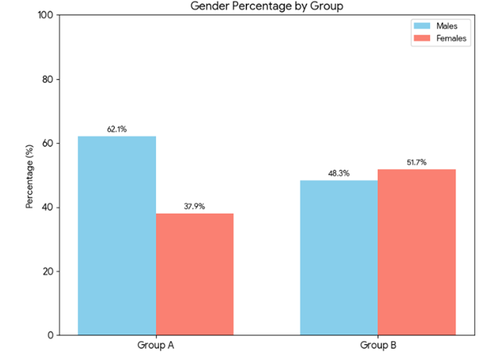
Table 3: Mean age, gestational age, birth weight and APGAR score in study groups
|
VARIABLES |
GROUPS |
Mean |
Std. Dev |
P-value t-test |
|
AGE (HOURS) |
GROUP A |
5.71 |
1.31 |
0.393 |
|
GROUP B |
5.51 |
1.29 |
||
|
GESTATIONAL AGE (Weeks) |
GROUP A |
38.5 |
1.8 |
0.104 |
|
GROUP B |
37.7 |
1.1 |
||
|
BIRTH WEIGHT (Kg) |
GROUP A |
2.97 |
0.42 |
0.473 |
|
GROUP B |
2.91 |
0.40 |
||
|
APGAR SCORE |
GROUP A |
2.83 |
0.38 |
0.612 |
|
GROUP B |
2.86 |
0.35 |
Other baseline clinical characteristics were also similar between the two groups (p>0.05, Table 3). Regarding the severity of HIE at baseline, in Group A, 1.7% (n=1/58) of neonates demonstrated grade I, 37.9% (n=22/58) grade II, and 60.3% (n=35/58) grade III HIE. In Group B, the corresponding percentages were 5.2% (n=3/58), 46.6% (n=27/58), and 48.3% (n=28/58), respectively. No statistically significant difference was observed in the distribution of HIE grades between the two groups (p=0.319, Table 4).
Table 4: Clinical characteristics at baseline
|
VARIABLES |
GROUP |
TOTAL |
P-VALUE CHI-SQUARE |
||
|
GROUP A |
GROUP B |
||||
|
FITS |
PRESENT |
53 |
49 |
102 |
0.254 |
|
91.4% |
84.5% |
87.9% |
|||
|
ABSENT |
5 |
9 |
14 |
||
|
8.6% |
15.5% |
12.1% |
|||
|
ACIDOSIS |
PRESENT |
47 |
51 |
98 |
0.305 |
|
81.0% |
87.9% |
84.5% |
|||
|
ABSENT |
11 |
7 |
18 |
||
|
19.0% |
12.1% |
15.5% |
|||
|
VENTILATED |
PRESENT |
43 |
45 |
88 |
0.664 |
|
74.1% |
77.6% |
75.9% |
|||
|
ABSENT |
15 |
13 |
28 |
||
|
25.9% |
22.4% |
24.1% |
|||
Figure 2: Clinical characteristics at baseline
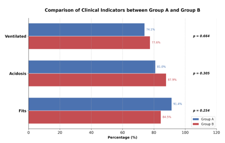
Table 5: Baseline HI grades in both groups
|
BASELINE HI EGRADES |
GROUP |
TOTAL |
|
|
GROUP A n(%) |
GROUP B n(%) |
||
|
GRADE I |
1 (1.7%) |
3 (5.2%) |
4 (3.4%) |
|
GRADE II |
22 (37.9%) |
27 (46.6%) |
49 (42.2%) |
|
GRADE III |
35 (60.3% |
28 (48.3%) |
63 (54.3%) |
At baseline, mean Thompson scores were comparable between Group A (melatonin plus standard care) and Group B (standard care alone), with no statistically significant difference observed (p>0.05). Similarly, at 24 hours (p=0.896) and day 3 (p=0.242), the mean Thompson scores remained statistically indistinguishable between the two groups. However, by day 7, a marked and statistically significant divergence emerged: the mean Thompson score in Group A was 4.4 (±3.1 SD), which was substantially lower than that in Group B, which measured 7.8 (±3.9 SD), yielding a p-value of 0.001 (Table 5). This indicates a superior neuroprotective effect of the melatonin-containing regimen over standard care alone by the end of the first week. The efficacy of treatment, defined as improvement in HIE grade from baseline, was observed in 82.1% (n=48/58) of patients in Group A, compared to only 51.7% (n=30/58) in Group B, and this difference was highly significant (p=0.001, Table 6). These findings demonstrate that the addition of melatonin to standard care significantly enhances clinical recovery in terms of HIE severity. Regarding mortality, the proportion of neonates who expired within the first seven days was 15.5% (n=9/58) in Group A, which was less than half the mortality rate observed in Group B, where 37.9% (n=22/58) of patients expired. This difference was also statistically significant (p=0.001, Table 7). Collectively, these results indicate that melatonin as an adjunct to standard care not only improves neurological outcomes but also substantially reduces early mortality in neonates with moderate to severe HIE.
Table 6: Mean Thompson score at baseline and at different time intervals
|
THOMPSON SCORE |
GROUPS |
Mean |
Std. Dev |
P-value t-test |
|
BASELINE |
GROUP A |
17.4 |
3.6 |
0.103 |
|
GROUP B |
15.9 |
3.5 |
||
|
24 HOURS |
GROUP A |
15.3 |
4.2 |
0.896 |
|
GROUP B |
15.2 |
4.1 |
||
|
DAY 3 |
GROUP A |
11.1 |
4.3 |
0.242 |
|
GROUP B |
12.1 |
4.3 |
||
|
DAY 7 |
GROUP A |
4.4 |
3.1 |
0.001 |
|
GROUP B |
7.8 |
3.9 |
Table 7: Improvement in HIE grades
|
IMPROVEMENT IN HI GRADES |
GROUP |
TOTAL |
P-VALUE CHI-SQUARE |
|
|
GROUP A |
GROUP B |
|||
|
PRESENT |
48 |
30 |
78 |
<0.001 |
|
82.8% |
51.7% |
67.2% |
||
|
ABSENT |
10 |
28 |
38 |
|
|
17.2% |
48.3% |
32.8% |
||
DISCUSSION :
Giant congenital melanocytic nevi (GCMN) represent Hypoxic‑ischemic encephalopathy (HIE) arises from inadequate antioxidant defence in the brain, with free radical formation representing one of the most important contributing factors to brain injury [17]. It has been hypothesized that excessive free radical production may diminish the synthesis of neurotrophic factors, thereby directly influencing neurogenesis [18]. Melatonin may serve as a potential therapeutic free radical scavenger and a broad‑spectrum antioxidant [19,20]. The present study was designed to determine the efficacy of melatonin in the management of neonates with HIE. In this trial, the mean Thompson score demonstrated a significant decline in Group A (melatonin plus standard care) compared to Group B (standard care alone) at seven days of therapy (4.4±3.1 versus 7.8±3.9; p=0.001). The efficacy of treatment, defined as improvement in HIE grade from baseline, was observed in 82.1% (n=48/58) of patients in Group A versus 51.7% (n=30/58) in Group B (p=0.001). Furthermore, our results showed better survival in the adjunctive melatonin group: 15.5% (n=9/58) of neonates expired within seven days in Group A compared to 37.9% (n=22/58) in Group B (p=0.001). Comparable findings have been reported in several other studies [21,22,23]. An initial observation indicated that in asphyxiated newborns with HIE, oral administration of melatonin (80 mg in eight doses) reduced serum malondialdehyde and nitrite/nitrate concentrations and improved survival [21].
Subsequent reports from the same group of investigators indicated that in preterm infants with respiratory distress, treatment with a high dose (100 mg/kg in ten infusions) improved outcome [22]. In a randomized trial involving 30 HIE newborns treated with 50 mg of melatonin as five daily enteral doses, the melatonin/hypothermia group exhibited a greater increase in melatonin levels, a decline in circulating oxidants, fewer seizures, and less white matter injury [23]. Moreover, a study by Ahmad et al. (2018) demonstrated improved survival in asphyxiated neonates [24], a finding that is consistent with the improved survival observed in Group A of the present study. Regarding the melatonin dose employed, the high peak plasma concentration and long half‑life of melatonin in newborns indicate that, in the neonatal clinical setting, it is possible to achieve and maintain high concentrations of melatonin using a single administration repeated every 12 or 24 hours [25]. A remarkable number of melatonin effects strongly suggest that it has important therapeutic implications in the management of HIE. Its strong safety profile, ability to cross the blood–brain barrier, and diverse pleiotropic properties have led to a large number of preclinical animal studies supporting its neuroprotective efficacy in reducing brain injury [26,27]. Whilst the preclinical data are compelling, clinical studies in infants with neonatal encephalopathy (NE) remain limited. Ahmed et al., in their meta‑analysis, described the available clinical evidence for melatonin as a potential therapy for NE [28]. They analyzed five randomized controlled trials comprising 215 neonates and found that long‑term developmental outcome data were lacking in all except one small study, which reported significantly higher composite cognition scores at 18 months. One study reported intermediate favorable development at six‑month follow‑up. A meta‑analysis of mortality in the combined hypothermia plus melatonin group versus hypothermia alone (two studies, 54 participants) demonstrated no significant reduction, with a relative risk (RR) of 0.42 (95% CI, 0.99–1.12) [28]. The authors concluded that the clinical data supporting the neuroprotective effects of melatonin in neonates are limited, and that larger, well‑designed, adequately powered multicenter clinical trials are urgently needed to define the neuroprotective role of melatonin in optimizing outcomes of NE [28].
The present study utilized the Thompson score to predict neonatal improvement following hypoxic insult. The Thompson score comprises clinical parameters with an average score range of 0–3, providing high predictability for improvement or deterioration in HIE grades. Shrestha et al. employed this score in a previous study [29]. In another study, Aoki et al. compared the Sarnat and Thompson scores for predicting short‑term outcomes in infants with mild NE, demonstrating that although the Sarnat score is most widely used to assess NE, the Thompson score at admission is a more useful predictor of short‑term outcome in infants with mild NE [30]. The results of the present study, together with several other studies cited in the literature, demonstrate that hypoxic‑ischemic insult leads to devastating neurological consequences. The advantages of melatonin its efficacy, low toxicity, and ability to readily cross the blood–brain barrier make it a promising molecule. However, studies focusing on the neuroprotective potential of melatonin and its possible therapeutic application after perinatal asphyxia in humans remain scarce, and its potential therapeutic role in humans needs to be validated in randomized trials. A major strength of the current study is its randomized controlled design with stringent enrolment criteria. Nevertheless, the present study has several limitations. First, we did not perform serum melatonin analysis as a baseline measurement of endogenous melatonin production. Second, superoxide dismutase activity was not measured to detect baseline oxidative stress with and without HIE, nor to examine the effect of melatonin on oxidative stress. Third, we did not measure serum nitric oxide to assess free radical concentration. Fourth, we did not follow up our study participants for a longer duration to evaluate neurological development. Finally, as we assessed a single dose of melatonin, we are unable to comment on the dose‑dependent nature of the neuroprotective action of melatonin, as suggested by some studies [25,26].
CONCLUSION :
The efficacy of treatment, defined as improvement in HIE grade, was significantly better in patients treated with melatonin plus standard care compared to those treated with standard care alone. Furthermore, a significantly lower number of neonates expired in the adjunctive melatonin therapy group than in the standard care alone group. These findings support the use of melatonin as an effective adjunctive therapy in the management of moderate to severe HIE.
Conflict of interest
The authors declared no conflict of interest.
Author Contribution
· Concept & Design of the study: Asma & Tahira Yasmeen
· Drafting: Sadia Riaz
· Data analysis: Muhammad Azhar & Khizar Ilyas
· Critical Review & Final approval: Hina Khan & Asma
All authors reviewed the results and approved the final version of the manuscript. They are also accountable for the study's integrity.
BIBLIOGRAPHY:
1. Glass HC. Hypoxic-Ischemic Encephalopathy and Other Neonatal Encephalopathies. Continuum (MinneapMinn). 2018;24(24(1, Child Neurology):57-71.
2. Executive summary:Neonatal encephalopathy and neurologic outcome, second edition. Report of the American College of Obstetricians and Gynecologists' Task Force on Neonatal Encephalopathy. Obstet Gynecol. 2014;123:896. Reaffirmed. 2020.
3. Namusoke H, Nannyonga MM, Ssebunya R, Nakibuuka VK. Incidence and short term outcomes of neonates with hypoxic ischemic encephalopathy in a Peri Urban teaching hospital, Uganda: a prospective cohort study. Matern Health Neonatol Perinatol. 2018;4:6-9.
4. Zanelli SA. Hypoxic-ischemic encephalopathy.emedicine. Available from: https://emedicine.medscape.com/article/973501-overview. [Accessed on April01,. 2022].
5. Memon S, Shaikh S, Bibi S. To compare the outcome (early) of neonates with birth asphyxia in-relation to place of delivery and age at time of admission. J Pak Med Assoc. 2012;62:1277-81.
6. Martinez-Biarge M, Ferriero DM, Cowan FM. Perinatal arterial ischemic stroke. HandbClinNeurol. 2019;162:239–266.
7. Wyckoff MH, Aziz K, Escobedo MB, Kapadia VS, Kattwinkel J, Perlman JM. Part 13:Neonatal Resuscitation: 2015 American Heart Association Guidelines Update for Cardiopulmonary Resuscitation and Emergency Cardiovascular Care. Circulation. 2015;132:S543-7.
8. Perlman JM, Wyllie J, Kattwinkel J. Neonatal Resuscitation:2015 International Consensus on Cardiopulmonary Resuscitation and Emergency Cardiovascular Care Science With Treatment Recommendations. Circulation. 2015;132:S204-8.
9. Paprocka J, Kijonka M, RzepkaB,Sokół M. Melatonin in Hypoxic-Ischemic Brain Injury in Term and Preterm Babies. Int J Endocrinol. 2019;2019:9626715.
10. D’Angelo G, Chimenz R, Reiter RJ, Gitto E. Use of Melatonin in Oxidative Stress Related Neonatal Diseases. Antioxidants (Basel). 2020;9(6):477-82.
11. Robertson NJ, Lingam I, Meehan C. High-Dose Melatonin and Ethanol Excipient Combined with Therapeutic Hypothermia in a Newborn Piglet Asphyxia Model. SciRep. 2020;10:3898-903.
12. Cardinali DP. An Assessment of Melatonin’s Therapeutic Value in the Hypoxic-Ischemic Encephalopathy of the Newborn Front. Synaptic Neurosci. 2019;11:34-9.
13. Antonio JC, Cuenca S, Teresa S. Hypothermia Plus Melatonin in AsphycticNewborns:A Randomized-Controlled Pilot Study. Ped Critical Care Med. 2020;21(7):647-55.
14. Ahmad QM, Chishti AL, Waseem N. Role of melatonin in management of hypoxic ischaemic encephalopathy in newborns:A randomized control trial. J Pak Med Assoc. 2018;68:1233-7.
15. Aly H, Elmahdy H, El-Dib M, Rowisha M, Awny M, El-Gohary T. Melatonin use for neuroprotection in perinatal asphyxia:a randomized controlled pilot study. J Perinatol. 2015;35:186–91.
16. Chandrasekaran M, Chaban B, Montaldo P, Thayyil S. Predictive value of amplitude-integrated EEG (aEEG) after rescue hypothermic neuroprotection for hypoxic ischemic encephalopathy: a meta-analysis. J Perinatol. 2017;37:684-9.
17. Ramaswamy V, Horton J, Vandermeer B Systematic review of biomarkers of brain injury in term neonatal encephalopathy. Pediatr Neurol. 2009;40:215-9.
18. Massaro AN, Jeromin A, Kadom N Serum biomarkers of MRI brain injury in neonatal hypoxic ischemic encephalopathy treated with whole-body hypothermia: a pilot study. Pediatr Crit Care Med. 2013;14:310-7.
19. Chalak LF, Sánchez PJ, Adams-Huet B Biomarkers for severity of neonatal hypoxic-ischemic encephalopathy and outcomes in newborns receiving hypothermia therapy. J Pediatr. 2014;164:468-73.
20. Martinello K., Hart A. R., Yap S., Mitra S., Robertson N. J. Management and investigation of neonatal encephalopathy: 2017 update. Archives of Disease in Childhood - Fetal and Neonatal Edition. 2017;102(4):F346–58.
21. Tordjman S, Chokron S, Delorme R. Melatonin: pharmacology, functions and therapeutic benefits. Current Neuropharmacology. 2017;15(3):434–43
22. Fulia F, Gitto E, Cuzzocrea S. Increased levels of malondialdehyde and nitrite/nitrate in the blood of asphyxiated newborns: reduction by melatonin. J. Pineal Res. 2001;31:343–9
23. Hu Y, Wang Z, Liu Y, Pan S. Melatonin reduces hypoxic-ischaemic (HI) induced autophagy and apoptosis: an in vivo and in vitro investigation in experimental models of neonatal HI brain injury. Neurosci. Lett. 2017;653:105–12
24. Revuelta M, Arteaga O, Alvarez A. Characterization of gene expression in the rat brainstem after neonatal hypoxic-ischemic injury and antioxidant treatment. Mol. Neurobiol. 2017;54:1129–43
25. Carloni S, Riparini G, Buonocore G, Balduini W. Rapid modulation of the silent information regulator 1 by melatonin after hypoxia-ischemia in the neonatal rat brain. J. Pineal Res. 2017;63:e12434.
26. Ramos E, Patino P, Reiter RJ, Gil-Martin E. Ischemic brain injury: New insights on the protective role of melatonin. Free Radic. Biol. Med. 2017;104:32–53.
27. Pang R, Avdic-Belltheus A, Meehan C, Martinello K, Mutshiya T. Melatonin and/or erythropoietin combined with hypothermia in a piglet model of perinatal asphyxia. Brain Commun. 2021;3:1-6
28. Ahmed J, Pullattayil S , Robertson NJ. Melatonin for neuroprotection in neonatal encephalopathy: A systematic review & meta-analysis of clinical trials. Eur J Paediatr Neurol. 2021;31:38-45.
29. Shrestha S. Correlation of thompson score in predicting early outcome of newborn with birth asphyxia. J Nepal Health Res Counc. 2020;18(3):406-10.
30. Aoki Y, Kono T, Enokizono M, Okazaki K. Short‐term outcomes in infants with mild neonatal encephalopathy: a retrospective, observational study BMC Pediatrics. 2021;12: 224