Management of a Sickle Cell Anaemia Patient Undergoing Total Hip Arthroplasty Complicated by Intraoperative Pulmonary Embolism A Case Report
- Salahulddin Abuljadail , Assistant Professor , Orthopaedic Department , College of medicine , King Faisal University, 31982, Al-Ahasa , Saudi Arabia.
Article Information:
Abstract:
Hip avascular necrosis is a well known complication secondary to recurrent vaso-occlusive crisis among sickle cell diseased patients. Advanced avascular necrosis of the hip necessitates total hip replacement procedure. Preoperative optimal management of sickle cell diseased Patients is crucial to avoid perioperative complications. Case Presentation: A 40-year-old sickle cell diseases female patient presented with worsening right hip pain and limited mobility, was diagnosed with advanced AVN of the right hip. Total hip replacement was indicated. Although meticulous preoperative measures were performed, an intraoperative pulmonary embolism was encountered just after acetabular cup implantation. The procedure was halted promptly and the patient was transferred to the intensive care unit for further investigation and management. A second surgery was performed 4 weeks later (after starting therapeutic anticoagulation regime) to complete the surgery. Conclusion: Sickle cell anemia patients undergoing total hip arthroplasty require meticulous preoperative preparation to reduce the risk of perioperative complications.
Keywords:
Article :
INTRODUCTION :
Sickle cell anaemia (SCA) is a hereditary haemoglobinopathy characterized by recurrent vaso-occlusive crises and various complications, including avascular necrosis (AVN) of the femoral head 1. AVN often necessitates surgical intervention such as total hip replacement (THR) to restore function and relieve pain. However, patients with SCA face an increased risk of perioperative complications, including thromboembolic events. This report presents a case of a patient with SCA undergoing THR who experienced an intraoperative pulmonary embolism (PE), leading to cessation of surgery and delayed reoperation 2.
SCA is a hereditary haemoglobinopathy caused by a mutation in the β-globin gene, resulting in the production of abnormal haemoglobin S (HbS). When deoxygenated, HbS polymerizes, leading to the characteristic sickling of red blood cells (RBCs). Sickled cells are rigid, prone to hemolysis, and have a shortened lifespan, resulting in chronic haemolytic anaemia. The sickling process also contributes to vaso-occlusion, causing tissue ischemia, pain crises, and multi-organ damage over time. SCA primarily affects individuals of African, Mediterranean, Middle Eastern, and Indian ancestry, with the highest prevalence in sub-Saharan Africa.
The disease has systemic implications, including chronic inflammation, endothelial dysfunction, and hypercoagulability, which predispose patients to thrombotic events. Advances in management, such as hydroxyurea, blood transfusion protocols, and early diagnosis, have significantly improved life expectancy. However, complications like AVN continue to affect the quality of life in many patients 1 3 4 5.
AVN, also known as osteonecrosis, is a debilitating complication of SCA, occurring due to repeated episodes of vaso-occlusion in the microcirculation of bones. The femoral head is most commonly affected because of its tenuous blood supply, making it particularly vulnerable to ischemic injury. AVN leads to the progressive collapse of bone structure, resulting in joint dysfunction, chronic pain, and significant impairment in mobility 2.
In patients with SCA, AVN can develop as early as adolescence and progresses more rapidly compared to non-sickle cell patients. The pathophysiology involves repeated cycles of ischemia and reperfusion injury, endothelial damage, and increased intraosseous pressure due to microvascular obstruction by sickled cells. Radiographic findings may include subchondral lucency (crescent sign) and eventual joint space narrowing, necessitating surgical intervention when conservative measures fail 1 2.
There are several preoperative risk factors in SCA that should be taken into consideration. Surgical interventions in SCA patients, such as THR for AVN, pose unique challenges due to the systemic effects of the disease. Among those are haematological and cardiopulmonary risk factors, bone and joint considerations, renal dysfunction, and infectious risk.
Regarding haematological risk factors, sickle cell crises might arise intra-operatively due to the stress of surgery, including hypoxia, acidosis, and dehydration, which can precipitate vaso-occlusive crises (VOC) or acute chest syndrome (ACS), a life-threatening complication involving pulmonary vaso-occlusion 1. Moreover, chronic haemolysis leads to baseline anaemia, which may exacerbate tissue hypoxia during surgery 1 5. On the other hand, patients with SCA are at an increased risk of thromboembolic events due to hypercoagulability, including deep vein thrombosis (DVT) and PE, due to heightened activation of coagulation pathways 1,5.
Among cardiopulmonary risk factors, pulmonary hypertension is common in SCA, it increases the risk of perioperative complications such as right heart strain 4. Impaired Oxygenation due to chronic pulmonary dysfunction and prior ACS episodes may lead to baseline hypoxia, increasing susceptibility to intraoperative complications 4.
Regarding bone and joint considerations, osteoporosis and fragility is a known risk factor in SCA patients, especially those who are under chronic steroid use which predisposes to fractures and delayed healing 3,6. In the advanced SCA, severe joint deformities in advanced AVN complicate surgical procedures, increasing intraoperative challenges 3,6.
Moreover, SCA can lead to renal impairment, affecting fluid management, medication clearance, and overall surgical outcomes 7. Functional asplenia increases susceptibility to infections, necessitating perioperative prophylactic antibiotics and vaccinations 1,7.
There are several risk factors in the perioperative period which include intraoperative and postoperative risk factors. Among intraoperative risk factors, hypoxia and acidosis during anaesthesia may trigger sickling crises. Thromboembolic events, including PE, may occur due to hypercoagulability and venous stasis 8-10.
Regarding postoperative complications, VOC or ACS is a potential complication which can be triggered by surgical stress or inadequate pain control. On the other hand, delayed wound healing, increased risk of infection as well as implant-related complications due to suboptimal bone quality are known postoperative complications among SCA patients 8-10.
Optimal management of SCA patients undergoing surgery requires meticulous preoperative planning, including haematological optimization, multidisciplinary coordination, and vigilant perioperative monitoring. Strategies such as preoperative blood transfusion to reduce HbS levels, hydration, and prophylactic anticoagulation play critical roles in reducing complications. Understanding the interplay between SCA and AVN is essential to improving surgical outcomes and ensuring long-term functional recovery 3-5,8,9.
Material and Methods:
A 40-year-old female with a history of homozygous sickle cell anaemia presented with worsening right hip pain and limited mobility. Radiographic imaging confirmed advanced AVN of the right femoral head Ficat stage 3 [Figure 1, 2]. Conservative management had failed, and she was scheduled for a THR. Her medical history included frequent vaso-occlusive crises. She had no known history of venous thromboembolism.
Preoperatively, the patient was optimized preoperatively with a transfusion protocol to reduce her haemoglobin S level to below 30%, achieving a haemoglobin level of 10.2 g/dL. Hydration, pain control, and avoidance of hypoxia and acidosis were prioritized. Prophylactic anticoagulation with low molecular weight heparin was initiated 24 hours before surgery.
The surgery commenced uneventfully under general anaesthesia. After finishing the implantation of the acetabular cup using an anterior minimal invasive approach to yhe right hip (AMIS) [Figure 3], the patient developed sudden hypoxia (SpO₂ dropped to 82%), hypotension (BP 76/48 mmHg), and tachycardia (HR 135 bpm). End-tidal CO₂ decreased, and clinical suspicion of a PE was raised. The surgery was immediately halted, neglecting femoral canal preparation and femoral stem implantation, and resuscitation efforts were initiated. The patient was stabilized with high-flow oxygen, intravenous fluids, and vasopressors. An intraoperative echocardiogram revealed signs of right ventricular strain, confirming the diagnosis of PE.
Postoperatively, the patient was transferred to the intensive care unit (ICU) for close monitoring and management. She was started on therapeutic anticoagulation with intravenous heparin after stabilization. A multidisciplinary team, including haematologists, orthopaedic surgeons, and anaesthesiologists, coordinated her care.
After four weeks, during which she remained clinically stable and completed anticoagulation therapy for the acute PE, the surgical team deemed her fit for a second attempt at THR. Preoperative optimization was repeated, including transfusion to reduce haemoglobin S levels, continuation of anticoagulation, and ensuring adequate hydration.
The second surgery was performed 4 weeks later under regional anaesthesia to minimize systemic stress. The aim of the second surgery was to complete the surgery with preparation and implantation of the femoral stem through the same approach. The procedure was completed successfully without complications postoperatively [Figure 4]. The one-year follow up was satisfactory with improving hip function and no further complications [Figure 5].
FIGURES
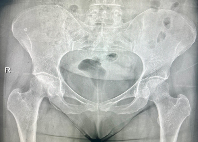
Figure 1: AP radiograph of the pelvis showing subchondral collapse of the right femoral head.
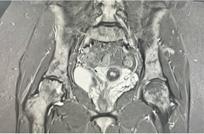
Figure 2: A coronal cut of MRI of the pelvis showing AVN of the right femoral head Ficat stage 3 with subchondral collapse.
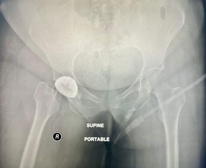
Figure 3: Post-operative AP radiograph of the pelvis (post 1st procedure) showing the implantation of the acetabular cup.
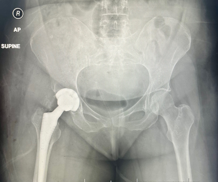
Figure 4: AP radiograpgh of the pelvis after the second surgery after procedure completion with implantation of the femoral stem.
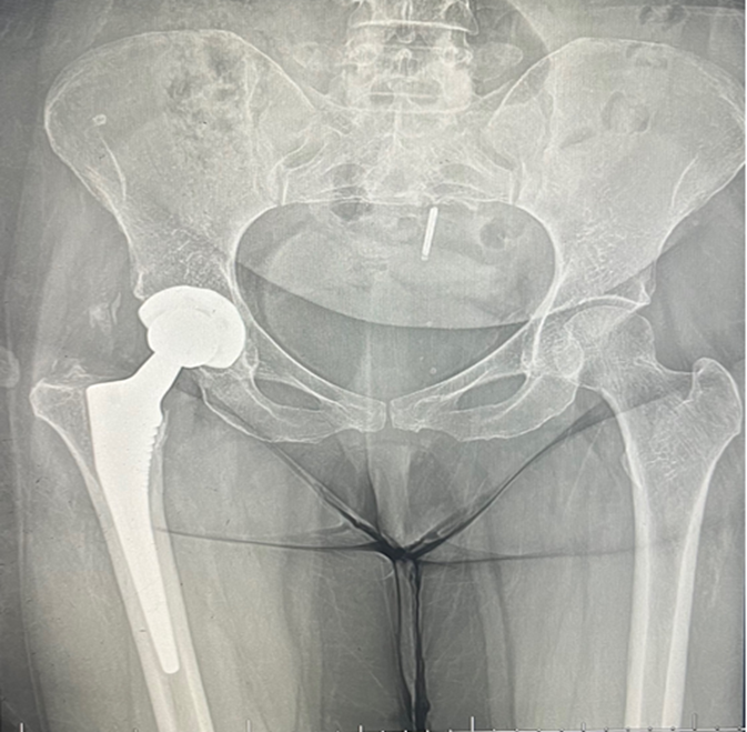
Figure 5: AP radiograph of the pelvis in one-year follow up
DISCUSSION:
This case highlights the heightened perioperative risks in patients with SCA, including the propensity for thromboembolic events such as PE. Factors such as hypercoagulability, hypoxia, and venous stasis contribute to this risk. The occurrence of an intraoperative PE emphasizes the importance of vigilance, early recognition, and prompt management.
Thromboemblic events in SCD-Patients are common, yet an underdiagnosed complication, this is supported by a study that showed incidental pulmonary thrombus in computertomography (CT) scan in 17% of patients with ACS 11.
Vichinsky et al. analyzed the seriuos and life-threatning postoperative complications in 138 orthopedic surgical procedures in sickle cell patients within the national sickle cell surgery study group. Sixty eight percent of the procedures involved the hip joint, most commonly hip joint replacement and decompression coring procedures for avascular necrosis. The preoperative hemoglobin S percentage varied significantly between the study subjects. Seventy-nine percent of the patient received preoperative hydration and about 97% of the patients were well monitored intraoperatively. The most common intraoperative complications is the excessive blood loss. In 17% of the cases was there an involvement of sickel-cell-related events (acute chest syndrome and/or vaso-occlusive crisis). Hypothermia was evident in 11% of the cases and transfusion-related complications in 9% of the patients 8,9.
Preoperative blood transfusion with the goal to decrease the HgbS to < 30% was investigated in several randomized controlled trials which showed lowered rate of complications such as ACS and VOC in low- to medium- risk procedures 10,12.
Regarding our case, delayed reoperation allowed time for recovery and optimization, ultimately leading to a successful outcome. Preoperative planning, multidisciplinary collaboration, and tailored perioperative protocols are essential for minimizing complications and achieving favorable outcomes in this high-risk population.
Anticoagulation was continued for three months, and the patient underwent intensive physiotherapy. At a six-month follow-up, she reported significant pain relief and improved mobility, with no recurrence of thromboembolic events or vaso-occlusive crises.
CONCLUSION:
Patients with SCA undergoing THR for AVN require meticulous perioperative management due to their elevated risk of complications. This case underscores the importance of individualized care, highlighting the challenges and successful management of a serious intraoperative complication.
Level of Evidence: This manuscript provides Level 4 evidence
REFERENCES:
1. disease. Lancet. 2010;376(9757):2018-31. doi:10.1016/S0140-6736(10)61029-X
2. Milner PF, Kraus AP, Sebes JI, et al. Sickle cell disease as a cause of osteonecrosis of the femoral head. N Engl J Med. 1991;325(21):1476-81. doi:10.1056/NEJM199111213252104
3. Hernigou P, Zilber S, Filippini P, et al. Total THA in adult osteonecrosis related to sickle cell disease. Clin Orthop Relat Res. 2008;466(2):300-8. doi:10.1007/s11999-007-0069-3
4. Gladwin MT, Sachdev V, Jison ML, et al. Pulmonary hypertension as a risk factor for death in patients with sickle cell disease. N Engl J Med. 2004;350(9):886-95. doi:10.1056/NEJMoa035477
5. Naik RP, Streiff MB, Haywood C, Jr., et al. Venous thromboembolism in adults with sickle cell disease: a serious and under-recognized complication. Am J Med. 2013;126(5):443-9. doi:10.1016/j.amjmed.2012.12.016
6. P H. Total hip replacement in sickle cell disease: complications and outcomes. Clinical Orthopaedics and Related Research. 2008;
7. Pham PT, Pham PC, Wilkinson AH, et al. Renal abnormalities in sickle cell disease. Kidney Int. 2000;57(1):1-8.
doi:10.1046/j.1523-1755.2000.00806.x
8. Vichinsky EP, Neumayr LD, Haberkern C, et al. The perioperative complication rate of orthopedic surgery in sickle cell disease: report of the National Sickle Cell Surgery Study Group. Am J Hematol. 1999;62(3):129-38. doi:10.1002/(sici)1096-8652(199911)62:3<129::aid-ajh1>3.0.co;2-j
9. Vichinsky EP, Haberkern CM, Neumayr L, et al. A comparison of conservative and aggressive transfusion regimens in the perioperative management of sickle cell disease. The Preoperative Transfusion in Sickle Cell Disease Study Group. N Engl J Med. 1995;333(4):206-13. doi:10.1056/NEJM199507273330402
10. Reed WF, Vichinsky EP. Transfusion practice for patients with sickle cell disease. Curr Opin Hematol. 1999;6(6):432-6. doi:10.1097/00062752-199911000-00014
11. Mekontso Dessap A, Deux JF, Abidi N, et al. Pulmonary artery thrombosis during acute chest syndrome in sickle cell disease. Am J Respir Crit Care Med. 2011;184(9):1022-9. doi:10.1164/rccm.201105-0783OC
12. Khurmi N, Gorlin A, Misra L. Perioperative considerations for patients with sickle cell disease: a narrative review. Can J Anaesth. 2017;64(8):860-869. Considerations perioperatoires pour les patients atteints d'anemie falciforme : un compte rendu narratif. doi:10.1007/s12630-017-0883-3