Toxicological evaluation of Diospyros buxifolia (Blume) Heirn." Bark extract: Acute and sub-acute studies in mice
- Bhargavi G , DoS and R in Biochemistry, Karnataka State Open University, Mysuru, Karnataka, India.
- Sapna M , DoS and R in Biochemistry, Karnataka State Open University, Mysuru, Karnataka, India.
- K.N. Ravindra , DoS and R in Botany, Karnataka State Open University, Mysuru, Karnataka, India.
- Suprada Rao M.S , DoS in Biochemistry, SBRR Mahajana First Grade College, Mysuru, Karnataka, India.
- Rajesh R , Chromed Biosciences Pvt. Ltd., Hirehalli, Tumkur, Karnataka, India.
- Natraju Angaswamy. , DoS and R in Biochemistry, Karnataka State Open University, Mysuru, Karnataka, India.
Article Information:
Abstract:
The abundance of bioactive phytochemicals in medicinal plants has a variety of applications in human health and pharmacological therapy. Diospyros buxifolia (DB, Ebenaceae) is a less-recognised source of secondary metabolites, nutraceuticals, and antioxidants, with potential therapeutic effects on intestinal disorders. Our previous publications identified more than 25 bioactive phytochemicals with a promising multitargeted approach targeting Wnt-β-catenin pathway proteins in colorectal cancer through molecular docking analysis. Further, our published results have demonstrated in vitro antiproliferative activity against colon cancer cells, thereby supporting the extract's effectiveness. However, no detailed investigation towards the possible toxicity of DB has been conducted. Therefore, this study aimed to evaluate the acute and sub-acute toxicity of DB bark methanol extract in Balb/c mice. Eight groups of mice (8-12 weeks, 25 ± 3g), comprising female and male groups (n = 5), were designed. Following OECD guidelines 423, 425, and 407, a 14-day (single oral dose) and a 28-day (daily doses) study was conducted at 500, 750, 1000, and 2000 mg/kg of DB bark methanol extract to investigate acute and sub-acute toxicity, respectively. No mice showed lethality over the administration of 2000 mg/kg b.w. The body weight and biochemical parameters were evaluated. The livers, lungs, heart, spleen and kidneys were examined histologically. No variations were observed in food intake, water consumption, mortality, or body and organ weights. Similarly, DB, even at higher doses, did not significantly alter haematological and serological parameters, with no significant signs of abnormalities or toxicity, all within the normal range, consistent with the CCSEA compendium. Additionally, histological examinations showed no significant difference compared to control animals. The present study demonstrated no acute or sub-acute toxicity following the oral administration of DB extracts. Therefore, DB extract is non-toxic at the tested doses and safe for food and pharmaceutical applications.
Keywords:
Article :
INTRODUCTION :
Phytochemical preparations and bioactive phytochemicals play a significant role in the treatment of human diseases. About 40% of the therapeutic agents currently used by locals in the Western Ghats of India are derived from natural resources, particularly medicinal plants. Based on the various descriptions in the literature, multiple parts of the genus Diospyros have been used to treat several infectious diseases, including antibacterial, antifungal, anthelminthic, and antiviral; urogenital (anti-hemorrhagic); skin diseases that include dermatitis, fresh wounds, bedsores, and rashes; and musculoskeletal disorders related to body pain, bruises, painful fractures, and rheumatism. Furthermore, the bark, fruit, and root are the most commonly employed components of Diospyros in traditional medicine [1, 2].
Diospyros buxifolia (DB) has a pantropical distribution and belongs to the genus Diospyros, which comprises around 500-600 species. DB, commonly known as Kunchigana mara, is a traditional plant with ethnopharmacological value for the treatment of diseases related to oral cavity health (oral wounds, bad breath, and toothaches) and gastrointestinal disorders. Triterpenoids, tannins, and naphthoquinones are among the active phytocompounds found in Diospyros species. The methanolic extracts of the leaves demonstrated significant antibacterial activity, while the stem extracts exhibited notable antioxidant potential. Thus, DB is a proven source of natural antimicrobial and antioxidant agents, which could address the challenges posed by drug-resistant microorganisms and oxidative stress in global health [3-5].
Although plant extracts have been extensively studied for their therapeutic potential, their toxicological effects in animal models are crucial for establishing safety profiles before clinical trials. Toxicological research plays a pivotal role in public health. These studies identify potential adverse effects, establish safe dosage ranges, and evaluate the impact on vital organs such as the liver, kidneys, and heart. This information is crucial for designing clinical trials that prioritise participant safety and for ensuring that only safe and effective drugs are approved for public use. Research papers provide insights into both the potential benefits and risks associated with their use. For example, Solanum elaeagnifolium extracts showed toxicity at high doses (2000 mg/kg) in Swiss albino mice, leading to death, while lower doses did not significantly affect biochemical markers or organ histology [6-8].
DB has been used as an herbal medicine with a range of health benefits. Our previous publications identified more than 14 potential GC-MS phytochemicals and 11 potential LCMS-MS compounds with a promising multitargeted approach targeting Wnt-β-catenin pathway proteins in colorectal cancer through molecular docking analysis. Further, our published results of in vitro antiproliferative activity against colon cancer cells and molecular docking studies h have demonstrated the effectiveness of the extract. Based on our literature review, the potential toxicity of this medicinal plant has not been examined [8, 9, 23]. Conducting acute and sub-acute toxicity studies in accordance with OECD (Organisation for Economic Co-operation and Development) guidelines is important for several reasons, including standardisation, regulatory acceptance, scientific rigour, comparability, compliance, and animal welfare. The toxicity studies conducted in accordance with OECD guidelines provide reliable, accepted, and ethically sound safety data crucial for protecting human health and the environment. Therefore, the current study was designed to evaluate the acute and subacute toxicity of the DB stem bark methanol extract in accordance with the OECD guidelines 407, 423 and 425, respectively. After oral administration, body weight, organ weight, histopathology, haematological parameters, and biochemical parameters were analysed.
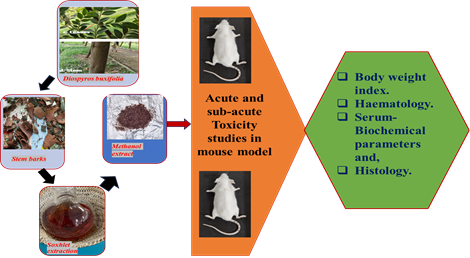
Figure 1: Graphical representation of the experimental design for acute and sub-acute toxicity studies of DB extract in mice.
Materials and methods:
Plant material: DB tree samples were collected from the Western Ghats of Karnataka, India. The Herbarium was submitted, and authenticated by Prof. Niranjan Raj S. The herbarium number (BOTIDB01) for the DB sample was obtained from the Department of Botany, KSOU, Mysuru. The stem bark of DB was surface-sterilised (3-4 times washed with tap water, then treated with 5% sodium hypochlorite solution for 15 minutes, rinsed with sterile water 3-4 times), air-dried, and finely powdered using a mixer-grinder. [8, 9].
Soxhlet extraction and preparation of methanol extract: The powder from the DB stem bark was defatted overnight (n-hexane), followed by methanol extraction using the Soxhlet apparatus (Labmatrix, Karnataka, India). The extract was concentrated under reduced pressure at 60 ± 1°C in a rotary vacuum evaporator (Steroglass, strike 300, Italy) until a solid mass was obtained, and stored in an air-tight container in a refrigerator (at 4 °C) [8, 9]. Stock solutions for mouse treatment were prepared in phosphate-buffered saline.
Experimental animal: Balb/c mice (8-12 weeks, 25 ± 3g) were procured from Chromed Biosciences Pvt. LTD., Tumkur, Karnataka, India, with an ethical clearance dated 10-12-2024 (Reference number-CBPL/CL/002/1012). The mice were housed individually in sterile cages and used for toxicity studies. Mice were maintained under standard conditions (22±3 °C; 12 h light/dark cycle; 40-50% humidity) with free access to water and food [10, 14, 21].
Oral acute and sub-acute toxicity studies: To establish the acute toxicity model, a single oral dose of DB extract at 500, 1000, and 2000 mg/kg b.w. was administered to groups 2, 3, and 4 of mice, respectively, by oral gavage. The mice were sacrificed after 14 days to analyse the toxicity in accordance with OECD guidelines 423 and 425.
Among Sub-acute studies- A daily dose of DB extract at 500, 750, or 1000 mg/kg b.w. was administered to groups 2, 3, and 4 of the mice, respectively, by oral gavage. In the study, the mice were sacrificed after 28 days to analyse the toxicity of the DB extract in accordance with OECD guideline 407. In both acute and sub-acute toxicity analyses, the animal grouping is given in Table 1.
The general behaviour of mice and signs of toxicity were observed continuously for 1 h following oral treatment, then intermittently for 4 h, and thereafter continuously for 24 h [16-20]. The mice were further observed once a day for the following treatments for behavioural changes, signs of toxicity and/or death and the latency of death. Organ weights and toxicity-associated parameters, including biochemical indicators, haematological and histological analyses, were measured and recorded upon sacrifice. [19-22].
Evaluation of relative body weight: The vital organs were dissected and weighed carefully. The relative organ weight of each animal was calculated by the formula:
Relative organ weight (g/100g) = organ weight(g)/body weight (g)×100
Haematological and serum analysis: The blood samples collected in EDTA tubes and serum samples for biochemical parameters were analysed for the parameters listed in Tables 3.2 and 3.3. For serum analysis, blood samples in non-EDTA-coated tubes were allowed to clot for 5 minutes, then immediately centrifuged at 3000 rpm for 10 minutes to separate the serum. Protein concentrations in the serum samples were analysed using the Lowery et al. method, with bovine serum albumin as the protein standard. All other haematological and biochemical parameters were determined using an automated haematology and serum biochemical analyser [10, 14, 16, 21].
Histopathological analysis by Harris hematoxylin & eosin technique: The tissues (kidney, liver, heart, lungs, and spleen) were fixed in buffered formalin (10% formaldehyde) overnight at room temperature and submitted for histological staining (Harris hematoxylin & eosin technique). Representative sections of the liver, spleen, and kidney were prepared and fixed in tissue cassettes, then processed in a tissue processor for 12 hours. The cassettes were embedded in molten paraffin wax and allowed to cool on a cold plate, forming blocks. These paraffin blocks were mounted on a rotary microtome to obtain 4 µm-thick sections. The tissue sections were carefully removed from the microtome knife and placed in a water bath to remove folds. The best sections were mounted on labelled slides and incubated in an oven at 58 °C overnight to fix. The slides were then stained with Harris hematoxylin and eosin. A mounting agent (Dibutyl phthalate in xylene) was applied, and the specimens were finally covered with a cover slip to prevent bubble formation [9-16]. The slides were examined microscopically using a standard compound microscope at 10X and 40X magnification and recorded for histopathological changes.
Statistical analysis
The computer-guided statistical programme GraphPad Prism, version 8.0.2, was used for analysis. The analysis involved summarising the data as the mean ± SD using descriptive statistics. Next, one-way and two-way ANOVA were performed to assess significant differences in mean values between the treatment groups, with p < 0.05 considered statistically significant [11-13].
Results and discussion::
Secondary metabolite-rich plants are known to have positive effects on human and animal health and serve as a foundation for all ethnopharmacological applications in medicine. Further, designing human trials depends on determining a safe dosage range in animal models. For example, the clinical potential of Caesalpinia spinosa is supported by the absence of severe toxicity in long-term investigations. Clinical trial design and patient population targeting can be influenced by knowledge of therapeutic effects and practices [8, 9]. For example, curcumin and other polyphenols derived from medicinal plants can have a dual role; they act as antioxidants at lower doses but can cause oxidative stress through ROS generation when consumed in excess. The balance between beneficial and toxic effects often depends on doses.
In the present study, experimental animals showed no changes in water or food intake or behaviour after oral administration of the DB extract in the acute and sub-acute groups 2, 3, and 4, compared to the respective controls. The weight gain of control/group-1 (2.8%), group-3 (2.6%) and group-4 (3.8%) mice was comparable to their initial weight in the acute 14-day study. However, the group-2 mice treated with 500 mg/kg gained only 0.5% to its initial weight (Figures 2A and 2B). Similarly, in the sub-acute treatment, groups 1, 3, and 4, treated with saline, 750, and 1000 mg/kg of DB, showed 5.6, 6.3, and 5.8% increases in body weight over 28 days (Figures 2C and 2D). As observed in acute treatment group-2 (500mg/kg b.w.) mice, the mice administered gained comparatively less (4.7%) than in other groups. In our toxicity study, a decrease in body weight of mice after administering a plant extract can indicate potential adverse effects of the extract on the animals. Some possible reasons for this weight loss include stress or toxicity to organs, leading to reduced appetite or reduced water and food intake. The reasons could also be gastrointestinal discomfort, diarrhoea, and excessive urination. However, mice treated with higher doses of DB did not show the effect, ruling out the extract's toxicity. Furthermore, detailed observations, biochemical assays, and histopathological studies are typically needed to confirm the cause [16-22].
The relative weights of the important organs of the test groups, such as the liver, heart, kidneys, and spleen, after sacrifice showed no significant change compared with those of the control mouse groups. Interestingly, the group 2 mice with comparatively less weight gain also did not show abnormal organ weights. Furthermore, there were minimal to insignificant differences in the aforementioned parameters between male and female mice among both acute and sub-acute treatments (Figures 3A, 3B, 3C and 3D).
The haematological results, with underlying parameters, showed no significant differences between the treatment groups and the control animals, except for an increased lymphocyte count of 12.1 and 16.6 /103/μl in Group 4 of both acute and sub-acute mice, compared to the control (7.0 and 6.5 /103/μl, respectively). Further, the haematological parameters such as Haemoglobin, RBC, WBC, Platelets, Granulocyte, Lymphocyte, Monocyte, MCV and MCH did not exceed the normal range for Balb/c mice as specified by the CCSEA (Committee for Control and Supervision of Experiments on Animals) rules (Tables 2 and 3) [19-22].
Further, serum biochemical parameters showed no significant differences between the treatment groups and the control animals, except for cholesterol levels, which were 174.19 and 149.26 U/L in groups 2 for acute toxicity and subacute toxicity, respectively, indicating a mild rise compared to the control groups (149.46 and 134.73 U/L, respectively). The serum glucose levels of 170.80 and 153.04 U/L in groups 2 and 4 for acute toxicity and 146.16 U/L in group 4 for sub-acute toxicity respectively, which showed a mild increase when compared to the control groups (glucose= 123.04 and 120.16 U/L for acute and sub-acute toxicity respectively), nevertheless, they did not stray from the normal range of Balb/c mice as directed by CCSEA rules (Tables 4 and 5) [19-22].
Performing histopathology is important in mouse toxicity studies because it provides detailed insights into the microscopic structural changes in tissues and organs caused by the extracts. Histology detects tissue and cellular damage to identify target organs and the mechanism of tissue damage and potential risks. In the current investigation, as shown in Figures 5 and 6, histological examination of the spleen, liver, and kidneys at the end of the treatment period showed a structural architecture similar to that of the control animals, with no significant lesions or other histopathological abnormalities [19, 20].
Although plant extracts have potential health benefits, their use in clinical trials requires thorough evaluation of safety and efficacy data from animal studies. From the literature, several plants used as herbal medicines have not consistently demonstrated safety during and after administration in pre-clinical and clinical assessments [9, 13, 14, 23]. To ensure human safety, extensive preclinical testing is required, given the heterogeneity of plant composition and the potential for adverse effects at high doses. Adhering to guidelines such as those from the OECD (407, 423, and 425) and the CCSEA compendium ensures that preclinical studies provide reliable data for regulatory approval and clinical trial progression [16-23]. Our experiments in Balb/c mice for acute and sub-acute toxicological parameters, following standard guidelines, indicate that the administration of DB bark-methanol extracts at dosages up to 2000 mg/kg in single-dose acute and up to 1000 mg/kg in repeated-dose sub-acute toxicity assessments did not demonstrate noticeable toxicity. Therefore, Diospyros buxifolia extract is safe for long-term use at these specific dosages and has established health benefits. However, due to resource limitations, the toxicity of DB was not evaluated in other organs, such as the nervous, reproductive, and immune systems. Further, the typical pre-chronic studies (90 days) with longer exposure periods may yield different results.
Table 1: Division of groups for the treatment of DB extract for acute and sub-acute toxicity studies using Balb/c mice.
|
Gender |
Acute toxicity studies (mg/kg body weight, single dose) |
Sub-acute toxicity studies (mg/kg body weight, daily dose) |
||||||
|
Group 1 |
Group 2 |
Group 3 |
Group 4 |
Group 1 |
Group 2 |
Group 3 |
Group 4 |
|
|
Male n=5/ group |
Control-saline |
500 |
1000 |
2000 |
Control-saline |
500 |
750 |
1000 |
|
Female n=5/ group |
Control-saline |
500 |
1000 |
2000 |
Control-saline |
500 |
750 |
1000 |
Note: n = Number of mice.
Figure 2: Body weight measurements (gms) of male and female mice in the acute [Figures A and B] and sub-acute [Figures C & D] toxicity study of the methanol extract of DB. Results are represented as the mean ± SD for n=5/group.
Figure 3: Organ weights (kidney, spleen, and liver in grams) of male and female mice in the acute [Figures A and B] and sub-acute [Figures C & D] toxicity study of the methanol extract of DB. Results are represented as the mean ± SD for n=5/group.
Figure 4: Representative morphology of various
organs of treated and control mice in acute toxicity studies [Left] and sub-acute toxicity studies [Right].
Table 2: Haematological parameters of Balb/c mice for acute toxicity studies. Values are expressed as the mean ± SD for n=5.
|
Acute toxicity- Haematological parameters |
||||
|
Parameters |
Control |
DB-500 mg/kg |
DB-1000 mg/kg |
DB-2000 mg/kg |
|
Haemoglobin (g/dl) |
13.1±2.24 |
11.4±0.8 |
16.3±1.8 |
15.7±1.85 |
|
RBC (106 /μl) |
7.73±2.2 |
7.5±0.2 |
9.5±1.2 |
9.49±0.24 |
|
WBC (103 /μl) |
11.5±0.1 |
12.9±1.85 |
13.0±1.85 |
12.8±1.4 |
|
Platelet (103 /μl) |
967±28.5 |
821±26 |
773±21 |
860±20 |
|
Granulocyte (103 /μl) |
8.6±0.15 |
7.8±0.15 |
7.8±0.15 |
8.3±0.15 |
|
Lymphocyte (103 /μl) |
7.0±0.25 |
6.4±1.2 |
7.4±0.4 |
12.1±1.0 |
|
Monocyte (103 /μl) |
1.2±0.01 |
1.3±0.2 |
0.78±0.2 |
0.9±0.1 |
|
Mean corpuscular volume (MCV)(fL) |
52±1.5 |
46.4±0.3 |
50.4±0.1 |
48.7±0.85 |
|
Mean corpuscular haemoglobin (MCH)(pg) |
15.3±0.35 |
15.2±0.15 |
16.0±0.1 |
15.7±0.1 |
TABLE3:Haematological parameters of Balb/c mice for sub-acute toxicity studies. Values are expressed as the mean ± for n=5.
|
Sub-acute toxicity- Haematological parameters |
||||
|
Parameters |
Control |
DB-500 mg/kg |
DB-750mg/kg |
DB-1000mg/kg |
|
Haemoglobin (g/dl) |
13.7±0.1 |
12.4±0.8 |
12.8±0.15 |
14.7±0.85 |
|
RBC (106 /μl) |
8.23±0.135 |
7.5±0.2 |
7.9±0.15 |
9.37±0.315 |
|
WBC (103 /μl) |
9.5±0.1 |
10.9±1.8 |
9.8±0.25 |
10.8±1.4 |
|
Platelet (103 /μl) |
948±28 |
931±26 |
911±31 |
840±50 |
|
Granulocyte (103 /μl) |
10.5±0.15 |
9.7±0.65 |
8.8±0.45 |
9.3±0.2 |
|
Lymphocyte (103 /μl) |
6.5±0.25 |
6.0±0.5 |
7.2±0.5 |
16.6±1.5 |
|
Monocyte (103 /μl) |
1.0±0.15 |
0.9±0.2 |
0.86±0.2 |
1.2±0.1 |
|
Mean corpuscular volume (MCV)(fL) |
52.0±1.5 |
46.4±0.3 |
46.4±0.3 |
47.0±0.8 |
|
Mean corpuscular haemoglobin (MCH)(pg) |
15.3±0.35 |
15.2±0.15 |
15.2±0.15 |
15.0±0.1 |
Table 4: The serum biochemical parameters of Balb/c mice for acute studies. Values are expressed as the mean ± SD for n=5.
|
Acute toxicity- Serum biochemical parameters |
||||
|
Parameters |
Control |
DB-500 mg/kg |
DB-1000 mg/kg |
DB-2000 mg/kg |
|
Alkaline Phosphatase (U/L) |
11.69±0.79 |
14.44±0.79 |
10.94±0.79 |
12.69±0.79 |
|
Bilirubin (U/L) |
4.70±0.001 |
5.95±0.001 |
4.82±0.001 |
5.20±0.001 |
|
Cholesterol (U/L) |
149.46±12.5 |
174.19±0.001 |
147.04±0.001 |
159.6±0.001 |
|
Creatinine (mg/dL) |
0.29±0.01 |
0.22±0.01 |
0.19±0.05 |
0.32±0.02 |
|
Glucose (U/L) |
123.04±14.5 |
148.24±0.001 |
170.80±0.001 |
153.04±0.001 |
|
Lactate dehydrogenase (LDH) (U/L) |
639.06±8.5 |
746.64±10.0 |
607.36±13.0 |
637.06±6.5 |
|
SGOT (U/L) |
78.09±5.54 |
88.56±3.53 |
85.07±2.52 |
93.36±4.32 |
|
SGPT, (U/L) |
24.43±2.01 |
21.38±0.504 |
26.61±3.52 |
24.40±1.51 |
|
Total protein (U/L) |
5.2±0.001 |
6.7±0.001 |
5.32±0.001 |
6.4±0.001 |
Table 5: The serum biochemical parameters of Balb/c mice for sub-acute toxicity studies. Values are expressed as the mean ± SD for n=5.
|
Sub-acute toxicity-Serum biochemical parameters |
||||
|
Parameters |
Control |
DB-500 mg/kg |
DB-750 mg/kg |
DB-1000 mg/kg |
|
Alkaline Phosphatase (U/L) |
6.78±0.00 |
12.69±0.79 |
10.6±0.79 |
9.25±4.76 |
|
Bilirubin (U/L) |
5.94±0.001 |
4.70±0.001 |
3.9±0.001 |
3.81±0.00 |
|
Cholesterol (U/L) |
134.73±0.00 |
149.26±0.001 |
139.46±0.001 |
134.45±0.001 |
|
Creatinine (mg/dL) |
0.29±0.01 |
0.20±0.05 |
0.18±0.05 |
0.30±0.02 |
|
Glucose (U/L) |
120.16±0.001 |
124.04±0.001 |
143.04±0.001 |
146.16±0.001 |
|
Lactate dehydrogenase (LDH) (U/L) |
591.20±8.5 |
623.23±8.0 |
620.0±10.0 |
657.01±6.5 |
|
SGOT (U/L) |
74.13±1.51 |
78.09±5.54 |
79.5±2.5 |
80.36±3.02 |
|
SGPT, (U/L) |
23.43±2.01 |
18.4±1.5 |
17.5±1.5 |
20.5±2.5 |
|
Total protein (U/L) |
5.5±0.001 |
5.71±0.001 |
6.02±0.001 |
5.62±0.001 |
|
SPLEEN |
|
LIVER |
|
KIDNEY |
Figure 5: Representative histopathology of different organs of treated and control mice among the acute toxicity studies.
|
SPLEEN |
|
LIVER |
|
KIDNEY |
|
SPLEEN |
|
LIVER |
|
KIDNEY |
|
CONTROL |
Figure 6: Representative histopathology of different organs of treated and control mice among the sub-acute toxicity studies.
%MCEPASTEBIN%%MCEPASTEBIN%%MCEPASTEBIN%%MCEPASTEBIN%
CONCLUSION :
Diospyros buxifolia has therapeutic potential as an alternative medicine for the treatment of many oral diseases and infections. Our previous in silico and in vitro studies have demonstrated promising anticancer and antioxidant activities. In the present work, the acute and sub-acute toxicities of DB bark methanol extract were examined through the oral route in Balb/c mice. The results of acute and sub-acute toxicity showed no significant toxic effects following examinations of water and food intake, behaviour, body weight, or organ weights compared to control animals. Haematological parameters, serum reports, and histopathology also showed values similar to controls, with no significant abnormalities or toxicity, all within the normal range. Therefore, DB bark extract is safe for use at these dosages and has established health benefits. These results provide valuable data on the non-toxic nature of DB extracts. Furthermore, assessments such as genotoxicity, subchronic toxicity, and reproductive toxicity are required to proceed to clinical studies of this medicinally important plant.
ACKNOWLEDGMENT: We acknowledge DOS&R in Biochemistry, KSOU, Mysuru, for the infrastructure and laboratory facilities. Further, the authors thank Dr Rajesh R, Chromed Biosciences Pvt. Ltd., Hirehalli, Tumkur, Karnataka, India, for providing the animal house facility.
CONFLICT OF INTEREST: The Authors declare no conflict of interest.
ETHICAL STATEMENT: Our experiments involved Balb/c mice for acute and sub-acute toxicity assessment, which were procured from the animal house facility, Chromed Biosciences Pvt. Ltd., Hirehalli, Tumkur, Karnataka, with a reference number CBPL/CL/002/1012 and were handled in accordance with institutional guidelines.
FUNDING: No external funding was provided for the present work.
DATA AVAILABILITY: All data are available as part of the article
BIBLIOGRAPHY:
1. Abdur Rauf, et al., Anticancer therapeutic potential of genus Diospyros: From phytochemistry to clinical applications—A review, Food Sci Nutr. 2024 Aug. 13;12(10):7033–7047.
2. Rauf, A. et al., Diospyros, an underutilised, Multi-Purpose Plant Genus: A Review. Biomed. Pharmacother. 2017; 91, 714–730.
3. Praveen Kumar Basavi, et al., Unveiling the antibacterial and antioxidant capabilities of Diospyros buxifolia leaf and stem extracts via GC-MS analysis. Nat Prod Res. 2024:1-7.
4. Dolatabadi S et al., A comprehensive review of medicinal plants and their beneficial roles in alleviating bisphenol A-induced organ toxicity. Naunyn Schmiedebergs Arch Pharmacol. Published online February 11, 2024.
5. Donglan T., et al., Number of species and geographical distribution of Diospyros L. (Ebenaceae) in China. Horticultural Plant Journal. 2019;5(2):59-69.
6. Wanda I.F., et al. Ex situ conservation of Diospyros spp. (Ebenaceae) in the Bogor botanic gardens, Indonesia. IOP Conf Ser: Earth Environ Sci. 2019;308(1):012080.
7. Arezoo Jondoaghleboob, et al., Callus induction, growth, and metabolite variations in two Taxus spp. under in vitro conditions. Indian Journal of Horticulture,2024, 81(03), 223–230.
8. Bhargavi Gangadhar, et al., A Comprehensive Review on the Ethnopharmacological Importance of Diospyros buxifolia (Blume) Hiern. Bioscience biotechnology Research Asia, 2025;1.
9. Bhargavi G, et al., Phytochemical analysis of Diospyros buxifolia bark extract via LC-MS/MS and evaluation of its antiproliferative activity against human colon carcinoma cells. Afr J Biomed Res. 2025;28(3s):352-359
10. Ribeiro A., et al., The genus Diospyros: A review of novel insights into the biological activity and species of Mozambican Flora. Plants (Basel). 2023;12(15):2833.
11. Rao, M. et al., Comparative evaluation of bioactive phytochemicals and cytotoxic activity of unripe and ripe aril extracts of Pithecellobium dulce (Roxb.) Benth. Annals of Phytomedicine. 2023;12.2.99.
12. Waterborg, J.H et al. The Lowry Method for Protein Quantitation. In: Walker, J.M. (eds) Proteins. Methods in Molecular Biology™, 1984. vol 1. Humana Press.
13. Benzie, I.F. et al., Herbal Medicine. In Herbal Medicine: Biomolecular and Clinical Aspects, 2nd ed.; CRC Press-Taylor & Francis: Boca Raton, FL, USA, 2011.
14. Ekor, M. The Growing Use of Herbal Medicines: Issues Relating to Adverse Reactions and Challenges in Monitoring Safety. Front. Pharmacol. 2014, 4.
15. Chalut, D.S. Toxicological risks of herbal remedies. Paediatr. Child Health 1999, 4, 536–538.
16. Test no. 423: acute oral toxicity-acute toxic class method. (2002). Organisation for Economic Co-operation and Development. (2002). Test No. 423: Acute Oral Toxicity—Acute Toxic Class Method. OECD Publishing.
17. Kharchoufa L, et al., Profile on medicinal plants used by the people of North Eastern Morocco: Toxicity concerns. Toxicon. 154:90-113. doi: 10.1016/j.toxicon.2018.09.003. Epub 2018 Sep 19.
18. Green BT, et al., Plant alkaloids that cause developmental defects through the disruption of cholinergic neurotransmission. Birth Defects Res C Embryo Today. 2013 Dec;99(4):235-46. doi: 10.1002/bdrc. 21049.
19. Ballesteros-Ramírez, et al. “Assessment of Acute and Chronic Toxicity in Wistar Rats (Rattus Norvegicus) and New Zealand Rabbits (Oryctolagus Cuniculus) of an Enriched Polyphenol Extract Obtained from Caesalpinia Spinosa.” Journal of Toxicology, Apr. 2024.
20. Bouslamti, M., et al., Phytochemical Analysis and Acute Toxicity of Solanum elaeagnifolium extract in Swiss Albino Mice. Scientific African. 2024.
21. Barrios, M., et al. Comparative hemostatic parameters in BALB/c, C57BL/6 and C3H/He mice. Thrombosis research, 124(3), 338–343. 2009.
22. Patel, S. et al. - Comparative Analysis of the Effect of Sex and Age on the Hematological and Biochemical Profile of BALB/c and C57BL/6 Inbred Mice. Journal of the American Association for Laboratory Animal Science: 2025, JAALAS, 64(1), 132–145.
23. Bhargavi Gangadhar et al. Molecular Docking Analysis of GC-MS Identified Phytochemicals from Diospyros buxifolia Targeting Wnt/β-catenin Pathway Proteins for Colorectal Cancer Prevention, Journal of Applied Bioanalysis, Dec. 2025, p. 140-150, Vol 11, No. 7.