A Comprehensive Comparative Evaluation to Determine the Effectiveness of Bioactive (Bio Dentine) and Resin Bonded Sealers, Focusing on Sealing Ability, Marginal Adaptation, and Dentinal Tubule Penetration as Analyzed by Scanning Electron Microscopy: An Original Research Study
- Shahji Arora , Private Dental Practitioner at Dr Shahji’s Dental Clinique, 111/74, Harsh Nagar, Kanpur City, UP, India
- Pragya Thakur , Reader, Department of Conservative Dentistry & Endodontics, Hazaribag College of Dental Sciences and Hospital, Hazaribag, Jharkhand, India
- Uddipan Kumar , Private Dental Practitioner, Smile World Dental Hospital & Implant Centre, Hazaribag, Jharkhand, India
- Gunjan Kedia , Reader, Department of Orthodontics and Dentofacial Orthopedics, Buddha Institute of Dental Sciences and Hospital, Patna, Bihar, India
- Gurkirat Singh , Senior Lecturer, Department of Conservative Dentistry & Endodontics, Gian Sagar Dental College and Hospital, Rajpura, Patiala, Punjab, India
- Pratibha Sharma , Senior Lecturer, Department of Conservative Dentistry and Endodontics, Mansarovar Dental College, Bhopal, MP, India
Article Information:
Abstract:
Aim: This study aims to determine the effectiveness of bioactive (bio-dentine) and resin-bonded sealers, focusing on sealing ability, marginal adaptation, and dentinal tubule penetration as analyzed by scanning electron microscopyMaterials and Methods: This study investigated the properties of sealing materials in endodontics using a sample of 80 extracted mandibular first premolars collected over three months, which were free from carious lesions and extracted due to periodontal complications. After extraction, the teeth were quickly immersed in distilled water and meticulously cleaned with an air-rotor hand piece and scaling instruments. Canal shaping employed ProTaper Gold rotary files (F2 size) combined with 5.25% sodium hypochlorite irrigation. Canals were rinsed, dried with absorbent paper points, and then obturated using gutta-percha with two types of sealers: Biodentine in Group 1 and AH Plus in Group 2. The sealing effectiveness was evaluated using scanning electron microscopy (SEM) at 1,000x magnification, focusing on the coronal, middle, and apical thirds. Two examiners analysed the SEM images for smear layer presence using a standardised scoring system. The study aimed to assess sealing ability, marginal adaptation, and dentinal tubule penetration of the sealing materials, providing insights into endodontic practices.Statistical Analysis and Results: The sample was divided into two groups: Group 1 (40 teeth) used gutta percha with the bioactive sealer Biodentine (BIO-C SEALER), while Group 2 (40 teeth) used gutta percha with the resin-bonded sealer AH Plus. Sealing efficacy was assessed using scanning electron microscopy (SEM) at magnification 1,000X across three root canal sections: coronal, middle, and apical. Group 1 showed permeability rates of 32.14% ± 24.56% (coronal), 34.56% ± 23.85% (middle), and 38.86% ± 24.85% (apical). In contrast, Group 2 had lower rates: 3.51% ± 5.68% (coronal), 5.45% ± 3.25% (middle), and 7.32% ± 4.56% (apical). Bone strength was also evaluated through SEM, revealing 2.11 ± 0.56 MPa (coronal), 2.15 ± 0.75 MPa (middle), and 2.17 ± 0.86 MPa (apical) for Group 1, while Group 2 yielded 1.85 ± 0.56 MPa (coronal), 2.13 ± 0.75 MPa (middle), and 2.15 ± 0.86 MPa (apical), indicating slightly lower strength. One-way ANOVA highlighted significant differences in permeability and bone strength between sealer types.Conclusion: This study concluded that bioactive sealers generally provide better adaptation with narrower gaps at the bonding interface. Bio-dentine's flowability and smaller particle size enhance penetration into dentinal tubules, especially in the middle and apical thirds of the root canal.
Keywords:
Article :
INTRODUCTION :
Root canal treatment, often referred to as endodontic therapy, is a commonly performed dental procedure designed to save a tooth that has suffered severe infection or extensive decay. This intricate process is typically painless and involves several meticulous steps aimed at restoring the tooth's health. The procedure begins with the careful removal of the damaged pulp tissue, which is the soft core of the tooth harbouring nerves and blood vessels.1,2 Once the pulp is removed, the dentist meticulously cleans and disinfects the complex network of canals within the tooth, ensuring that all traces of infection are eliminated. This thorough cleaning is essential for promoting healing and preventing future complications. After the intricate canals are cleaned, they are filled with a rubber-like material known as gutta-percha. This filling material is crucial in preventing further infection and protecting the tooth from damage.3,4 To enhance the tooth's functionality and durability after the procedure, it is often restored with a crown, providing a natural appearance and strength. Central to the success of root canal treatment are root canal sealers. These specialized materials work hand in hand with gutta-percha to create a secure and hermetic seal within the tooth's canals. Their importance cannot be overstated, as they are responsible for filling any voids or irregularities that may exist, as well as addressing lateral canals that could be susceptible to reinfection.5,6 Root canal sealers play multiple critical roles in the treatment process. They serve as binding agents to firmly secure the gutta-percha in place, offer lubrication during the filling process, and possess antibacterial properties that further safeguard against infection. Additionally, these sealers adhere securely to the dentin of the root, ensuring the integrity of the treatment. A variety of sealers are available, ranging from traditional Zinc Oxide-Eugenol formulations to innovative bioceramic options, each specifically designed to cater to the unique needs of different clinical scenarios.7,8 Biodentine is one notable material in this category; it is a bioactive cement composed primarily of tricalcium silicate. Renowned for its excellent sealing capabilities and high biocompatibility, Biodentine also promotes the formation of hydroxyapatite crystals at the dentin interface, fostering hard tissue mineralization.9,10 This makes it an excellent choice for procedures such as pulp capping, perforation repair, and apexification. Biodentine is frequently distinguished from its counterpart, BioRoot RCS, which is specifically designed as a resin-free, bioactive root canal sealer. When exploring the variety of endodontic sealers, resin-based options emerge as standout materials due to their high performance. These sealers, often composed of epoxy resin or methacrylate formulations, are widely recognized for their impressive adhesion to dentin, low solubility, and remarkable sealing capabilities.11 In clinical practice, they typically outperform traditional sealers, thanks to their exceptional long-term dimensional stability and high radio-opacity. Among these materials, AH Plus is frequently regarded as the benchmark or gold standard against which other sealers are compared within the field of endodontics. In addition to the advancements in sealing materials, Scanning Electron Microscopy (SEM) has become an invaluable imaging technique in dental research and clinical applications. This sophisticated method employs a precisely focused electron beam to intricately scan the surfaces of various samples, generating high-resolution greyscale images that offer a detailed, three-dimensional perspective of surface topography and composition. Through this advanced imaging technology, researchers and dental professionals are afforded a comprehensive view of the morphology, structural characteristics, and compositional complexities of materials at both micro and nanoscale levels, deepening their understanding of material behaviour and interaction in clinical settings.12,13 This study aims to determine the effectiveness of bioactive (bio-dentine) and resin-bonded sealers, focusing on sealing ability, marginal adaptation, and dentinal tubule penetration as analyzed by scanning electron microscopy.
Materials and Methods:
This comprehensive study delved into the intricate properties of sealing materials used in endodontics, focusing on a carefully selected sample of 80 extracted mandibular first premolars. These teeth were sourced over a dedicated three-month period, chosen specifically for reasons unrelated to any traumatic dental issues. Each premolar exhibited remarkable structural integrity, completely devoid of carious lesions, and was extracted due to significant periodontal complications that required intervention. To maintain the teeth's natural properties immediately after extraction, they were swiftly immersed in pristine distilled water. The outer surfaces of the teeth underwent a meticulous cleaning procedure, utilizing an air-rotor hand piece along with precision scaling instruments to ensure that every detail was attended to. This careful handling and preparation set the stage for a thorough analysis of the sealing materials in endodontic practices. This cleaning procedure ensured that the teeth were in optimal condition for subsequent detailed analysis. Careful consideration was taken in selecting the specimens; any teeth exhibiting multiple foramina, signs of infection, or those that had previously undergone endodontic treatments were systematically excluded from the study to avoid potential confounding variables. For stability and ease of analysis, the root surfaces of the selected teeth were embedded in acrylic resin. Additionally, the occlusal surfaces were uniformly flattened using diamond rotary instruments to standardize access cavities across all specimens. To establish the working lengths of the root canals, a No. 15 K-file was employed, carefully positioned to be 1 mm short of the root apex, thereby ensuring accurate canal length determination. Canal instrumentation was carried out using ProTaper Gold rotary files, specifically the F2 size, to facilitate efficient cleaning and shaping. This instrumentation process was supplemented by continuous irrigation with a 5.25% sodium hypochlorite solution, which is known for its efficacious antibacterial properties. Following the instrumentation phase, the canals were thoroughly rinsed with distilled water and dried meticulously using absorbent paper points to prepare them for obturation. The obturation of the canals was conducted utilizing two distinct types of sealers in conjunction with gutta percha, and the specimens were divided into two groups for comparative analysis. Group 1 comprised 40 extracted mandibular first premolars where the canals were obturated using gutta percha paired with a bioactive sealer identified as Biodentine (bio-C sealer). Group 2 also consisted of 40 extracted mandibular first premolars; however, in this case, the canals were obturated with gutta percha along with a resin-bonded sealer, specifically the AH Plus, known for its superior adhesion properties. The effectiveness of the two sealing materials—bioactive (Biodentine) and resin-bonded (AH Plus)—was rigorously evaluated through scanning electron microscopy (SEM) at a magnification of 1,000x. The assessment focused on three distinct regions of the root canals: the coronal, middle, and apical thirds. Two experienced examiners conducted a meticulous analysis of the SEM images to evaluate the presence of the smear layer, utilizing a standardized scoring system to ensure consistency and reliability. The primary objective of this study was to ascertain the sealing ability, marginal adaptation, and the extent of dentinal tubule penetration of both sealing materials, as critically assessed through advanced microscopy techniques, thereby contributing valuable insights into endodontic practices.
Statistical Analysis
In this study, we conducted all statistical analyses utilizing SPSS version 31.0 software, a robust and versatile tool specifically designed for statistical computing and data analysis in the field of social sciences. This approach enabled us to execute a thorough and meticulous comparison of categorical data, providing us with confidence that our findings truly capture the intricate trends and relationships present within the dataset. By leveraging the advanced features of SPSS, we were able to ensure that our analyses were both comprehensive and reliable, ultimately enhancing the credibility of our research outcomes.
Results:
This study embarks on a meticulous evaluation of 80 extracted mandibular first premolar teeth, all of which were systematically collected over a period of three months. Importantly, each extraction was carried out for strictly non-traumatic reasons, ensuring a controlled environment for the research. The primary aim of the study was to investigate and compare the efficacy of two distinct obturation techniques applied in the field of endodontics, with a specific focus on the performance of different sealers used alongside gutta percha for filling the root canals. To facilitate a thorough comparative analysis, the sample was systematically divided into two groups. Group 1 comprised 40 extracted mandibular first premolar teeth, each of which underwent canal obturation with gutta percha paired with the bioactive sealer known as Biodentine, which will be referred to throughout this study as BIO-C SEALER. On the other hand, Group 2 also consisted of 40 premolars, but here, the process involved the use of the resin-bonded sealer known as AH Plus for canyon obturation. To assess the sealing efficacy of these two innovative materials, scanning electron microscopy (SEM) was applied at a magnification of 1,000x, providing an intricate view of the microscopic features. The examination was focused on three critical sections of the root canals: the coronal third, the middle third, and the apical third. This comprehensive approach aimed to evaluate the sealing integrity and permeability of the materials, ultimately shedding light on their effectiveness in endodontic procedures. Table 1 provides an overview of the total number of teeth categorized into the two groups, clearly delineating their respective methodologies. In Table 2, the details for Group 1 (N=40) are described further. Each extracted mandibular first premolar underwent a thorough evaluation, where the canals were filled with gutta-percha in conjunction with the bioactive sealer Biodentine. The permeability of the root canal filling was assessed through scanning electron microscopy at the coronal, middle, and apical levels. Statistical analysis was performed using the Pearson Chi-Square test to gauge the significance of the results. The findings indicated that the permeability rates were 32.14% ± 24.56% for the coronal third, 34.56% ± 23.85% for the middle third, and 38.86% ± 24.85% for the apical third. Table 3 outlines the results for Group 2 (N=40), where each premolar’s canals were filled using gutta-percha combined with the resin-bonded sealer AH Plus. Similar analysis methods were utilized, with scanning electron microscopy evaluating permeability at the same three levels. The statistical assessment via the Pearson Chi-Square test revealed permeability rates of 3.51% ± 5.68% for the coronal third, 5.45% ± 3.25% for the middle third, and 7.32% ± 4.56% for the apical third, illustrating a notable disparity between the two groups. Table 4 presents findings for Group 1 (N=40) in terms of bone strength evaluation, where the canals were again filled with gutta-percha and Biodentine. The SEM analysis was conducted at the coronal, middle, and apical levels to evaluate the integration and strength of the sealers with the surrounding bone. The statistical assessment using the Pearson Chi-Square test yielded results of 2.11 ± 0.56 MPa for the coronal section, 2.15 ± 0.75 MPa for the middle section, and 2.17 ± 0.86 MPa for the apical section, indicating an overall satisfactory bone strength performance. Table 5 provides a detailed account for Group 2 (N=40), where the effectiveness of AH Plus as a sealer was assessed in terms of its contribution to bone strength. Following the same methodological approach, the SEM analysis presented findings of 1.85 ± 0.56 MPa for the coronal level, 2.13 ± 0.75 MPa for the middle level, and 2.15 ± 0.86 MPa for the apical level. These results illustrated a slightly lower bone strength performance compared to Group 1. Lastly, Table 6 presents a comprehensive estimation among all studied groups through one-way ANOVA, analyzing the overall effects and interactions between the sealer types and canal obturation methods employed across the different groups. This statistical approach enables a robust comparison of the findings, highlighting significant differences in both permeability and bone strength between the materials tested.
Table 1: The total number of teeth has been categorised into two distinct groups
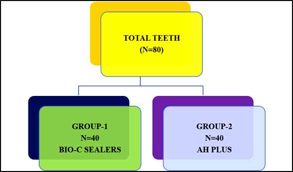
Table 2: Group 1 (N=40) an extracted mandibular first premolar was evaluated, with its canals filled using gutta-percha and the bioactive sealer Biodentine (BIO-C SEALER). The evaluation was conducted using scanning electron microscopy at the coronal, middle, and apical levels to determine the permeability. A statistical assessment was performed using the Pearson Chi-Square test to determine the level of significance
|
Anatomical Divisions |
Value |
Stat. Mean |
Std. Dev. |
Std. Error |
95% CI |
Pearson Chi-Square Value |
df |
p value |
|
Coronal |
32.14%±24.56% |
1.15 |
0.621 |
0.268 |
1.97 |
1.082 |
1.0 |
0.09 |
|
Middle |
34.56%±23.85% |
1.17 |
0.794 |
0.655 |
1.76 |
2.381 |
1.0 |
0.02* |
|
Apical |
38.86%±24.85% |
1.19 |
0.989 |
0.979 |
1.88 |
3.278 |
1.0 |
0.16 |
|
*p<0.05 significant |
||||||||
Table 3: Group 2 (N=40) an extracted mandibular first premolar was evaluated, with its canals filled using gutta-percha and the bioactive sealer resin bonded (AH PLUS). The evaluation was conducted using scanning electron microscopy at the coronal, middle, and apical levels to determine the permeability. A statistical assessment was performed using the Pearson Chi-Square test to determine the level of significance
|
Anatomical Divisions |
Value |
Stat. Mean |
Std. Dev. |
Std. Error |
95% CI |
Pearson Chi-Square Value |
df |
p value |
|
Coronal |
3.51%±5.68% |
1.07 |
1.056 |
0.013 |
0.012 |
1.034 |
1.0 |
0.06 |
|
Middle |
5.45%±3.25% |
1.08 |
1.026 |
0.016 |
0.034 |
1.008 |
1.0 |
0.13 |
|
Apical |
7.32%±4.56% |
1.19 |
1.055 |
0.018 |
0.098 |
0.056 |
1.0 |
0.76 |
|
*p<0.05 significant |
||||||||
Table 4: Group 1 (N=40) an extracted mandibular first premolar was evaluated, with its canals filled using gutta-percha and the bioactive sealer Biodentine (BIO-C SEALER). The evaluation was conducted using scanning electron microscopy at the coronal, middle, and apical levels to determine the bone strength. A statistical assessment was performed using the Pearson Chi-Square test to determine the level of significance
|
Anatomical Divisions |
Value |
Stat. Mean |
Std. Dev. |
Std. Error |
95% CI |
Pearson Chi-Square Value |
df |
p value |
|
Coronal |
2.11±0.56Mpa |
1.13 |
0.034 |
0.055 |
1.34 |
1.381 |
1.0 |
0.02* |
|
Middle |
2.15±0.75Mpa |
1.17 |
0.056 |
0.019 |
0.036 |
1.078 |
1.0 |
0.06 |
|
Apical |
2.17±0.86Mpa |
1.18 |
0.066 |
0.078 |
0.057 |
1.928 |
1.0 |
0.08 |
|
*p<0.05 significant |
||||||||
Table 5: Group 2 (N=40) an extracted mandibular first premolar was evaluated, with its canals filled using gutta-percha and the bioactive sealer resin bonded (AH PLUS). The evaluation was conducted using scanning electron microscopy at the coronal, middle, and apical levels to determine the bone strength. A statistical assessment was performed using the Pearson Chi-Square test to determine the level of significance
|
Anatomical Divisions |
Value |
Stat. Mean |
Std. Dev. |
Std. Error |
95% CI |
Pearson Chi-Square Value |
df |
p value |
|
Coronal |
1.85±0.56Mpa |
0.07 |
0.056 |
0.049 |
0.036 |
1.035 |
1.0 |
0.06 |
|
Middle |
2.13±0.75Mpa |
0.05 |
0.035 |
0.016 |
0.037 |
1.038 |
1.0 |
0.55 |
|
Apical |
2.15±0.86Mpa |
0.09 |
0.098 |
0.067 |
0.088 |
0.076 |
2.0 |
0.06 |
|
*p<0.05 significant |
||||||||
Table 6: Estimation amongst all studied groups using one-way ANOVA
|
Variables |
Degree of Freedom |
Sum of Squares ∑ |
Mean Sum of Squares m∑ |
F |
Level of Sig. (p) |
|
Between Groups |
5 |
2.782 |
2.572 |
1.4 |
0.01* |
|
Within Groups |
25 |
2.563 |
0.623 |
_ |
|
|
Cumulative |
671.12 |
5.831 |
*p<0.05 significant |
||
DISCUSSION :
Immanuel KG et al. reported in their study that Root canal treatment is necessary when the pulp of a tooth becomes infected due to decay, trauma, or repeated dental work. Common symptoms of infection include severe pain, swelling, sensitivity to temperature changes, and infections in the gums. The procedure begins with a thorough diagnosis and the administration of local anaesthesia. A rubber dam is placed around the tooth to keep it dry, and a small hole is created to access the pulp chamber.14,15 Ng YL et al showed in their study that infected tissue is removed, and the canals are shaped and disinfected using solutions such as sodium hypochlorite. After
cleaning, the canals are filled with gutta-percha and sealed, often requiring the placement of a permanent crown afterward. Recovery may involve mild soreness, which can usually be managed with medication. Patients are advised to avoid chewing on the treated tooth until it is fully restored.16,17 van der Sluis LW et al included in their study that root canal sealers play a crucial role in the success of the procedure by filling any gaps to prevent fluid leakage and providing antimicrobial properties. Common sealers include bioceramic sealers (such as BioRoot RCS), epoxy resin-based sealers (like AH Plus), zinc oxide eugenol, calcium hydroxide sealers, and silicone-based sealers (like GuttaFlow). Key factors in selecting a sealer include biocompatibility, ease of retreatment, and the method of application. Endodontic sealers offer various advantages: epoxy resin-based sealers (e.g., AH Plus), bioceramic sealers (e.g., iRoot SP), zinc oxide-eugenol sealers (e.g., Tubli-Seal), calcium hydroxide-based sealers (e.g., Sealapex), silicone-based sealers (e.g., RoekoSeal), and glass ionomer-based sealers (e.g., Ketac Endo).18,19 Dos Santos Costa FM et al included in their study that biodentine stands out in the realm of endodontics due to its exceptional purity and bioactive properties, making it an ideal choice for crucial procedures such as pulp capping and the repair of root perforations. This advanced bioceramic material is prized for its relatively rapid setting time, which is approximately 12 minutes, although it requires a longer duration for complete hardening to achieve optimal strength.20,21 KomabayashiT in their study in recent developments, innovative formulations like Biodentine XP have emerged, designed to offer enhanced handling characteristics that improve user experience during application. One of the key advantages of Biodentine is its water-based chemistry, which reduces stickiness compared to traditional resin sealers. This feature not only facilitates easier manipulation but also significantly boosts biocompatibility, fostering a more favourable environment for healing. However, there are some considerations to keep in mind.22 Primus CM et al reviewed in their study that while Biodentine is generally recognised for its stability, there have been occasional reports of discolouration, particularly in specialised revascularisation scenarios. These instances highlight the need for careful selection and application in specific dental treatments to ensure the best outcomes for patients. Resin-based root canal sealers, on the other hand, demonstrate superior adhesion to root canal walls and gutta-percha.23 Badawy RE et al included in their study that they provide low solubility and minimal polymerisation shrinkage, ensuring a reliable seal over time. These sealers possess good flow characteristics, facilitating penetration into lateral canals and dentinal tubules. However, freshly mixed epoxy resins can be cytotoxic; once set, they are typically well-tolerated.24 Augusto CM et al showed in their study that common examples include epoxy resin-based sealers like AH Plus and RC Seal, favoured for their low shrinkage and high bond strength, as well as methacrylate and polyvinyl resin-based sealers. Despite their advantages, resin-based sealers have limitations. Their hydrophobic nature makes them less effective in moist environments compared to bioceramic alternatives. Additionally, the strong adhesion of resin-based sealers can create difficulties during endodontic retreatment. Scanning Electron Microscopy (SEM) is a powerful imaging technique that uses a focused beam of electrons to examine samples at high resolution. This method surpasses traditional light microscopy by utilising the shorter wavelength of electrons, resulting in detailed three-dimensional-like images of micro- and nanostructures, providing valuable insights into the properties and interactions of materials.25,26
CONCLUSION :
The authors conducted an in-depth analysis of the effectiveness of bioactive sealers, such as bio-dentine, compared to resin-bonded sealers, focusing on sealing ability, marginal adaptation, and dentinal tubule penetration through scanning electron microscopy. The study concluded that bioactive sealers generally exhibit superior adaptation, evidenced by narrower gaps at the bonding interface. Bio-dentine's enhanced flowability and smaller particle size allow for better penetration into dentinal tubules, especially in the middle and apical thirds of the root canal. A notable advantage of bioactive sealers is their zero shrinkage upon setting, which contributes to a more hermetic seal. Overall, bioactive sealers outperform resin-based alternatives in sealing reliability. The authors emphasize the need for further research to validate these findings.
BIBLIOGRAPHY:
1. Mushtaq A, Alsanafi S, Elmsmari F, et al. Effect of root canal filling techniques and materials on endodontic treatment outcomes: a systematic review and meta-analysis. Sci Rep. 2026;16(1):95-98.
2. Üstün Y, Eraslan Akyüz İ, Manolya Özdemir İ. Effects of Different Root Canal Sealers and Irrigation Agitation Methods on Postoperative Pain in the Treatment of Teeth with Chronic Apical Periodontitis: A Randomized Clinical Trial. J Endod. 2026;52(2):227-238.
3. Siqueira JF Jr. Aetiology of root canal treatment failure: why well-treated teeth can fail. Int Endod J. 2001;34(1):1-10.
4. McCabe PS, Dummer PM. Pulp canal obliteration: an endodontic diagnosis and treatment challenge. Int Endod J. 2012;45(2):177-197.
5. van der Sluis LW, Versluis M, Wu MK, Wesselink PR. Passive ultrasonic irrigation of the root canal: a review of the literature. Int Endod J. 2007;40(6):415-426.
6. Khandelwal A, Janani K, Teja K, et al. Periapical Healing following Root Canal Treatment Using Different Endodontic Sealers: A Systematic Review. Biomed Res Int. 2022;35(6);92-97.
7. Cardinali F, Camilleri J. A critical review of the material properties guiding the clinician's choice of root canal sealers. Clin Oral Investig. 2023;27(8):4147-4155.
8. Zamparini F, Prati C, Taddei P, Spinelli A, Di Foggia M, Gandolfi MG. Chemical-Physical Properties and Bioactivity of New Premixed Calcium Silicate-Bioceramic Root Canal Sealers. Int J Mol Sci. 2022;23(22):13-19.
9. Sabharwal S, Kumar P, Chugh VK, Gupta A, Pathak K, Patnana A. A Comparative Evaluation of Post-operative Pain in Patients Undergoing Root Canal Treatment With Four Different Types of Sealers: A Randomized Controlled Trial. Cureus. 2025;17(5):84-86.
10. Aminoshariae A, Primus C, Kulild JC. Tricalcium silicate cement sealers: Do the potential benefits of bioactivity justify the drawbacks?. J Am Dent Assoc. 2022;153(8):750-760.
11. Carvalho MA, Lazari-Carvalho PC, Maffra PET, et al. Immediate Pre-Endodontic Dentin Sealing (IPDS) Improves Resin-Dentin Bond Strength. J Esthet Restor Dent. 2025;37(1):39-47.
12. Kim YK, Grandini S, Ames JM, et al. Critical review on methacrylate resin-based root canal sealers. J Endod. 2010;36(3):383-399.
13. Risnes S, Saeed M, Sehic A. Scanning Electron Microscopy (SEM) Methods for Dental Enamel. Methods Mol Biol. 2019;192(2):293-308.
14. Immanuel KG, Anju P, Sanap S, et al. Evaluation of the Efficacy of Smear Layer Removal Using Different Irrigation Activation Methods during Root Canal Treatment: An In Vitro Study. J Contemp Dent Pract. 2026;27(1):55-58.
15. Gomes BPFA, Aveiro E, Kishen A. Irrigants and irrigation activation systems in Endodontics. Braz Dent J. 2023;34(4):1-33.
16. Ng YL, Mann V, Gulabivala K. A prospective study of the factors affecting outcomes of nonsurgical root canal treatment: part 1: periapical health. Int Endod J. 2011;44(7):583-609.
17. Căpută PE, Retsas A, Kuijk L, Chávez de Paz LE, Boutsioukis C. Ultrasonic Irrigant Activation during Root Canal Treatment: A Systematic Review. J Endod. 2019;45(1):31-44.
18. van der Sluis LW, Versluis M, Wu MK, Wesselink PR. Passive ultrasonic irrigation of the root canal: a review of the literature. Int Endod J. 2007;40(6):415-426.
19. Coşar M, Kandemir Demirci G, Çalışkan MK. The effect of two different root canal sealers on treatment outcome and post-obturation pain in single-visit root canal treatment: A prospective randomized clinical trial. Int Endod J. 2023;56(3):318-330.
20. Dos Santos Costa FM, Fernandes MH, Batistuzzo de Medeiros SR. Genotoxicity of root canal sealers: a literature review. Clin Oral Investig. 2020;24(10):3347-3362.
21. Coşar M, Kandemir Demirci G, Çalışkan MK. The effect of two different root canal sealers on treatment outcome and post-obturation pain in single-visit root canal treatment: A prospective randomized clinical trial. Int Endod J. 2023;56(3):318-330.
22. Komabayashi T, Colmenar D, Cvach N, Bhat A, Primus C, Imai Y. Comprehensive review of current endodontic sealers. Dent Mater J. 2020;39(5):703-720.
23. Primus CM, Tay FR, Niu LN. Bioactive tri/dicalcium silicate cements for treatment of pulpal and periapical tissues. Acta Biomater. 2019;99(6):35-44.
24. Badawy RE, Mohamed DA. Evaluation of new bioceramic endodontic sealers: An in vitro study. Dent Med Probl. 2022;59(1):85-92.
25. Augusto CM, Cunha Neto MA, Pinto KP, et al. Influence of the use of chelating agents as final irrigant on the push-out bond strength of epoxy resin-based root canal sealers: A systematic review. Aust Endod J. 2022;48(2):347-363.
26. Nguyen JNT, Harbison AM. Scanning Electron Microscopy Sample Preparation and Imaging. Methods Mol Biol. 2017;16(6):71-84.