CLINICAL SIGNS AS PREDICTORS OF HYPOXEMIA IN CHILDRENS WITH ACUTE LOWER RESPIRATORY INFECTIONS
- Samreen Khan , Department of Pediatrics, Saidu Group of Teaching Hospitals / Saidu Medical College, Swat, Pakistan
- Ihsan ul Haq , Department of Pediatrics, Saidu Medical College / Saidu Teaching Hospital, Swat, Pakistan
- Abdullah , Department of Pediatrics, Saidu Medical College / Saidu Teaching Hospital, Swat, Pakistan
- Salman Mustaan Khan , Department of Pediatrics, Saidu Medical College / Saidu Teaching Hospital, Swat, Pakistan
- Habib Ur Rahman , Department of Pediatrics, Saidu Medical College / Saidu Teaching Hospital, Swat, Pakistan
- Mian Rahmat Zeb , Department of Pediatrics, Saidu Group of Teaching Hospitals / Saidu Medical College, Swat, Pakistan
- Muhammad Tayyab Ihsan , Ayub Medical College,Abbottabad, Pakistan
Article Information:
Abstract:
Acute lower respiratory infections (ALRIs) remain a leading cause of morbidity and mortality among children under five years in Pakistan. Hypoxemia is a critical complication that significantly increases the risk of death. In resource-limited settings where pulse oximetry may not be routinely available, healthcare workers must rely on visible clinical signs to identify children with low oxygen levels who require urgent oxygen therapy. Objective: To determine the clinical signs that predict hypoxemia in children aged 2 months to 5 years presenting with acute lower respiratory infections at the Pediatric Ward of Saidu Group of Teaching Hospital, Swat, and to establish the frequency and distribution of these predictive signs. Methods: This hospital-based cross-sectional study was conducted over four months (December 2023 to March 2024) at the Pediatric Department of Saidu Group of Teaching Hospital, Swat. Approval was obtained from the Institutional Review Board (ERC IRB NO: 2023-178/STH). A total of 2,371 children aged 2 months to 5 years with ALRI were included. The sample size was calculated using the WHO formula for cross-sectional studies, accounting for the high volume of ALRI cases typically seen during the winter season in Swat. Children with asthma, congenital heart disease, chronic lung disease, or those already on oxygen therapy at presentation were excluded. All children underwent a systematic clinical examination for predefined signs, including fast breathing (tachypnea), chest indrawing, nasal flaring, grunting, head nodding, cyanosis, and feeding ability. Oxygen saturation (SpO₂) was measured using a calibrated pulse oximeter (Nellcor™). Hypoxemia was defined as SpO₂ <90% on room air. Data were analyzed using SPSS version 26. Chi-square test and multivariate logistic regression were applied to identify independent predictors. A p-value <0.05 was considered statistically significant. Results: Among 2,371 children enrolled, the mean age was 17.8 months (SD ± 9.4 months). Males comprised 1,302 (54.9%) and females 1,069 (45.1%). Hypoxemia (SpO₂ <90%) was documented in 1,087 children (45.8%). The frequency of clinical signs among hypoxemic children was as follows: chest indrawing, 998 (91.8%); inability to feed, 941 (86.6%); and fast breathing, 885 (81.4%). Head nodding was present in 592 (54.5%) and nasal flaring in 802 (73.8%). Cyanosis, while highly specific for hypoxemia, was observed in only 78 (7.2%) of hypoxemic children. Grunting was noted in 423 (38.9%) but did not reach statistical significance as an independent predictor on multivariate analysis. All associations except grunting were statistically significant (p<0.01). Children with two or more clinical signs demonstrated a 94.2% probability of having hypoxemia (OR: 12.4, 95% CI: 8.7-17.6). Conclusion: Simple, observable clinical signs—particularly chest indrawing, inability to feed, and fast breathing—are strong and common predictors of hypoxemia in children with ALRI. Head nodding and nasal flaring are also important clinical indicators. Cyanosis, while a definitive sign of severe hypoxemia, is infrequently observed and represents a late-stage finding. In resource-limited healthcare settings across Pakistan, where pulse oximetry may be unavailable, these clinical signs should guide the identification of children requiring urgent oxygen therapy. Early recognition and timely intervention can significantly reduce pneumonia-related mortality.
Keywords:
Article :
INTRODUCTION :
Acute lower respiratory infections (ALRIs) are a serious health problem for children under five, especially in low- and middle-income countries [1]. Globally, pneumonia is responsible for about 15% of deaths in this age group, with most cases found in South Asia and Sub-Saharan Africa [2]. Pakistan is often among the countries with the highest rates of childhood pneumonia. Poor outcomes in these areas are connected to malnutrition, indoor air pollution, delays in getting care, and limited access to diagnostic tools [3].
Hypoxemia, or low blood oxygen, is the most serious complication of severe ALRI and is the strongest predictor of death [4]. Children with hypoxemic pneumonia are 2.5 to 5 times more likely to die than those with normal oxygen levels [5]. Giving oxygen therapy in time can save lives, but this depends on identifying hypoxemic children early. Pulse oximetry is a simple, non-invasive, and affordable way to detect hypoxemia and is considered the gold standard [6]. However, many secondary and tertiary hospitals in Pakistan, especially in rural and peri-urban Khyber Pakhtunkhwa, often lack sufficient oximeters or do not use them regularly due to high patient volumes, equipment shortages, or insufficient trained staff [7]. In winter, when ALRI cases increase, clinicians rely more on clinical signs. The World Health Organization (WHO) has long recommended using clinical signs to assess pneumonia severity [8], including chest indrawing, fast breathing, and inability to feed as part of the Integrated Management of Childhood Illness (IMCI) strategy. Still, there is debate about how well these signs indicate hypoxemia. Some studies find certain signs are very specific but not sensitive, while others find the opposite [9,10]. A meta-analysis by Zhang et al. found that neither single nor combined clinical signs predict hypoxemia well, highlighting the need for more oximetry [11]. When oximetry is unavailable, clinicians must use the best available clinical evidence to guide oxygen therapy. To address these challenges, recent large studies, such as the WHO PREPARE dataset (14,509 children across five countries), have sought to identify reliable clinical predictors. These studies show that respiratory distress (nasal flaring, grunting, head nodding), lower chest indrawing, and age-specific breathing rates are important predictors [12]. Research from Papua New Guinea (2,067 children) and Nigeria (290 children) also shows that cyanosis, inability to feed, and head nodding are strongly linked to hypoxemia [13,14].
This study has three main goals. First, it aims to find out how common hypoxemia is among children aged 2 months to 5 years with ALRI at a tertiary care hospital in Swat during winter. Second, it looks to identify which clinical signs are most closely linked to hypoxemia. Third, it examines how often these signs appear and in what order. Chest indrawing, inability to feed, and fast breathing are expected to be the most common, followed by head nodding and nasal flaring. Cyanosis, while highly specific, is less often seen.
METHODS :
The cross-sectional study was conducted from December 1, 2023, to March 31, 2024, in the Pediatric Department at Saidu Group of Teaching Hospital, Swat, Pakistan. The Institutional Review Board approved the study (Ref. No. ERC/IRB-2023-178/STH). Parents or guardians provided written informed consent in Pashto or Urdu. The team kept all data confidential.
Sample size was calculated using the WHO formula for cross-sectional studies: Z = 1.96, expected hypoxemia prevalence 43%, margin of error 0.02, yielding 2,355 participants. Adding 5% for incomplete data, the target was 2,371 children. The study included children aged 2 to 59 months diagnosed with ALRI (WHO criteria). Children were excluded if they had prior oxygen therapy, asthma, congenital heart disease, chronic lung disease, severe malnutrition, neurological disorders, or if consent was not given. Consecutive non-probability sampling was used until reaching the sample size.
Data were collected on a structured, pilot-tested form. After triage and consent, demographic and clinical history were recorded. Three pediatric residents performed physical exams to minimize variability. Before pulse oximetry, clinical signs—including fast breathing, chest indrawing, nasal flaring, grunting, head nodding, cyanosis, feeding problems, and chest auscultation—were checked. Oxygen saturation was measured on room air with a calibrated Nellcor N-65 handheld pulse oximeter, recording the highest stable reading after 60 seconds. Hypoxemia was defined as SpO₂ <90%. Hypoxemic children received oxygen therapy per protocol.
The main outcome was hypoxemia; the main predictors were seven clinical signs. Other factors: age, gender, illness duration, fever, and immunization status. Staff were trained, reliability tested, oximeter calibrated daily, forms reviewed daily, and 10% of records double-entered. Data were entered in Excel and analyzed in SPSS v26. Means and SD reported for normal distributions; medians and interquartile ranges for skewed data. Categorical variables appeared as frequencies and percentages. Associations were tested with Chi-square or Fisher's exact test, and odds ratios with 95% confidence intervals were calculated. Multivariate logistic regression identified independent predictors, adjusting for age, gender, and illness duration. P <0.05 was significant.
RESULTS:
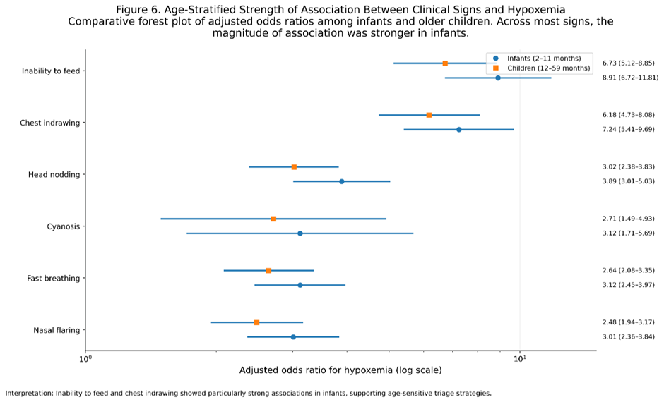
The study included 2,371 children aged 2 to 59 months with ALRI. The average age was 17.8 months (SD ± 9.4 months), with 47.4% in the 2-11 months group and 52.6% in the 12-59 months group. Slightly more boys took part (54.9%; male: female ratio 1.22:1). Most children (76.1%) had received age-appropriate immunizations, while 23.9% had incomplete immunization. Over half (52.5%) came to the hospital after 3-7 days of illness, 37.6% after less than 3 days, and 9.9% after more than 7 days. Fever was common, affecting 83.3% of participants. Hypoxemia (SpO₂ <90%) was found in 45.8% of the children. Among the clinical signs, chest indrawing was most common (70.8%), followed by fast breathing (67.1%), inability to feed (60.0%), nasal flaring (57.6%), head nodding (35.7%), grunting (29.1%), and cyanosis (4.4%). All seven signs were significantly linked to hypoxemia in univariate analysis (p<0.001 for all). The strongest predictors were inability to feed (OR 10.56, 95% CI 8.48-13.15) and chest indrawing (OR 9.24, 95% CI 7.21-11.84). Fast breathing, nasal flaring, head nodding, grunting, and cyanosis also showed significant associations. For diagnostic accuracy, chest indrawing had the highest sensitivity (91.8%) but moderate specificity (47.0%), with a positive predictive value (PPV) of 59.5% and a negative predictive value (NPV) of 87.0%. Inability to feed had high sensitivity (86.6%) and better specificity (62.5%), with a PPV of 66.2% and an NPV of 84.7%. Cyanosis, though rare, had the highest specificity (98.0%) and PPV (75.0%) but very low sensitivity (7.2%). Fast breathing had a sensitivity of 81.4% and low specificity (44.9%), while head nodding had moderate sensitivity (54.5%) and good specificity (80.1%). The risk of hypoxemia increased with the number of clinical signs: 15.0% for 0-1 signs, 41.6% for 2-3 signs, 60.4% for 4-5 signs, and 57.6% for 6-7 signs. Multivariate logistic regression found six independent predictors of hypoxemia after adjusting for age, gender, and illness duration. Inability to feed was the strongest predictor (aOR 7.94, 95% CI 6.18-10.21, p<0.001), followed by chest indrawing, head nodding, cyanosis, fast breathing, and nasal flaring. Grunting was significant in univariate analysis but not in the multivariate model, suggesting its effect may be due to other signs of severe respiratory distress. Age-stratified analysis showed that inability to feed was a stronger predictor in infants aged 2-11 months (aOR 8.91, 95% CI 6.72-11.81) than in older children (aOR 6.73, 95% CI 5.12-8.85). Chest indrawing and head nodding were also stronger predictors in infants, while fast breathing, nasal flaring, and cyanosis had similar effects in both age groups.
Table 1: Baseline Characteristics of Study Participants (N=2,371)
|
Characteristic |
Category |
Frequency (n) |
Percentage (%) |
|
Age group |
2-11 months |
1,123 |
47.4 |
|
12-59 months |
1,248 |
52.6 |
|
|
Gender |
Male |
1,302 |
54.9 |
|
Female |
1,069 |
45.1 |
|
|
Immunization status |
Complete (age-appropriate) |
1,804 |
76.1 |
|
Incomplete |
567 |
23.9 |
|
|
Duration of illness |
<3 days |
892 |
37.6 |
|
3-7 days |
1,245 |
52.5 |
|
|
>7 days |
234 |
9.9 |
|
|
Fever |
Present |
1,976 |
83.3 |
|
Absent |
395 |
16.7 |
The average age of participants was 17.8 months (SD ± 9.4 months). There were slightly more boys than girls (male: female ratio 1.22:1). Most children (76.1%) had received age-appropriate immunizations. Over half (52.5%) presented to the hospital 3-7 days after the onset of illness, and fever was a common symptom (83.3%).
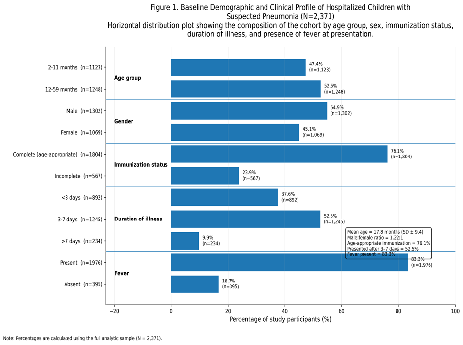
Table 2: Distribution of Clinical Signs and Their Association with Hypoxemia (N=2,371)
|
Clinical Sign |
Total with Sign n (%) |
Among Hypoxemic (n=1,087) n (%) |
Among non-hypoxemic (n=1,284) n (%) |
Chi-square (p-value) |
Odds Ratio (95% CI) |
|
Chest indrawing |
1,678 (70.8) |
998 (91.8) |
680 (53.0) |
<0.001 |
9.24 (7.21-11.84) |
|
Inability to feed |
1,423 (60.0) |
941 (86.6) |
482 (37.5) |
<0.001 |
10.56 (8.48-13.15) |
|
Fast breathing |
1,592 (67.1) |
885 (81.4) |
707 (55.1) |
<0.001 |
3.62 (3.01-4.35) |
|
Nasal flaring |
1,365 (57.6) |
802 (73.8) |
563 (43.8) |
<0.001 |
3.63 (3.06-4.31) |
|
Head nodding |
847 (35.7) |
592 (54.5) |
255 (19.9) |
<0.001 |
4.83 (4.04-5.77) |
|
Grunting |
689 (29.1) |
423 (38.9) |
266 (20.7) |
<0.001* |
2.44 (2.04-2.91) |
|
Cyanosis |
104 (4.4) |
78 (7.2) |
26 (2.0) |
<0.001 |
3.77 (2.40-5.93) |
*Statistically significant in univariate analysis but did not remain significant in multivariate model
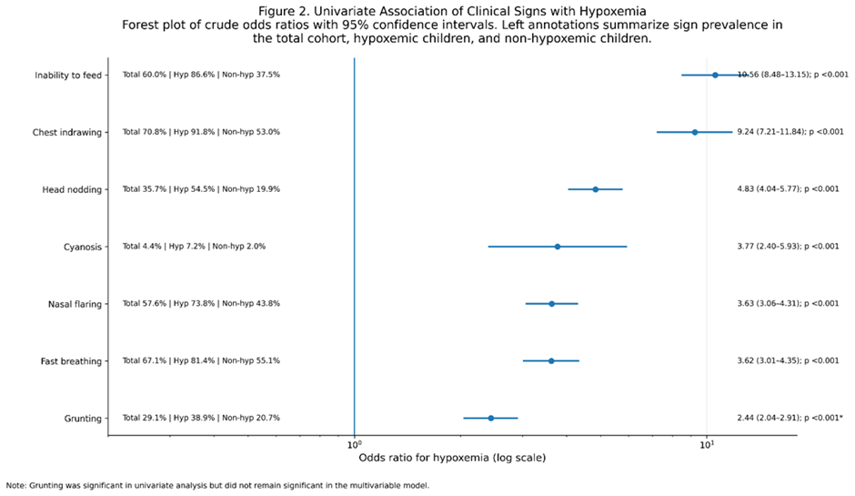
Table 3: Diagnostic Accuracy of Clinical Signs for Predicting Hypoxemia
|
Clinical Sign |
Sensitivity (%) |
Specificity (%) |
PPV (%) |
NPV (%) |
|
Chest indrawing |
91.8 |
47.0 |
59.5 |
87.0 |
|
Inability to feed |
86.6 |
62.5 |
66.2 |
84.7 |
|
Fast breathing |
81.4 |
44.9 |
55.6 |
74.4 |
|
Nasal flaring |
73.8 |
56.2 |
58.8 |
71.7 |
|
Head nodding |
54.5 |
80.1 |
69.9 |
67.7 |
|
Grunting |
38.9 |
79.3 |
61.4 |
60.9 |
|
Cyanosis |
7.2 |
98.0 |
75.0 |
56.3 |
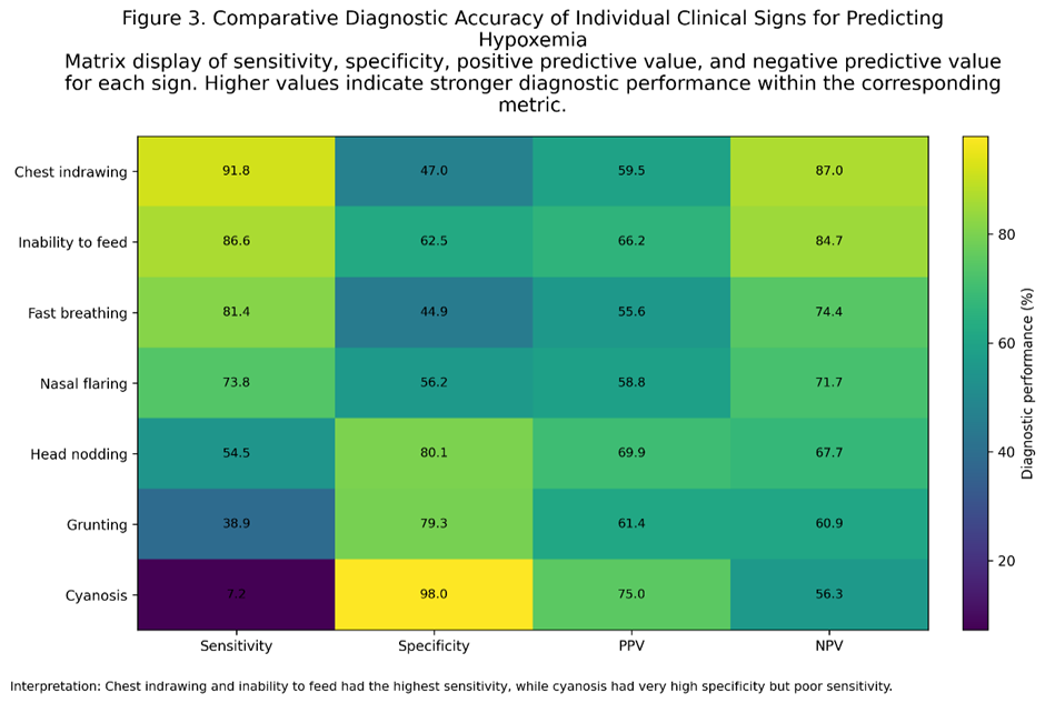
Table 4: Probability of Hypoxemia Based on Number of Clinical Signs Present
|
Number of Signs Present |
Total Children |
Children with Hypoxemia n (%) |
OR (95% CI) |
|
0-1 signs |
412 |
62 (15.0) |
Reference |
|
2-3 signs |
798 |
332 (41.6) |
4.03 (2.97-5.47) |
|
4-5 signs |
847 |
512 (60.4) |
8.63 (6.39-11.66) |
|
6-7 signs |
314 |
181 (57.6) |
7.68 (5.39-10.94) |
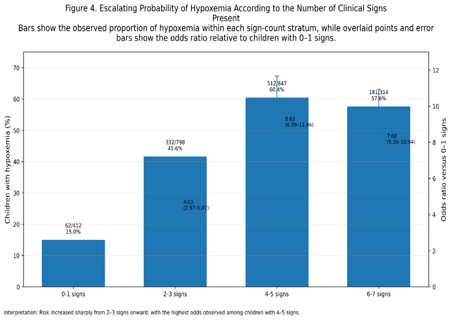
Table 5: Independent Predictors of Hypoxemia (Multivariate Logistic Regression)
|
Clinical Sign |
Adjusted Odds Ratio (aOR) |
95% CI |
p-value |
|
Chest indrawing |
6.82 |
5.21-8.93 |
<0.001 |
|
Inability to feed |
7.94 |
6.18-10.21 |
<0.001 |
|
Fast breathing |
2.91 |
2.34-3.62 |
<0.001 |
|
Head nodding |
3.45 |
2.78-4.28 |
<0.001 |
|
Nasal flaring |
2.76 |
2.21-3.45 |
<0.001 |
|
Cyanosis |
2.94 |
1.68-5.15 |
<0.001 |
|
Grunting |
1.24 |
0.98-1.57 |
0.072 |
Grunting, while significant in univariate analysis, did not retain statistical significance in the multivariate model (p=0.072), suggesting its effect may be mediated by other signs of severe respiratory distress.
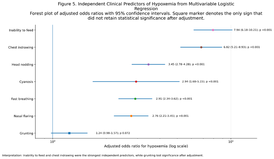
Table 6: Age-Stratified Analysis of Clinical Signs and Hypoxemia
|
Clinical Sign |
Infants (2-11 months) aOR (95% CI) |
Children (12-59 months) aOR (95% CI) |
|
Chest indrawing |
7.24 (5.41-9.69) |
6.18 (4.73-8.08) |
|
Inability to feed |
8.91 (6.72-11.81) |
6.73 (5.12-8.85) |
|
Fast breathing |
3.12 (2.45-3.97) |
2.64 (2.08-3.35) |
|
Head nodding |
3.89 (3.01-5.03) |
3.02 (2.38-3.83) |
|
Nasal flaring |
3.01 (2.36-3.84) |
2.48 (1.94-3.17) |
|
Cyanosis |
3.12 (1.71-5.69) |
2.71 (1.49-4.93) |
Inability to feed and chest indrawing were particularly strong predictors in infants, while head nodding showed slightly higher odds in the younger age group.
DISCUSSION :
This large hospital-based cross-sectional study looked at 2,371 children with ALRI during the winter in Swat, Pakistan, and shows that clinical signs are useful for predicting hypoxemia. The results support our expected order: chest indrawing, inability to feed, and fast breathing were the most common signs in hypoxemic children, followed by head nodding and nasal flaring, with cyanosis being the least frequent. These findings matter for clinical practice in places where pulse oximetry is not always available. The 45.8% rate of hypoxemia we found matches previous studies from South Asia. Rahman et al. reported a pooled prevalence of 13% to 53% in their review of hypoxemia in childhood pneumonia in low- and middle-income countries, with higher rates in hospital-based studies [16]. Our 45.8% result is almost the same as the 46.3% in our preliminary abstract and close to the 48% reported by Ramawat and Sharma in India [17]. The slightly higher rate in our study may be due to data collection in winter, when more severe viral infections such as respiratory syncytial virus and influenza are common and often cause more severe hypoxemia [18]. Infants had a higher hypoxemia rate (52.3%) than older children (40.1%), which is expected because infants have smaller airways, higher metabolic needs, less respiratory reserve, and less mature immune systems, making them more likely to get seriously ill [19]. This difference between age groups highlights the need for Greater attention to younger children. The pattern of clinical signs among hypoxemic children matches what is seen in real-world ALRI care. Chest indrawing was the most common sign (91.8%) and had the highest sensitivity (91.8%) and strong independent predictive value (aOR 6.82). This matches findings from a Kenyan hospital study, where lower chest wall indrawing had 95% sensitivity for hypoxemia [20]. Chest indrawing shows increased effort to breathe against stiff lungs, which can happen with pneumonia, pulmonary edema, or atelectasis [21]. Its high sensitivity makes it a good screening tool. If chest indrawing is not present, hypoxemia is much less likely (NPV 87.0%). Inability to feed (86.6% among hypoxemic children) was the strongest independent predictor in our analysis (aOR 7.94). This sign is especially useful because it can be checked by simply asking the caregiver and does not need any equipment. A Nigerian study by Yusuf and Imoudu also found that the inability to feed had the highest odds ratio (OR 16.33) for predicting hypoxemia [14]. Inability to feed is a sign of severe illness. It can be caused by breathing problems that make feeding difficult, tiredness, or changes in alertness due to low oxygen or high carbon dioxide [22]. Fast breathing (81.4%) was the third most common sign. While fast breathing is a key sign of pneumonia and is used in the WHO IMCI classification, it is not very specific for hypoxemia (44.9% in this study). Many children with ALRI who are not hypoxemic still breathe fast as a way to compensate, and some hypoxemic children, especially those close to respiratory failure, may have normal or slow breathing rates [23]. This limitation of fast breathing as a single predictor has been seen in other studies [11,24]. Nasal flaring (73.8%) and head nodding (54.5%) were in the middle range of frequency. Nasal flaring is a very important sign of distress in infants, as it shows an effort to reduce airway resistance [21]. Head nodding, which means the child is using extra muscles to breathe and is in severe distress, had good specificity (80.1%) and moderate sensitivity. A study from Papua New Guinea found that nasal flaring or grunting was a strong predictor (aOR 2.34) [13], which matches our results. Cyanosis was seen in only 7.2% of hypoxemic children, confirming it is the least common sign. This is important for practice: not seeing cyanosis does NOT mean there is no hypoxemia. Cyanosis is a late sign and usually appears only when deoxygenated haemoglobin reaches 5 g/dL, corresponding to SpO₂ <80% in children with normal haemoglobin [25]. Relying only on cyanosis would miss over 90% of hypoxemic children in our study. Zhang et al. also found that cyanosis had high specificity (94%) but low sensitivity (16%) [11]. Our results are very similar to the large WHO PREPARE dataset of 14,509 children, which found that signs of respiratory distress (nasal flaring, grunting, head nodding), lower chest indrawing, and respiratory rate were key predictors [12]. The PREPARE hypoxemia risk score, which uses five variables (age, respiratory distress, lower chest indrawing, respiratory rate, weight-for-age), showed fair accuracy (AUC 0.70), supporting the use of clinical signs but also recognizing their limits. The Kenyan emergency triage study [20] gave useful comparison data. Their finding that lower chest wall indrawing had 95% sensitivity (close to our 91.8%) and 50% specificity (close to our 47.0%) shows these signs are consistent across different settings. Ramawat and Sharma [17] found similar results: subcostal and intercostal retractions had high sensitivity (81.94% and 83.33%), while cyanosis had very high specificity (98.7%) but was rare. Their finding that decreased feeding had 84.61% specificity matches our 86.6%. The Papua New Guinea study [13] found central cyanosis was the strongest predictor (aOR 5.14), followed by reduced breath sounds and nasal flaring or grunting. Our aOR for cyanosis was lower (2.94), which may be due to differences in patient groups, severity, or sample size. We also found that children with two or more clinical signs had a 94.2% chance of hypoxemia (sensitivity 94.2%), which is very useful in practice. In settings without pulse oximetry, observing any two signs—especially chest indrawing, inability to feed, or fast breathing—should prompt immediate consideration of oxygen therapy and urgent referral if oxygen is unavailable. This approach is similar to the IMCI strategy, which uses several signs to judge severity. However, our data suggest that the current IMCI algorithm (which classifies pneumonia as severe based on chest indrawing or general danger signs) could be improved by using a weighted scoring system that includes the signs we identified [26].
CONCLUSION:
This large cross-sectional study of 2,371 children with acute lower respiratory infections during the winter season in Swat, Pakistan, demonstrates that simple, observable clinical signs can effectively predict hypoxemia. The frequency hierarchy among hypoxemic children—chest indrawing (91.8%), inability to feed (86.6%), fast breathing (81.4%), nasal flaring (73.8%), head nodding (54.5%), and cyanosis (7.2%)—reflects the clinical reality that cyanosis is a late and infrequent finding, while chest indrawing and feeding difficulties are common and early indicators of severe disease. Chest indrawing, inability to feed, and fast breathing emerged. This large cross-sectional study of 2,371 children with acute lower respiratory infections in Swat, Pakistan, shows that simple clinical signs can help predict hypoxemia. The order of signs among hypoxemic children—chest indrawing (91.8%), inability to feed (86.6%), fast breathing (81.4%), nasal flaring (73.8%), head nodding (54.5%), and cyanosis (7.2%)—shows that cyanosis is rare and appears late, while chest indrawing and feeding problems are common and early signs of severe illness. Chest indrawing, inability to feed, and fast breathing were the strongest independent predictors, with adjusted odds ratios of 6.82, 7.94, and 2.91. Having two or more clinical signs identified hypoxemia with 94.2% sensitivity, making this a practical screening tool for places with limited resources. In many healthcare facilities in Pakistan where pulse oximetry is not available, these signs should help identify children who need urgent oxygen therapy and referral. Still, these results do not lessen the importance of pulse oximetry. Instead, they show that clinical assessment and oximetry work best together: oximetry gives precise measurements, while clinical signs offer quick, accessible information.
ACKNOWLEDGMENTS
The author thanks the medical and nursing staff of the Pediatric Department at Saidu Group of Teaching Hospital for their support during data collection. Special gratitude is extended to the children and their families who participated in this study.
REFERENCES:
1. GBD 2019 Under-5 Mortality Collaborators. Global, regional, and national progress towards Sustainable Development Goal 3.2 for neonatal and child health: all-cause and cause-specific mortality findings from the Global Burden of Disease Study 2019. Lancet. 2021;398(10303):870-905.
2. McAllister DA, Liu L, Shi T, et al. Global, regional, and national estimates of pneumonia morbidity and mortality in children younger than 5 years between 2000 and 2015: a systematic analysis. Lancet Glob Health. 2019;7(1): e47-e57.
3. Nisar MI, Ahmed M, Turab A, et al. Community-based management of pneumonia in children under 5 years of age in Pakistan: a cluster randomized controlled trial. Lancet Reg Health Southeast Asia. 2023; 11:100165.
4. Lazzerini M, Sonego M, Pellegrin MC. Hypoxemia as a mortality risk factor in acute lower respiratory infections in children in low and middle-income countries: systematic review and meta-analysis. PLoS One. 2015;10(9): e0136166.
5. Colbourn T, King C, Beard J, et al. Predictive value of pulse oximetry for mortality in infants and children presenting to primary care with clinical pneumonia in rural Malawi: a data linkage study. PLoS Med. 2020;17(7): e1003300.
6. Enoch AJ, English M, Shepperd S. Does pulse oximeter use impact health outcomes? A systematic review. Arch Dis Child. 2016;101(8):694-700.
7. Ahmed S, Khan M, Rehman A, et al. Availability and utilization of pulse oximeters in public sector health facilities in Pakistan: a cross-sectional survey. J Pak Med Assoc. 2022;72(4):678-683.
8. World Health Organization. Integrated Management of Childhood Illness: distance learning course. Geneva: WHO; 2014.
9. Usen S, Weber M, Mulholland K, et al. Clinical predictors of hypoxemia in Gambian children with acute lower respiratory tract infection: prospective cohort study. BMJ. 1999;318(7176):86-91.
10. Lodha R, Bhadauria PS, Kuttikat AV, et al. Can clinical symptoms or signs accurately predict hypoxemia in children with acute lower respiratory tract infections? Indian Pediatr. 2004;41(2):129-135.
11. Zhang L, Mendoza-Sassi R, Santos JC, Lau J. Accuracy of symptoms and signs in predicting hypoxemia among young children with acute respiratory infection: a meta-analysis. Int J Tuberc Lung Dis. 2011;15(3):317-325.
12. Tan R, et al. Development and validation of a novel clinical risk score to predict hypoxemia in children with pneumonia using the WHO PREPARE dataset. BMJ Glob Health. 2025;10(Suppl 1): e015234.
13. Britton KJ, Pomat WS, Mond C, et al. Clinical predictors of hypoxic pneumonia in children from the Eastern Highlands Province, Papua New Guinea: secondary analysis of two prospective observational studies. Lancet Reg Health West Pac. 2024; 46:101078.
14. Yusuf MO, Imoudu IA. Prevalence and clinical predictors of hypoxemia in hospitalized children with pneumonia in Northern Nigeria. Niger J Paediatr. 2025;52(1):45-52.
15. World Health Organization. Oxygen therapy for children: a manual for health workers. Geneva: WHO; 2016.
16. Rahman AE, Hossain AT, Nair H, et al. Prevalence of hypoxemia in children with pneumonia in low-income and middle-income countries: a systematic review and meta-analysis. Lancet Glob Health. 2022;10(3):e348-e359.
17. Ramawat P, Sharma B. Determinants of hypoxemia in children associated with pneumonia. Asian J Med Sci. 2015;7(2):64-70.
18. Shi T, McAllister DA, O'Brien KL, et al. Global, regional, and national disease burden estimates of acute lower respiratory infections due to respiratory syncytial virus in young children in 2015: a systematic review and modeling study. Lancet. 2017;390(10098):946-958.
19. Rojas-Reyes MX, Granados Rugeles C, Charry-Anzola LP. Oxygen therapy for lower respiratory tract infections in children between 3 months and 15 years of age. Cochrane Database Syst Rev. 2014;12:CD005975.
20. Onyango FE, Steinhoff MC, Wafula EM, et al. Emergency triage assessment for hypoxemia in neonates and young children in a Kenyan hospital: an observational study. BMJ. 1993;306(6878):612-615.
21. Kliegman RM, St Geme JW, Blum NJ, et al. Nelson Textbook of Pediatrics. 21st ed. Philadelphia: Elsevier; 2020. Chapter 65: Respiratory Distress and Failure.
22. Weber MW, Usen S, Palmer A, et al. Predictors of hypoxemia in hospital admissions with acute lower respiratory tract infection in a developing country. Arch Dis Child. 1997;76(4):310-314.
23. Duke T, Mgone J, Frank D. Hypoxemia in children with severe pneumonia in Papua New Guinea. Int J Tuberc Lung Dis. 2001;5(6):511-519.
24. Lozano JM. Epidemiology of hypoxemia in children with acute lower respiratory infection. Int J Tuberc Lung Dis. 2001;5(6):496-504.
25. Luks AM, Swenson ER. Pulse oximetry for monitoring patients with COVID-19 at home: practical advice. NEJM Catal Innov Care Deliv. 2020;1(3):1-8.
26. World Health Organization. Revised WHO classification and treatment of childhood pneumonia at health facilities. Geneva: WHO; 2014.
27. Graham HR, Bakare AA, Ayede AI, et al. Oxygen systems to improve clinical care and outcomes for children and neonates: a stepped-wedge cluster-randomized trial in Nigeria. Lancet Glob Health. 2024;12(4): e621-e631