Pathology of Acute Appendicitis: Is It Vestigial Too?
- Bharati Rajgadkar , Assistant Professor, Department of Pathology, T. N. Medical College & BYL Nair Ch. Hospital, Mumbai, India.
- Aditi Raj , Associate Professor, Department of Pathology, MGM Medical College, Panvel, MGMIHS, Navi Mumbai, India.
- Pradeep Vaideeswar , Professor and Head of Department, Department of Pathology, Seth GS Medical College & KEM Hospital, Mumbai, India.
Article Information:
Abstract:
Acute appendicitis is one of the most common emergency abdominal surgeries performed worldwide and management in most cases is based on the clinical classification of simple (non-perforated) and complex (gangrenous or perforated) appendicitis. Histopathological examination (comprising “O” and “C” sections) remains the gold standard method for confirming the diagnosis, in which certain microscopic features may be overlooked. Aims: The aim of this study was to describe the histopathological findings of all appendectomy specimens and study the inter-observer variability in reporting of pathological features of acute appendicitis. Materials and Methods: A prospective study was performed on consecutive 250 appendicectomy specimens received with a clinical diagnosis of acute appendicitis in surgical pathology. The patient demographics and gross features were noted in all cases. The reports were initially issued after studying the routine sections, following which the appendices were serially sectioned and additional sections were processed. The initial and revised diagnoses were compared. Results: A comparison of the revised and initial (often reported by junior faculty) diagnoses was done in 250 appendicectomy specimens from 160 males and 90 females. Discordant diagnoses were noted in 167 cases (66.8 %), particularly for eosinophil-rich appendicitis, peri-appendicitis, appendicular perforations, parasitic inflammations, diverticulae, obliterations, and mucosal alterations. Conclusions: We suggest that even if junior faculty are reporting appendicectomy specimens, senior pathologists should intermittently review and supervise their reporting so that important diagnostic features are not missed. Appendix should be serially sectioned, and these additional sections can easily be accommodated in a single block. This would help to pick up important histopathology findings, which can alter the definitive treatment and future repercussions.
Keywords:
Article :
INTRODUCTION :
Acute appendicitis is the most frequent cause of emergency abdominal surgery and remains a significant contributor to surgical morbidity. The lifetime risk of developing appendicitis is approximately 7–8%, with a peak incidence in the second and third decades of life with a male predominance (Ferris et al., 2017).1 Although clinical evaluation and imaging aid diagnosis, definitive confirmation relies on histopathological examination of the resected appendix (Andersson, 2014).2
The widely accepted pathophysiology of acute appendicitis involves luminal obstruction of the appendix, most commonly by fecoliths, lymphoid hyperplasia, parasites, or neoplasms, leading to increased intraluminal pressure, compromised blood supply, bacterial invasion, and subsequent inflammation (Carr, 2000).3 Histologically, neutrophilic infiltration of the muscularis propria is considered the key diagnostic criterion (Rosai, 2011).4 Since histopathological confirmation (usually after “O” and “C” sections) takes time, the initial
therapeutic management is based on the intra-operative diagnosis of uncomplicated (simple) and complicated (complex) appendicitis.5 Nevertheless, a pathological diagnosis remains the gold standard, despite the clinico-pathological discrepancies.6 With the current emphasis on oncopathological practice, it may be presumed that reporting of appendicular histopathology “cuts no ice” and certain features may be overlooked.
Despite its clinical importance, the appendix has long been considered a vestigial organ. Emerging evidence contradicts this view, demonstrating that the appendix is rich in gut-associated lymphoid tissue (GALT) and plays a role in immune regulation and maintenance of normal gut flora (Kooij et al., 2016).7 The “safe-house” hypothesis suggests that the appendix acts as a reservoir for beneficial bacteria, aiding recolonization following enteric infections (Bollinger et al., 2007).8 Histopathological examination of appendectomy specimens frequently reveals a wide range of findings, including lymphoid hyperplasia, parasitic infestations, granulomatous inflammation, and neoplasms, even in clinically suspected cases of acute appendicitis (Jones et al., 2007).9
AIM & OBJECTIVES
Aims: The aim of this study was to describe the histopathological findings of all appendectomy specimens and study the inter-observer variability in reporting of pathological features of acute appendicitis.
Objectives
· To classify appendectomy specimens according to standardized histopathological criteria into uncomplicated, complicated, eosinophil-rich appendicitis, peri-appendicitis
· To analyse the overall histopathological discordance between initial diagnosis after routine “O” and “C” sections and revised histopathological findings of a senior pathologist with more than 21 years of experience after appendix was entirely processed.
· To assess the frequency of others pathological features, including fecoliths, lymphoid hyperplasia, parasitic infestations, diverticulae, mucosal alterations, and neoplastic lesions.
MATERIALS & METHODS:
Study Design
This study was a prospective study.
Study Population
The study population consisted of 250 consecutive appendectomy specimens received with a clinical diagnosis of acute appendicitis. Patients of all age groups and both sexes were included.
Study Place
The study was carried out in the Department of Pathology in a tertiary care centre in Mumbai
Study Period
The study was conducted over a period of 2 years.
Ethical Considerations
The study was conducted after obtaining approval from the Institutional Ethics Committee (IEC). Patient confidentiality was strictly maintained, and no personal identifiers were used. As this was a specimen-based observational study, informed consent waiver was granted as per institutional policy.
Inclusion Criteria
· All appendectomy specimens received with a initial diagnosis of acute appendicitis
· Specimens obtained following primary surgical intervention
· Specimens that were adequate for complete histopathological evaluation
Exclusion Criteria
· Appendices removed after initial conservative (non-operative) management
· Specimens that were inadequate or insufficient for detailed analysis
· Appendices received as part of other abdominal surgeries (e.g., colectomy, gynecological procedures)
Methodology
Gross Examination
All appendectomy specimens were fixed in 10% neutral buffered formalin. Patient demographics (age, sex) and gross features were recorded such as:
- Length and diameter of appendix
- Serosal congestion or exudates
- Presence of perforation, gangrene, fecolith, or luminal contents.
Histopathological Examination
Firstly routine “O” and “C” sections were examined by a junior pathologist with less than 5 years of experience of Histopathology reporting. Later entire appendices were serially sectioned at 2–3 mm intervals and completely submitted for histopathological processing and detailed microscopic examination of original and more sections was performed by a single senior surgical pathologist with over 21 years of experience. Paraffin-embedded tissue sections of 3–4 µm thickness were stained with Hematoxylin and Eosin (H&E). Special stains were used where indicated.
Histopathological Classification
250 cases were classified based on previously published criteria into the following categories:3,10-11
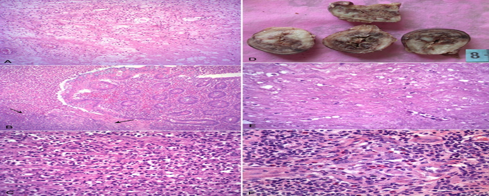
o Acute catarrhal (Figure 1A): Neutrophilic infiltration restricted to the mucosa or submucosa with or without luminal exudates and focal/multifocal epithelial ulceration
o Acute suppurative (phlegmonous) (Figure 1B): Transmural inflammation, luminal exudates, multi-focal/diffuse epithelial ulceration with serosal exudates
2. Complicated Appendicitis
o Acute necrotizing (Figure 1C): Features of Acute suppurative appendicitis along with formation of abscesses in one or more layers and/or destruction of the muscularis propria.
o Gangrenous (Figures 1D and 1E): Features of Acute suppurative appendicitis along with coagulative necrosis, hemorrhage, and vascular thrombosis
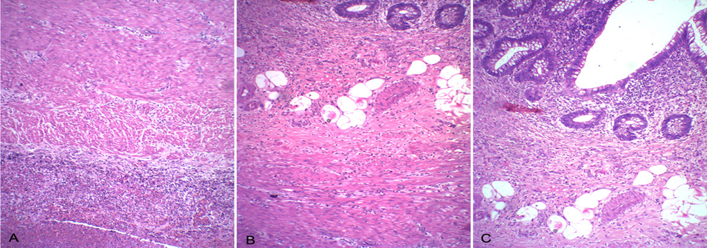
o Perforated appendicitis (Figure 2)
3. Eosinophil-rich Appendicitis (Figure 1F): Prominent or exclusive eosinophilic infiltration, especially involving the muscularis propria by eosinophils
4. Peri-appendicitis (Figure 3): Predominant serosal inflammation with extensive infiltration of neutrophils to muscularis propria and adjacent submucosa
5. Others(n=75)
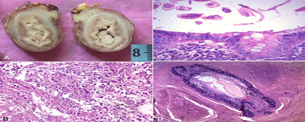
o Parasitic infestations (Figure 4)
o Diverticulae (Figure 5A)
o Appendiceal obliteration (Figure 5B)
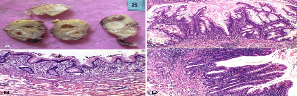
o Mucosal alterations or tumors (Figure 6)
o Appendix with only fecolith
o Appendix with only lymphoid hyperplasia
o Histologically normal appendix
Investigations
Clinical diagnosis was based on:
· History and physical examination
· Laboratory investigations (eg. leukocyte count)
· Radiological findings where available
However, clinical and radiological parameters were not analyzed, and emphasis was placed on histopathological diagnosis.
Outcome Measures
Primary outcome measures included:
- Histopathological spectrum of appendiceal lesions
- Frequency of uncomplicated versus complicated appendicitis
- Frequency of eosinophil-rich appendicitis and peri-appendicitis
- Degree of discordance between initial diagnosis and revised histopathological diagnosis
RESULTS:
Among the 250 patients with a clinical diagnosis of acute appendicitis, there were 160 males and 90 females. The age range was between 1.5 months to 66 years (mean age of 26 years); 37 of these were younger than 18 years of age. Comparisons of the revised and initial diagnoses have been tabulated (Tables 1 and 2).
Table 1: Appendicectomy - Types of Appendicitis (N=175)
|
Type of Appendicitis |
Revised Histopathological Diagnosis |
No. of Cases |
Initial Histopathological Diagnosis |
No. of Cases |
|
Uncomplicated Appendicitis (N = 75) |
Acute catarrhal appendicitis |
13 |
Acute catarrhal appendicitis |
8 |
|
Acute suppurative appendicitis |
2 |
|||
|
Chronic appendicitis |
3 |
|||
|
Acute suppurative appendicitis |
62 |
Acute catarrhal appendicitis |
2 |
|
|
Acute suppurative appendicitis |
58 |
|||
|
Chronic appendicitis |
2 |
|||
|
Complicated Appendicitis (N = 58) |
Acute necrotizing appendicitis |
32 |
Acute suppurative appendicitis |
30 |
|
Acute necrotizing appendicitis |
2 |
|||
|
Acute gangrenous appendicitis |
4 |
Acute gangrenous appendicitis |
4 |
|
|
Appendicular perforation in Acute catarrhal appendicitis 01
Acute suppurative appendicitis 09
Acute necrotizing appendicitis 10
Acute gangrenous appendicitis 02 |
22 |
Acute catarrhal appendicitis |
1 |
|
|
Acute suppurative appendicitis |
9 |
|||
|
Acute necrotizing appendicitis |
10 |
|||
|
Acute suppurative appendicitis with perforation |
2 |
|||
|
Eosinophil-rich Appendicitis (N = 21) |
Eosinophil-rich appendicitis |
21 |
Acute catarrhal appendicitis |
6 |
|
Acute suppurative appendicitis |
3 |
|||
|
Chronic appendicitis |
12 |
|||
|
Peri-appendicitis (N = 21) |
Peri-appendicitis |
21 |
Acute suppurative appendicitis |
18 |
|
Chronic appendicitis |
3 |
Table 1 summarizes the distribution of appendicectomy specimens according to the revised histopathological diagnosis and compares them with the initial histopathological interpretation in a total of 175 cases of acute appendicitis.
Uncomplicated appendicitis constituted the largest group, accounting for 75 cases. Within this category, acute catarrhal appendicitis was identified in 13 cases on revised histopathological examination; however, the initial diagnosis correlated in only 8 cases, while 2 cases were initially labelled as acute suppurative appendicitis and 3 as chronic appendicitis. Acute suppurative appendicitis formed the major subset of uncomplicated appendicitis, with 62 cases confirmed on revision. Of these, 58 cases were correctly diagnosed initially, whereas 2 cases were misinterpreted as acute catarrhal appendicitis and another 2 as chronic appendicitis.
Complicated appendicitis was observed in 58 cases. Acute necrotizing appendicitis was the most frequent entity in this group, comprising 32 cases; however, the majority of these cases (30) were initially reported as acute suppurative appendicitis, with only 2 cases correctly identified as necrotizing appendicitis.
Acute gangrenous appendicitis was relatively uncommon, with 4 cases, all of which showed complete (100%) concordance between initial and revised diagnoses. Additional 22 cases within the complicated appendicitis group exhibited appendicular perforation and amongst these only two cases were given a diagnosis of perforation by junior pathologist. Hence, 20 cases of perforation were completely missed.
Eosinophil-rich appendicitis was identified in 21 cases on revised histopathological examination. None of these cases were picked up initially; instead, 6 of these cases were initially labelled as acute catarrhal appendicitis, 3 as acute suppurative appendicitis, and 12 as chronic appendicitis, highlighting the ignorance of this entity on initial histopathology reporting. Peri-appendicitis was also observed in 21 cases. On initial histopathological evaluation, 18 of these cases were reported as acute suppurative appendicitis, while 3 were diagnosed as chronic appendicitis, indicating that appendicular perforation (Figure3) with peri appendicitis was missed by the junior pathologist.
Table 2: Appendicectomy – Other Histopathological Findings (N = 75)
|
Histopathological Findings |
Revised Histopathological Diagnosis |
No of cases |
Initial Histopathological Diagnosis |
No. of Cases |
|
Parasites and other specific Inflammation (N = 9) |
Amoebic appendicitis |
01 |
Acute suppurative appendicitis |
01 |
|
Enterobius vermicularis |
04 |
Normal appendix |
02 |
|
|
Fecolith |
01 |
|||
|
Chronic appendicitis |
01 |
|||
|
Enterobius / calcified parasite with appendicitis |
04 |
Acute catarrhal appendicitis |
01 |
|
|
Acute suppurative appendicitis |
01 |
|||
|
Acute necrotizing appendicitis |
02 |
|||
|
Diverticulae (N = 4) |
Acute suppurative appendicitis |
01 |
Acute suppurative appendicitis |
02 |
|
Acute suppurative appendicitis with perforation |
01 |
Acute suppurative appendicitis |
01 |
|
|
Eosinophil-rich appendicitis |
02 |
Chronic appendicitis |
01 |
|
|
Mucosal Alterations / Tumors (N= 4) |
Carcinoid tumor |
01 |
Carcinoid |
01 |
|
Low-grade mucinous neoplasm with acute suppurative appendicitis |
01 |
Acute suppurative appendicitis |
01 |
|
|
Epithelial metaplasia with acute suppurative appendicitis |
02 |
Acute suppurative appendicitis |
02 |
|
|
Other Findings |
Only fecolith |
15 |
Normal appendix |
01 |
|
Fecolith |
02 |
|||
|
Chronic appendicitis |
12 |
|||
|
Only lymphoid hyperplasia |
06 |
Acute catarrhal appendicitis |
02 |
|
|
Acute suppurative appendicitis |
01 |
|||
|
Chronic appendicitis |
03 |
|||
|
Appendiceal obliteration |
03 |
Chronic appendicitis |
03 |
|
|
Normal appendix |
34 |
Normal appendix |
04 |
|
|
Acute catarrhal appendicitis |
01 |
|||
|
Chronic appendicitis |
29 |
Table 2 depicts the spectrum of additional histopathological findings identified in 75 appendicectomy specimens on revised evaluation by a senior pathologist and compares them with the initial histopathological diagnoses. Parasites and other specific inflammatory conditions including Entamoeba histolytica and Enterobius vermicularis were picked up in 9 cases (Figure 4).
Diverticular disease (Figure 5A) was observed in 4 cases on revised diagnosis which were totally missed on initial diagnosis.
Out of 4 cases of Mucosal alterations including tumors which were picked up on revised diagnosis, only one case of carcinoid tumour showed complete concordance and was diagnosed correctly on initial evaluation.
Other incidental and non-inflammatory findings formed a substantial proportion of cases. Isolated fecolith without significant inflammation was identified in 15 cases of which the majority were labelled as chronic appendicitis (12 cases). Only lymphoid hyperplasia was noted in 6 cases, which were initially misdiagnosed as acute or chronic appendicitis
Appendiceal obliteration was observed in 3 cases, all of which were initially labelled as chronic appendicitis. A histologically normal appendix was identified in 34 cases on revised examination. Despite the absence of significant pathology, only 4 of these cases had been initially reported as normal. The remaining cases were all over diagnosed as acute or chronic appendicitis, highlighting a high rate of negative appendicectomy with inflammatory overinterpretation on histopathology.
Table 3: Clinicopathological Discordance in Appendectomy Specimens (N = 250)
|
Diagnostic correlation |
No. of cases |
Percentage (%) |
|
Concordant diagnosis |
83 |
33.2 |
|
Discordant diagnosis |
167 |
66.8 |
|
Total |
250 |
100 |
Table 3 highlights the overall concordance between the initial diagnosis and revised histopathological diagnosis was observed in 83 cases, accounting for 33.2% of the study population. A discordance was noted in 167 cases, representing 66.8% of cases
.
Table 4: Spectrum and Biological Activity of Appendiceal Pathology Supporting Functional Role of Appendix (N = 250)
|
Pathological Parameter |
Spectrum of Histopathological findings |
Frequency (n) |
Percentage (%) |
|
Histopathological findings |
Acute inflammatory lesions (catarrhal, suppurative, necrotizing) |
154 |
61.6 |
|
Parasites / specific infections |
9 |
3.6 |
|
|
Diverticular disease |
4 |
1.6 |
|
|
Neoplastic / premalignant lesions |
4 |
1.6 |
|
|
Normal appendix |
34 |
13.6 |
|
|
Fecolith |
15 |
6 |
|
|
Appendiceal obliteration |
03 |
1.2 |
|
|
Evidence of Immunological / Reactive Activity |
Lymphoid hyperplasia |
06 |
2.4 |
|
Eosinophil-rich inflammation |
21 |
8.4 |
Table 4 summarises the spectrum of appendiceal pathology observed in the study highlighting histopathological features indicative of biological and immunological activity in a total of 250 appendectomy specimens.
|
|
|
Figure 1: A. Acute catarrhal appendicitis showing edematous sub-mucosa with scattered neutrophils (H&E x 100); B. Acute suppurative appendicitis with luminal exudate and focal epitheial ulceration (arrows, H&E x 100); C. Acute necrotizing appendicitis showing diffuse and extensive neutrophilic iflitation with destruction of the muscularis propria (H&E x 100); Acute gangrenous appendicitis characterized by D. an enlarged appendix with hemorrhagic walls and E. muscularis propria with coagulative necrosis; F. Eosinophilic appendicitis – the smooth muscle cells of the muscularis propria splayed apart by eosinophils and lymphocytes (H&E x 400). |
|
|
|
Figure 2: Appendicular Perforation : A. The tip of the appensix is covered by thick exudate; B. The tip has been cut through the area of perforation (arrow); C. Histology showing the tract of perforation (H&E x 100). |
|
|
|
Figure 4: Parasitic infestations : A. Enlarged appendix with lumen filled with granular pale yellow material; B. Sparse inflammatory cells and trophozoites of E. histolytica in an edematous and necrotic muscularis propria (H&E x 400); C. Eggs of E. vermicularis in the appendicular lumen (H&E x 200); D. A calcified parasite fills up the lumen of the appendix (H&E x 100). |
|
|
Figure 6: A. The peri-appendicular fat is surrounded by mucin filled cystic spaces. Similar material is also present in the lumen of the appendix, well seen in the ‘C’ section. On histology, the appendix revealed B. undulating mucigenic epithelial growth pattern, loss of muscularis mucosae and lymphoid follicles – low-grade appendicular mucinous neoplasm (LAMN, H&E x 200); C. Another case of LAMN showing villiform epithelial growth pattern (H&E x 200); D. Hyperplastic polyp with a serrated morphology (H&E x 200). |
DISCUSSION:
Surgically resected appendices, following suspicion or diagnosis of acute appendicitis, are one of the most common specimens received for histopathology examination. However, such specimens do not get their due importance, and in few institutes, they may not even undergo histopathological evaluation, though the importance of histopathological evaluation has been often highlighted and have been subjects of clinico-pathological discrepancies.9,12 Popular textbooks in surgical pathology recommend only ‘O’ and ‘C’ sections, which are generally evaluated and signed out by histopathologists often positioned at a junior rank. This practice can potentially miss out on incidental, aberrant and/or significant pathological findings that may impact further patient management. This study evaluated an inter-observer variation in the pathological assessment where appendicectomy specimens after initial routine sectioning required for issue of reports, were subsequently serially sectioned and entirely processed (in most instances in a single block) and revised histopathological evaluation was performed by a senior pathologist. The revised results were compared with the initial diagnosis.
Luminal obstruction of the appendix has been considered to be the center point of acute appendicitis where a rise in intra-luminal pressure and supervening bacterial infection leads to varying stages of inflammation, designated as uncomplicated (catarrhal, phlegmonous and suppurative) and complicated (necrotizing, gangrenous and perforative).5 The present study demonstrates a substantial degree of histopathological discordance in appendectomy specimens, with only 33.2% of cases showing concordance between initial diagnosis and revised histopathological findings, while discordance was observed in 66.8% of cases. However, the marked pathological discordance observed in this study suggests that appendiceal disease encompasses a heterogeneous spectrum extending beyond overt acute inflammation.
On reviewing the cases in this series, no discrepancy was observed in cases of gangrenous appendicitis owing to its distinctive morphology. While minor discrepancy was noted in cases of uncomplicated appendicitis (9 of 75, 12 %). Cases of acute necrotizing inflammation (30 of 32, 93.6 %) and appendicular perforations (20 of 22, 90.9 %) had major discrepant diagnosis. Eosinophil-rich appendicitis or eosinophilic appendicitis, a term introduced by Arvindan, is said to be a rare allergic response in the appendicular milieu (incidence of only 0.03. to 1.2 %), associated with elevation of the eosinophilic cationic proteins even in serum. 13,14 It was as seen as a revised diagnosis in 21 (one of which was associated with appendicular diverticula) of our 250 cases (8.4 %); recognition is important as it may not respond to antibiotic therapy and can be a forerunner to other systemic allergic responses. A similar number of cases was re-designated as peri-appendicitis as the inflammatory exudate was chiefly found over the serosal aspect with variable extension into the muscularis propria.11 Since the inflammation is due to other causes within the abdomen, it would be important to be vigilant about the post-operative course after appendicectomy. Although, ours was a prospective study and diagnosis was made on re-assessment, we did not have follow-up on these patients.
We would like to highlight that parasitic infestations including acute amebiasis (9 cases) had not been diagnosed in the initial assessment. Amebic appendicitis has an incidence of 0.5 to 2.3 %, but mandates specific treatment and calls for further imaging assessment and stool examination for the colonic involvement.15 The same would apply to the presence of other nematode parasites, of which E. vermicularis is most common and found in 0.6% to 13% of resected appendices.16 We did not find any other specific organisms or microbial-specific inflammatory reactions. Appendiceal diverticulas are uncommon, and the reported incidence in appendectomy specimens has ranged from 0.004 % to 2.1 %.17 We discovered 4 such cases (1.6 %) during reassessment. Four variants of diverticular disease of the appendix are recognized: appendicular diverticula without inflammation, acute appendicitis with diverticula, acute appendicular diverticulitis with acute appendicitis, and isolated acute diverticulitis.18 Appendicular diverticulosis is usually asymptomatic but can present with complications like inflammation and perforation. Features of acute suppurative appendicitis and eosinophilic appendicitis had been present; one of the cases of diverticula exhibiting acute suppurative appendicitis also had perforation. The diverticulas can also harbor tumors, which were not seen in any of our 4 cases.19
Although carcinoid was picked up at initial diagnosis (also shown to the senior most cadre), neoplastic appendiceal mucinous lesions and other mucosal alterations (seen in 0.2 to 1.4 % of appendectomies) were missed in 3 cases (1.2 % among 250 cases). The classification of these lesions is complex and has important clinical implications; though in many cases appendicectomy without any other therapeutic modalities is indeed curative.20 Another important fact is that in cases where appendicectomies were performed based on clinical suspicion, we did not find evidence of appendicitis (negative appendicectomy or findings of lymphoid hyperplasia / fecolith) on thorough histopathological examination, but many had been signed out as acute catarrhal or chronic appendicitis. This trend should be discouraged so that clinicians work up patients for other causes of right iliac fossa pain and other pathological conditions are not overlooked. Histologically normal appendices were identified in 13.6% of cases, comparable to reported negative appendectomy rates.
The pathologists need to realize the importance of keen gross and microscopic examination of appendicectomy specimens. While grossing, such specimens should be serially sectioned to look for any gross abnormality. Besides confirming the diagnosis of appendicitis and categorizing it into various types, submitting additional transverse sections will allow detection of other histologic findings and enhance the diagnostic value to avoid minor or major repercussions in the follow-up of some patients.
Evidence of immunological and reactive activity was observed in a subset of specimens, with lymphoid hyperplasia (2.4%) and eosinophil-rich inflammation (8.4%). The appendix is a recognized component of gut-associated lymphoid tissue (GALT), rich in lymphoid follicles and involved in mucosal immune responses (Bollinger et al., 2007; Kooij et al., 2016). Eosinophil-predominant appendicitis, described by Lamps et al. (2001), represents a distinct immune-mediated process often associated with parasitic or hypersensitivity reactions.7-8,16 These immunological features provide a biological basis for appendiceal symptoms in the absence of classical acute inflammation and may partially explain the high number of negative appendicectomy specimens and clinicopathological discordance observed.
LIMITATIONS OF THE STUDY
· This was a single centre, tertiary hospital-based prospective study, which may limit the generalizability of the findings to other populations or healthcare settings.
· Clinical parameters such as symptom duration, Alvarado score, laboratory values, and radiological findings were not analysed or correlated statistically with histopathological outcomes.
· Long-term clinical follow-up of patients was not available, limiting assessment of clinical outcomes related to specific histopathological patterns.
CONCLUSION :
The present study demonstrates a wide and diverse histopathological spectrum in appendectomy specimens clinically diagnosed as acute appendicitis. Acute inflammatory lesions constituted the majority of cases; however, a substantial proportion of specimens revealed eosinophil-rich appendicitis, lymphoid hyperplasia, parasitic infestations, diverticular disease, neoplastic lesions, and histologically normal appendices. The frequent identification of reactive lymphoid hyperplasia and eosinophil-predominant inflammation highlights the immunologically active nature of the appendix. These findings challenge the traditional concept of the vermiform appendix as a purely vestigial organ and support its role as a biologically and immunologically functional structure. A high rate of pathological discordance was observed, with nearly two-thirds of cases showing mismatch between initial diagnosis and final histopathological findings. We suggest that even if junior pathologists are reporting appendicectomy specimens, senior pathologists should intermittently review and supervise their reporting so that important diagnostic features are not missed. Meticulous histopathological examination of all appendectomy specimens is therefore essential, not only for accurate diagnosis but also for identifying clinically significant incidental and immune-mediated pathologies.
ACKNOWLEDGEMENT
The authors sincerely thank the Departments of Pathology and Surgery of the participating institutions for their cooperation and support during the conduct of this study. We acknowledge the technical staff of the histopathology laboratory for assistance with specimen processing and slide preparation. We are also grateful to the junior faculty and residents whose initial reporting formed an integral part of this study and contributed to evaluating inter-observer variability.
Author Contributions and Declarations
· Each author has contributed to the creation and framing of this manuscript and also in designing, redrafting and final proof reading of the article
· There has been no conflict of interest.
· The manuscript has been read and approved by all the authors and all the names are mentioned in the order that is acceptable to all.
Funding
No external funding was received for this study.
REFERENCES :
1. Ferris M, Quan S, Kaplan BS, Molodecky N, Ball CG, Chernoff GW, et al. The global incidence of appendicitis: a systematic review of population-based studies. Ann Surg. 2017;266(2):237–241. doi:10.1097/SLA.0000000000002188.
2. Andersson RE. The natural history and traditional management of appendicitis revisited: spontaneous resolution and predominance of prehospital perforations imply that a correct diagnosis is more important than an early diagnosis. World J Surg. 2014;38(2):312–317. doi:10.1007/s00268-013-2246-y.
3. Carr NJ. The pathology of acute appendicitis. Ann Diagn Pathol. 2000;4(1):46–58. doi:10.1016/S1092-9134(00)90011-X.
4. Rosai J. Rosai and Ackerman’s surgical pathology. 10th ed. St. Louis: Elsevier Mosby; 2011. p. 712–719.
5. Bhangu A, Soreide K, Di Saverjo S, Assarsson JH, Drake FT. Acute appendicitis: modern understanding of pathogenesis, diagnosis, and management. Lancet 2015;386:1278-87.
6. Cho J, Lee D, Sung K, Baek J, Lee J. Clinical implication of discrepancies between surgical and pathological diagnoses of acute appendicitis. Ann Surg Treat Res 2017;93:43-9.
7. Kooij IA, Sahami S, Meijer SL, Buskens CJ, Te Velde AA. The immunology of the vermiform appendix: a review of the literature. Clin Exp Immunol. 2016;186(1):1–9. doi:10.1111/cei.12821.
8. Bollinger RR, Barbas AS, Bush EL, Lin SS, Parker W. Biofilms in the large bowel suggest an apparent function of the human vermiform appendix. J Theor Biol. 2007;249(4):826–831. doi:10.1016/j.jtbi.2007.08.032.
9. Jones AE, Phillips AW, Jarvis JR, Sargen K. The value of routine histopathological examination of appendicectomy specimens. BMC Surg. 2007;7:17. doi:10.1186/1471-2482-7-17.
10. Tayfur M, Balci MG. Pathological changes in appendectomy specimens including the role of parasites: A retrospective study of 2400 cases of acute appendicitis. Niger J Clin Pract 2019;22:270-5.
11. Chaudhary P, Nabi I, Arora MP. Periappendicitis: Our 13 year experience. Int J Surg 2014;12:1010-3.
12. Omiyale AO, Adjepong S. Histopathological correlations of appendectomies: A clinical audit of a single center. Ann Transl Med 2015;3:119
13. Aravindan KP. Eosinophils in acute appendicitis: possible significance. Indian J Pathol Microbiol 1997;40:491-8.
14. Carvalho N, Carolino E, Coelho H, Barreira AL, Moreira L, André M, et al. Eosinophil granule proteins involvement in acute appendicitis - An allergic disease? Int J Mol Sci 2023; 24: 9091.
15. Herrera GC, Herrera AC, Ponton P, Molina GA, Constante JE, Delgado JA. Amebiasis, a rare cause of acute appendicitis. J Surg Case Rep 2019; 2019; rjz076
16. Lamps LW. Infectious Causes of Appendicitis. Infect Dis Clin N Am 2010; 24: 995- 1018
17. Bonomo LD, Zago M, Quirico C, Ottinetti A, Panzera F, Galli A, et al. Surgical rarities: case report of appendicular diverticulitis and literature review. J Surg Case Rep 2022; 2022: rjac319.
18. Lipton S, Estrin J, Glasser I. Diverticular disease of the appendix. Surg Gynecol Obstet 1989;168:13-6.
19. Chan DL, Lim C, Bakhtiar A. Clinical significance of appendiceal diverticulum: a significant marker for appendiceal neoplasia in Australian patients. Int J Colorectal Dis 2018;33:1569-74.
20. Rossi A, Patel NM. Appendiceal neoplasms - A practical guide. J Surg Oncol 2023;127:1300-5.