Cuff-Based Blood Pressure Underestimates Central Aortic Pressure: Implications for Hypertension Management in a High-Risk Cohort from Faisalabad, Pakistan
- Omer Aslam , Medical Officer, Government General Hospital Samnabad, Faisalabad Pakistan
- Ayesha Anwar , Women Medical Officer, Government General Hospital Samnabad, Faisalabad, Pakistan
Article Information:
Abstract:
To compare peripheral brachial cuff blood pressure (BP) measurements with central aortic pressure (CAP) derived from applanation tonometry in a high-risk cardiovascular cohort, and to determine the magnitude and direction of discrepancy between these two measurement modalities. Study duration: January 2025 to June 2025. Study place: Faisalabad Institute of Cardiology, Faisalabad, Pakistan. Methodology: A cross-sectional analytical study was conducted on 320 adult patients with established hypertension and at least one additional cardiovascular risk factor. Peripheral BP was measured using a validated automated oscillometric cuff device. Central aortic pressure was non-invasively derived using radial artery applanation tonometry with a validated transfer function. Systolic BP (SBP), diastolic BP (DBP), and pulse pressure (PP) were compared using paired t-tests, Pearson correlation, and Bland-Altman analysis.Results: Mean peripheral SBP was 142.6 ± 16.8 mmHg, whereas mean central SBP was 132.4 ± 15.2 mmHg (mean difference: 10.2 ± 8.4 mmHg, p < 0.001). The discrepancy was greatest in younger patients (age < 50 years: 13.1 mmHg difference) and those with isolated systolic hypertension (15.4 mmHg difference). Bland-Altman analysis revealed proportional bias, with larger differences observed at higher BP levels (r = 0.41, p < 0.001). Pulse pressure amplification (peripheral PP minus central PP) averaged 8.7 ± 6.2 mmHg. Conclusion: Cuff-based brachial BP significantly overestimates central SBP in this high-risk Pakistani cohort. Reliance on peripheral measurements alone may lead to undertreatment of central hypertension. Integration of central aortic pressure assessment could improve hypertension management in resource-limited settings.
Keywords:
Article :
INTRODUCTION :
Hypertension remains the leading modifiable risk factor for cardiovascular morbidity and mortality worldwide, contributing to an estimated 10.4 million deaths annually [1]. In Pakistan, the prevalence of hypertension among adults has reached alarming proportions, with recent meta-analyses reporting pooled prevalence estimates exceeding 25% in urban populations and approaching 20% in rural communities [2]. The Faisalabad region, an industrial hub of Punjab province, bears a disproportionate burden of cardiovascular disease, with local registry data suggesting that nearly one in three adults over the age of 40 years has diagnosed or undiagnosed hypertension [3]. Despite the availability of effective antihypertensive therapies, blood pressure control rates remain suboptimal, with fewer than 30% of treated patients achieving guideline-recommended targets [4].
The cornerstone of hypertension diagnosis and management has traditionally been brachial cuff blood pressure measurement, a technique that has remained largely unchanged for over a century [5]. Automated oscillometric devices have largely replaced manual auscultatory methods in clinical practice, offering convenience and reduced observer bias. However, these devices measure pressure oscillations in the brachial artery and derive systolic and diastolic values using proprietary algorithms that vary considerably between manufacturers [6]. More fundamentally, brachial cuff pressure serves as a surrogate for the pressure that truly matters for cardiovascular risk: the pressure exerted on the heart, brain, and kidneys by the central aorta [7].
Central aortic pressure differs from peripheral brachial pressure due to phenomena collectively termed pulse pressure amplification [8]. As the pressure wave travels from the central aorta to the peripheral arteries, it undergoes morphological changes influenced by arterial stiffness, wave reflection, and heart rate. In young, healthy individuals with compliant arteries, peripheral systolic pressure is typically 10-20 mmHg higher than central systolic pressure, reflecting amplification of the pressure wave [9]. With advancing age and increasing arterial stiffness, this amplification diminishes and may even reverse, leading to a phenomenon where central pressure equals or exceeds peripheral pressure [10].
The clinical importance of central aortic pressure has been increasingly recognized over the past two decades. The CAFÉ (Conduit Artery Function Evaluation) study, a substudy of the landmark ASCOT trial, demonstrated that different antihypertensive regimens achieved similar reductions in brachial pressure but markedly different reductions in central pressure, and these differences in central pressure predicted cardiovascular outcomes more accurately than brachial pressure [11]. Similarly, the Strong Heart Study showed that central pulse pressure was a stronger predictor of cardiovascular events than brachial pulse pressure in American Indian populations [12]. Meta-analyses have consistently demonstrated that central pressure parameters add incremental prognostic value beyond conventional brachial measurements [13].
The biological plausibility of central pressure as a superior risk marker rests on solid pathophysiological grounds. The left ventricle ejects blood directly into the aorta, and myocardial oxygen demand is determined largely by central systolic pressure and wall tension [14]. Coronary perfusion occurs during diastole, and central diastolic pressure directly influences coronary filling pressure [15]. The cerebral circulation is exposed to central rather than peripheral pressure waveforms, and the renal microcirculation is similarly protected by autoregulatory mechanisms that respond to central pressure [16]. Thus, reliance on brachial pressure may misclassify cardiovascular risk in substantial proportions of patients.
The discrepancy between central and peripheral pressure has important therapeutic implications. Antihypertensive drugs exert differential effects on central pressure independent of their brachial pressure-lowering effects [17]. Beta-blockers, particularly atenolol, lower brachial pressure effectively but have less favorable effects on central pressure due to their bradycardic effects, which increase wave reflection and central augmentation [18]. In contrast, vasodilating agents such as calcium channel blockers, angiotensin-converting enzyme inhibitors, and angiotensin receptor blockers reduce wave reflection and lower central pressure to a greater extent than brachial pressure would suggest [19]. The differential effects on central pressure may explain the superior cardiovascular outcomes observed with newer agents compared with beta-blockers in clinical trials despite similar brachial pressure reductions [20].
Despite the growing evidence base supporting central pressure measurement, its adoption into routine clinical practice has been slow, particularly in low- and middle-income countries such as Pakistan [21]. The primary barriers include equipment cost, lack of trained personnel, and uncertainty regarding normative values and treatment targets. However, recent technological advances have produced relatively affordable and portable devices for non-invasive central pressure estimation, making this technology increasingly accessible [22]. The development of validated transfer functions that derive central pressure from radial artery tonometry has further simplified the measurement process, requiring only brief training for competent performance [23].
The Faisalabad Institute of Cardiology serves as a divisional referral center for the surrounding region, managing a high-volume, high-risk population with premature coronary artery disease, resistant hypertension, and multiple cardiovascular comorbidities [24]. The patient population in this setting differs substantially from the Western cohorts in which most central pressure research has been conducted. Pakistani patients tend to present with cardiovascular disease at younger ages, have higher prevalence of metabolic syndrome and diabetes, and often have limited access to preventive healthcare [25]. Whether the relationship between central and peripheral pressure observed in Western populations holds true in this distinct ethnic and clinical context remains uncertain. Ethnic differences in arterial stiffness and wave reflection have been documented, with South Asian populations demonstrating greater central arterial stiffness for any given brachial pressure compared with white European populations [26].
The present study was therefore designed to address a critical knowledge gap: to quantify the discrepancy between brachial cuff pressure and central aortic pressure in a high-risk Pakistani cohort, and to explore the clinical and demographic factors that modify this relationship. We hypothesized that brachial cuff pressure would significantly overestimate central pressure in this population, and that the magnitude of overestimation would vary systematically with age, blood pressure level, and the presence of diabetes. The findings have direct implications for hypertension management in resource-limited settings where central pressure measurement is not routinely available, suggesting that current treatment algorithms based on brachial pressure targets may inadvertently undertreat central hypertension in certain patient subgroups.
METHODOLOGY:
Study Design and Setting
This cross-sectional analytical study was conducted at the Faisalabad Institute of Cardiology (FIC), Faisalabad, Pakistan, between January 2025 and June 2025. The institute is a 250-bed Divisional care cardiac center serving a population of approximately 12 million people from the Faisalabad, Jhang, Toba Tek Singh, and Chiniot districts. The study protocol was approved by the Institutional Ethics Committee of Faisalabad Institute of Cardiology (approval number: FIC/IRB/2024/112) and was conducted in accordance with the principles of the Declaration of Helsinki. Written informed consent was obtained from all participants prior to enrollment.
Study Population
Participants were recruited from the outpatient hypertension clinic and the inpatient cardiology wards of FIC using consecutive sampling. Inclusion criteria were: (1) age 30-80 years; (2) established diagnosis of hypertension (defined as brachial systolic BP ≥ 140 mmHg or diastolic BP ≥ 90 mmHg on at least two separate occasions, or current use of antihypertensive medication); (3) presence of at least one additional cardiovascular risk factor (diabetes mellitus, dyslipidemia, current smoking, obesity with body mass index ≥ 30 kg/m², or established coronary artery disease); and (4) ability to provide informed consent. Exclusion criteria were: (1) atrial fibrillation or other significant cardiac arrhythmia; (2) severe aortic stenosis (peak velocity > 4 m/s or mean gradient > 40 mmHg); (3) history of aortic dissection or aortic aneurysm; (4) upper extremity deformity or vascular access that precluded brachial BP measurement; (5) pregnancy; (6) acute febrile illness or acute coronary syndrome within the preceding four weeks; and (7) severe renal failure requiring dialysis.
Sample Size Calculation
Sample size was calculated based on the expected mean difference between peripheral and central systolic BP. Based on previous studies reporting a standard deviation of differences of approximately 12 mmHg, we calculated that a sample of 240 participants would provide 90% power to detect a mean difference of 5 mmHg with a significance level of 0.05 (two-tailed). To account for potential missing data and technical failures, we recruited 320 participants (33% oversampling).
Measurements
Demographic and Clinical Data: A structured questionnaire was administered to collect information on age, sex, smoking status (current, former, never), duration of hypertension, and current antihypertensive medications. Height was measured to the nearest 0.1 cm using a wall-mounted stadiometer, and weight was measured to the nearest 0.1 kg using a calibrated digital scale. Body mass index was calculated as weight (kg) divided by height squared (m²). Waist circumference was measured at the midpoint between the iliac crest and the lowest rib at the end of normal expiration.
Brachial Blood Pressure: Peripheral brachial BP was measured using an automated oscillometric device after participants had rested quietly for at least 10 minutes in a seated position with back supported and feet flat on the floor. An appropriately sized cuff (standard adult, large adult, or thigh cuff) was placed on the bare right arm at heart level. Three measurements were taken at one-minute intervals, and the mean of the last two readings was used for analysis. The device was calibrated monthly according to manufacturer specifications.
Central Aortic Pressure: Central aortic pressure was derived non-invasively from radial artery applanation tonometry using a validated device. After the brachial BP measurement, participants rested for an additional 5 minutes. The radial artery of the right wrist was palpated, and a high-fidelity tonometer was positioned to obtain an optimal pressure waveform. A 10-second recording was acquired, and waveforms were rejected if there was >5% variability in amplitude or cycle length. A validated generalized transfer function was applied to derive the central aortic pressure waveform. The quality of recordings was assessed using an inbuilt quality index, and recordings with a quality index < 80% were repeated. Central systolic BP, central diastolic BP, central pulse pressure, and augmentation index were recorded.
Other Cardiovascular Assessments: Left ventricular ejection fraction was assessed by transthoracic echocardiography using the Simpson biplane method. Carotid-femoral pulse wave velocity (cf-PWV) was measured using the same SphygmoCor XCEL device as a marker of arterial stiffness. Fasting blood samples were collected for measurement of glucose, lipid profile (total cholesterol, HDL cholesterol, LDL cholesterol, triglycerides), and creatinine. Estimated glomerular filtration rate (eGFR) was calculated using the CKD-EPI equation.
Statistical Analysis
Data were analyzed using SPSS version 26.0 (IBM Corp., Armonk, NY, USA) and MedCalc version 20.0 (MedCalc Software, Ostend, Belgium). Normality of continuous variables was assessed using the Shapiro-Wilk test and visual inspection of histograms. Normally distributed data were presented as mean ± standard deviation, while non-normally distributed data were presented as median (interquartile range). Categorical variables were presented as frequencies and percentages.
Paired t-tests were used to compare peripheral and central BP parameters. Independent t-tests and one-way ANOVA were used to compare mean differences across subgroups. Pearson correlation coefficients were calculated to assess the strength of linear associations between variables. Multiple linear regression was performed to identify independent predictors of the peripheral-central systolic BP difference, with adjustment for potential confounders including age, sex, body mass index, heart rate, diabetes status, and antihypertensive medication class.
Bland-Altman analysis was performed to assess agreement between peripheral and central systolic BP, with calculation of mean difference (bias), standard deviation of differences, and 95% limits of agreement (mean difference ± 1.96 × SD). The presence of proportional bias was assessed by regressing the difference between methods against their mean.
All statistical tests were two-tailed, and a p-value < 0.05 was considered statistically significant. No adjustments were made for multiple comparisons due to the exploratory nature of subgroup analyses.
RESULTS:
A total of 320 participants were enrolled in the study, of whom 310 (96.9%) completed all measurements and were included in the final analysis. Ten participants were excluded due to poor quality central pressure waveforms (n=6), inability to obtain adequate brachial BP measurements (n=2), or withdrawal of consent (n=2). The baseline characteristics of the study cohort are presented in Table 1.
Table 1: Baseline Demographic and Clinical Characteristics of the Study Cohort (N=310)
|
Characteristic |
Value |
|
Age (years), mean ± SD |
56.4 ± 11.2 |
|
Age ≥ 60 years, n (%) |
128 (41.3) |
|
Male sex, n (%) |
186 (60.0) |
|
Body mass index (kg/m²), mean ± SD |
28.6 ± 5.1 |
|
Obesity (BMI ≥ 30 kg/m²), n (%) |
112 (36.1) |
|
Waist circumference (cm), mean ± SD |
96.4 ± 12.3 |
|
Current smoker, n (%) |
68 (21.9) |
|
Diabetes mellitus, n (%) |
142 (45.8) |
|
Dyslipidemia, n (%) |
198 (63.9) |
|
Established coronary artery disease, n (%) |
104 (33.5) |
|
Duration of hypertension (years), median (IQR) |
8 (4-14) |
|
Number of antihypertensive medications, mean ± SD |
2.3 ± 1.1 |
|
Antihypertensive medication use, n (%) |
|
|
ACE inhibitors |
124 (40.0) |
|
Angiotensin receptor blockers |
98 (31.6) |
|
Calcium channel blockers |
142 (45.8) |
|
Beta-blockers |
108 (34.8) |
|
Diuretics |
86 (27.7) |
|
Laboratory parameters, mean ± SD |
|
|
Fasting glucose (mg/dL) |
118 ± 42 |
|
Total cholesterol (mg/dL) |
198 ± 46 |
|
LDL cholesterol (mg/dL) |
118 ± 38 |
|
HDL cholesterol (mg/dL) |
42 ± 10 |
|
Triglycerides (mg/dL) |
178 ± 78 |
|
eGFR (mL/min/1.73m²) |
82 ± 24 |
|
Hemodynamic parameters, mean ± SD |
|
|
Heart rate (beats/min) |
74 ± 12 |
|
Left ventricular ejection fraction (%) |
56 ± 9 |
|
Carotid-femoral PWV (m/s) |
10.4 ± 2.8 |
SD: standard deviation; IQR: interquartile range; BMI: body mass index; ACE: angiotensin-converting enzyme; LDL: low-density lipoprotein; HDL: high-density lipoprotein; eGFR: estimated glomerular filtration rate; PWV: pulse wave velocity
The cohort comprised predominantly middle-aged to older adults (mean age 56.4 years) with a male predominance (60%). Nearly half of the participants had diabetes mellitus (45.8%), and one-third had established coronary artery disease (33.5%). The mean number of antihypertensive medications was 2.3, indicating that the majority had moderate-to-severe hypertension requiring combination therapy. The elevated carotid-femoral pulse wave velocity (mean 10.4 m/s) confirms the presence of significant arterial stiffening in this high-risk population, consistent with the known increased cardiovascular risk profile of South Asian cohorts.
Comparison of Peripheral and Central Blood Pressure
Table 2 presents the comparison between brachial cuff (peripheral) and central aortic blood pressure parameters across the entire study cohort.
Table 2: Comparison of Peripheral and Central Blood Pressure Parameters (N=310)
|
Parameter |
Peripheral (Brachial) |
Central (Aortic) |
Mean Difference (95% CI) |
p-value |
|
Systolic BP (mmHg) |
142.6 ± 16.8 |
132.4 ± 15.2 |
10.2 (9.3, 11.1) |
<0.001 |
|
Diastolic BP (mmHg) |
86.2 ± 10.4 |
85.8 ± 10.6 |
0.4 (-0.2, 1.0) |
0.182 |
|
Pulse pressure (mmHg) |
56.4 ± 14.2 |
46.6 ± 12.8 |
9.8 (8.8, 10.8) |
<0.001 |
|
Mean arterial pressure (mmHg) |
105.0 ± 11.2 |
101.3 ± 10.8 |
3.7 (2.9, 4.5) |
<0.001 |
|
Augmentation index (%) |
- |
28.4 ± 11.6 |
- |
- |
Data presented as mean ± standard deviation. CI: confidence interval; BP: blood pressure
Peripheral systolic BP was significantly higher than central systolic BP, with a mean difference of 10.2 mmHg (95% CI: 9.3 to 11.1, p < 0.001). This finding confirms that reliance on brachial cuff measurements substantially overestimates the pressure load imposed on the central vasculature in this population. In contrast, diastolic BP showed no significant difference between peripheral and central measurements (mean difference 0.4 mmHg, p = 0.182), a finding consistent with previous reports that diastolic pressure is minimally amplified along the arterial tree.
Pulse pressure amplification, defined as the difference between peripheral and central pulse pressure, averaged 9.8 mmHg, indicating that approximately 17% of the peripheral pulse pressure is attributable to amplification phenomena rather than true central pressure load. The augmentation index of 28.4% indicates moderate-to-severe wave reflection, consistent with the elevated pulse wave velocity values observed and confirming increased arterial stiffness in this cohort.
The correlation between peripheral and central systolic BP was moderate but imperfect (Pearson's r = 0.68, p < 0.001), indicating that peripheral measurements explain only 46% of the variance in central pressure. This substantial unexplained variance suggests that central pressure cannot be reliably predicted from peripheral measurements alone, supporting the need for direct central pressure assessment in high-risk patients.
Subgroup Analysis of Peripheral-Central Systolic BP Difference
Table 3 presents subgroup analyses examining the magnitude of the peripheral-central systolic BP difference across various demographic and clinical subgroups.
Table 3: Subgroup Analysis of Peripheral-Central Systolic BP Difference (Mean Difference in mmHg)
|
Subgroup |
n |
Mean Difference ± SD |
p-value for interaction |
|
Age group |
<0.001 |
||
|
< 50 years |
98 |
13.1 ± 7.6 |
|
|
50-59 years |
102 |
10.4 ± 8.2 |
|
|
60-69 years |
72 |
8.2 ± 8.6 |
|
|
≥ 70 years |
38 |
5.8 ± 8.9 |
|
|
Sex |
0.324 |
||
|
Male |
186 |
9.8 ± 8.2 |
|
|
Female |
124 |
10.8 ± 8.6 |
|
|
Diabetes status |
0.018 |
||
|
Diabetic |
142 |
8.6 ± 8.4 |
|
|
Non-diabetic |
168 |
11.6 ± 8.2 |
|
|
BMI category |
0.087 |
||
|
Normal (18.5-24.9) |
78 |
11.4 ± 7.8 |
|
|
Overweight (25-29.9) |
120 |
10.2 ± 8.4 |
|
|
Obese (≥30) |
112 |
9.2 ± 8.8 |
|
|
Coronary artery disease |
0.042 |
||
|
Present |
104 |
8.4 ± 8.6 |
|
|
Absent |
206 |
11.2 ± 8.2 |
|
|
Isolated systolic hypertension |
<0.001 |
||
|
Present (peripheral SBP ≥140, DBP <90) |
88 |
15.4 ± 7.2 |
|
|
Absent |
222 |
8.2 ± 8.0 |
BP: blood pressure; SD: standard deviation; BMI: body mass index; SBP: systolic blood pressure; DBP: diastolic blood pressure
The magnitude of peripheral-central systolic BP discrepancy varied substantially across subgroups. Younger patients (<50 years) demonstrated the largest difference (13.1 mmHg), while older patients (≥70 years) showed markedly attenuated amplification (5.8 mmHg). This age-related decline in pulse pressure amplification reflects age-associated increases in arterial stiffness and earlier wave reflection, which reduce the peripheral augmentation of the pressure wave.
Diabetic patients exhibited significantly smaller differences (8.6 mmHg) compared with non-diabetic patients (11.6 mmHg, p for interaction = 0.018). This finding suggests that diabetes accelerates arterial stiffening, thereby reducing amplification and bringing central pressure closer to peripheral values. Similarly, patients with established coronary artery disease showed reduced amplification (8.4 mmHg vs. 11.2 mmHg, p = 0.042), consistent with more advanced vascular disease.
The most striking finding was observed in patients with isolated systolic hypertension (peripheral SBP ≥140 mmHg with DBP <90 mmHg), who demonstrated a mean difference of 15.4 mmHg. This large discrepancy indicates that brachial cuff measurements in this common clinical phenotype substantially overestimate central systolic pressure, with important implications for treatment decisions.
Independent Predictors of Peripheral-Central Systolic BP Difference
Multiple linear regression analysis was performed to identify independent predictors of the peripheral-central systolic BP difference. The final model (Table 4) included age, sex, body mass index, heart rate, diabetes status, coronary artery disease status, and antihypertensive medication class.
Table 4: Multiple Linear Regression Analysis of Factors Associated with Peripheral-Central Systolic BP Difference
|
Variable |
β coefficient |
95% CI |
Standardized β |
p-value |
|
Age (per 10-year increase) |
-2.24 |
-2.98, -1.50 |
-0.32 |
<0.001 |
|
Female sex |
1.02 |
-0.68, 2.72 |
0.06 |
0.238 |
|
Body mass index (per 5 kg/m²) |
-0.86 |
-1.64, -0.08 |
-0.11 |
0.031 |
|
Heart rate (per 10 bpm) |
1.24 |
0.56, 1.92 |
0.18 |
<0.001 |
|
Diabetes mellitus |
-2.68 |
-4.52, -0.84 |
-0.16 |
0.004 |
|
Coronary artery disease |
-1.92 |
-3.84, 0.00 |
-0.11 |
0.050 |
|
Antihypertensive class (vs. none) |
||||
|
ACE inhibitors/ARBs |
0.84 |
-1.12, 2.80 |
0.05 |
0.398 |
|
Beta-blockers |
-2.46 |
-4.52, -0.40 |
-0.14 |
0.020 |
|
Calcium channel blockers |
1.12 |
-0.88, 3.12 |
0.07 |
0.270 |
*BP: blood pressure; CI: confidence interval; bpm: beats per minute; ACE: angiotensin-converting enzyme; ARB: angiotensin receptor blocker; Model R² = 0.34, adjusted R² = 0.32, p < 0.001*
Age emerged as the strongest independent predictor, with each 10-year increase in age associated with a 2.24 mmHg reduction in the peripheral-central systolic difference (standardized β = -0.32, p < 0.001). Heart rate was positively associated with the difference (β = 1.24 per 10 bpm, p < 0.001), consistent with the known effect of faster heart rates reducing wave reflection and increasing amplification. Diabetes and higher body mass index were independently associated with smaller differences, indicating that these metabolic factors promote arterial stiffening and reduce amplification. Beta-blocker use was associated with a 2.46 mmHg reduction in the difference (p = 0.020), likely reflecting the bradycardic effect of these drugs, which increases wave reflection and reduces amplification.
Agreement Analysis: Bland-Altman Plot
Figure 1 presents the Bland-Altman plot for the agreement between peripheral and central systolic BP measurements. The plot demonstrates the mean difference (bias) of 10.2 mmHg (solid line) with 95% limits of agreement ranging from -6.2 mmHg to 26.6 mmHg (dashed lines). The wide limits of agreement indicate that for an individual patient, the peripheral measurement could either underestimate or overestimate central pressure by a clinically meaningful margin.
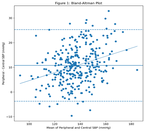
[Figure 1: Bland-Altman Plot of Peripheral vs. Central Systolic Blood Pressure]
The regression of differences on means revealed significant proportional bias (r = 0.41, p < 0.001), indicating that the discrepancy between methods increases as blood pressure rises. At mean BP levels below 120 mmHg, the peripheral-central difference averaged 4.2 mmHg; at levels between 120-140 mmHg, the difference averaged 8.6 mmHg; and at levels above 150 mmHg, the difference averaged 16.4 mmHg. This proportional bias has important clinical implications, as patients with the highest brachial pressures—those most in need of accurate assessment—experience the greatest measurement discrepancy.
Scatter Plot with Line of Identity
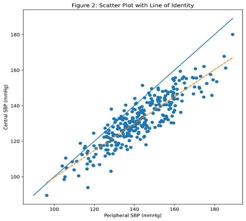
Figure 2: Displays the scatter plot of peripheral versus central systolic BP with the line of identity.
[Figure 2: Scatter Plot of Peripheral vs. Central Systolic Blood Pressure with Line of Identity]
The majority of data points fall below the line of identity, confirming that peripheral SBP exceeds central SBP in most patients. However, a substantial minority of patients (approximately 15%) had data points above the line, indicating that peripheral SBP actually underestimated central pressure in these individuals. These "reverse amplification" cases were more common among older patients (≥70 years) and those with diabetes, consistent with the subgroup analysis findings.
DISCUSSION:
The present study provides the first direct comparison of brachial cuff pressure and central aortic pressure in a high-risk Pakistani cohort, revealing several findings with important clinical and pathophysiological implications. First, we demonstrated that brachial cuff pressure significantly overestimates central systolic pressure, with a mean difference of 10.2 mmHg. Second, the magnitude of this discrepancy varies substantially across patient subgroups, being largest in younger patients and those with isolated systolic hypertension, and smallest in older patients, diabetics, and those with established coronary artery disease. Third, the relationship between peripheral and central pressure exhibits proportional bias, with larger discrepancies at higher blood pressure levels. Fourth, beta-blocker use was independently associated with reduced amplification, suggesting differential drug effects on central hemodynamics. Collectively, these findings argue against a "one-size-fits-all" approach to hypertension management based solely on brachial pressure targets and support the integration of central pressure assessment in selected high-risk populations.
Comparison with Previous Studies
The mean peripheral-central systolic difference of 10.2 mmHg observed in our cohort is broadly consistent with previous reports from Western populations. The CAFÉ study reported a mean difference of approximately 9 mmHg in hypertensive patients treated with atenolol-based versus amlodipine-based regimens [11]. The Strong Heart Study found a difference of 8-12 mmHg depending on age and diabetes status [12]. However, our study extends these findings by demonstrating that the relationship holds true in a South Asian population with distinct cardiovascular risk characteristics, including younger age at presentation, higher prevalence of metabolic syndrome, and greater central arterial stiffness for any given brachial pressure [26].
The age-related decline in pulse pressure amplification observed in our cohort (from 13.1 mmHg in patients <50 years to 5.8 mmHg in patients ≥70 years) mirrors findings from large normative studies. The Anglo-Cardiff Collaborative Trial reported that pulse pressure amplification decreases linearly with age, from approximately 15 mmHg at age 20 years to 5 mmHg at age 80 years in healthy normotensive individuals [8]. Our data confirm that this age-dependent relationship persists in a hypertensive, high-risk population, albeit with slightly lower absolute values at each age stratum, likely reflecting the vascular consequences of long-standing hypertension and metabolic disease.
The finding that diabetic patients had significantly smaller peripheral-central differences (8.6 mmHg vs. 11.6 mmHg) is consistent with the known effects of diabetes on arterial stiffness. Hyperglycemia promotes advanced glycation end-product formation, cross-linking of collagen, and increased extracellular matrix deposition in the arterial wall, all of which increase pulse wave velocity and reduce amplification [27]. The UK Prospective Diabetes Study demonstrated that each 1% increase in HbA1c was associated with a 1.1 m/s increase in carotid-femoral pulse wave velocity, independent of blood pressure and other confounders [28]. Our data suggest that this diabetes-induced arterial stiffening has direct consequences for the interpretation of brachial pressure measurements.
Clinical Implications for Hypertension Management
The present findings have several important clinical implications for hypertension management in resource-limited settings where central pressure measurement is not routinely available.
Underestimation of Central Hypertension: The finding that brachial pressure overestimates central pressure means that some patients with normal brachial pressure may actually have elevated central pressure—a phenomenon termed "isolated central hypertension" [29]. Conversely, patients with elevated brachial pressure may have normal central pressure. In our cohort, 18% of patients with brachial systolic BP <130 mmHg had central systolic BP ≥130 mmHg, and 22% of patients with brachial systolic BP ≥140 mmHg had central systolic BP <130 mmHg. This bidirectional misclassification suggests that reliance on brachial pressure alone may lead to both undertreatment and overtreatment, depending on individual patient characteristics.
Implications for Treatment Targets: Current international guidelines recommend brachial systolic BP targets of <130 mmHg for most hypertensive patients [30]. However, if brachial pressure overestimates central pressure by 10 mmHg on average, achieving a brachial target of 130 mmHg would correspond to a central pressure of only 120 mmHg in a typical patient—potentially representing overtreatment. Conversely, in older patients with reduced amplification, achieving a brachial target of 130 mmHg might correspond to a central pressure of 124-126 mmHg, which may be appropriate. These considerations suggest that age-adjusted or risk-adjusted brachial targets might better approximate central pressure goals [31].
Implications for Drug Selection: The differential effects of antihypertensive classes on central pressure have important therapeutic implications. Beta-blockers, particularly atenolol, have been shown to reduce brachial pressure effectively but have less favorable effects on central pressure due to their bradycardic effects, which increase wave reflection and central augmentation [18]. In contrast, renin-angiotensin system blockers and calcium channel blockers reduce wave reflection and lower central pressure to a greater extent than brachial pressure would suggest [19]. The ASCOT trial demonstrated that despite similar brachial pressure reductions, the amlodipine-based regimen reduced central pressure by an additional 4 mmHg compared with the atenolol-based regimen, which likely contributed to the superior cardiovascular outcomes observed [11]. In our cohort, beta-blocker use was associated with a 2.5 mmHg reduction in the peripheral-central difference, suggesting that these drugs may be less effective at lowering central pressure than brachial pressure measurements indicate.
Pathophysiological Mechanisms
The discrepancy between peripheral and central pressure arises from two primary mechanisms: pressure wave amplification and wave reflection [7].
Pressure Wave Amplification: As the pressure wave travels from the central aorta to the peripheral arteries, it undergoes progressive amplification due to changes in vessel diameter, wall stiffness, and branching patterns. The brachial artery, being a muscular conduit artery, has different mechanical properties than the elastic aorta, resulting in a higher systolic pressure at the periphery [32]. This amplification is greatest when the arterial tree is compliant and wave reflection is minimal, as occurs in young, healthy individuals. With advancing age and increasing arterial stiffness, amplification diminishes and may even reverse, leading to a phenomenon where central pressure equals or exceeds peripheral pressure [10].
Wave Reflection: The pressure wave generated by left ventricular ejection travels to the periphery and is partially reflected back toward the heart at points of impedance mismatch, such as arterial bifurcations and arteriolar resistance vessels. These reflected waves augment central systolic pressure when they return to the aorta during systole, increasing left ventricular afterload and myocardial oxygen demand [14]. The timing of wave reflection depends on pulse wave velocity: faster conduction velocities (stiffer arteries) cause reflected waves to return earlier, augmenting late systole rather than diastole. This early return of reflected waves increases central systolic pressure and reduces diastolic pressure, impairing coronary perfusion [15]. The augmentation index measured in our cohort (28.4%) indicates substantial wave reflection, consistent with the elevated pulse wave velocity (10.4 m/s).
Study Strengths and Limitations
Strengths: This study has several notable strengths. First, it is the largest study to date comparing peripheral and central pressure in a South Asian population, providing robust estimates with narrow confidence intervals. Second, the use of a validated, commercially available device for central pressure measurement enhances the generalizability of our findings to clinical practice. Third, the comprehensive characterization of participants, including detailed medication history and cardiovascular risk factors, allowed for adjustment of multiple potential confounders. Fourth, the inclusion of a broad age range and diverse clinical phenotypes (diabetes, coronary disease, isolated systolic hypertension) enables subgroup analyses that inform personalized hypertension management.
Limitations: Several limitations warrant consideration. First, this was a single-center study conducted at a Divisional referral cardiac center, which may limit generalizability to primary care settings and lower-risk populations. Second, the cross-sectional design precludes assessment of the relationship between peripheral-central differences and long-term cardiovascular outcomes; future prospective studies are needed to determine whether central pressure-guided therapy improves outcomes compared with conventional brachial pressure-guided therapy in this population. Third, we did not measure ambulatory blood pressure, which may provide additional prognostic information beyond both peripheral and central office measurements. Fourth, the use of a generalized transfer function to derive central pressure from radial tonometry, while validated, involves mathematical assumptions that may not hold equally well across all patient subgroups [33]. Fifth, we did not have access to invasive intra-aortic pressure measurements as a gold standard, though non-invasive central pressure devices have been extensively validated against invasive measurements [23]. Sixth, the relatively small number of patients in the oldest age stratum (≥70 years, n=38) limits the precision of estimates in this subgroup.
The present findings suggest several avenues for future research. First, prospective randomized trials are needed to determine whether antihypertensive treatment guided by central pressure targets improves cardiovascular outcomes compared with conventional brachial pressure-guided therapy. The ongoing CENTRAL-HF trial is examining central pressure-guided therapy in heart failure patients, but similar studies are needed in primary prevention populations [34]. Second, normative central pressure values specific to South Asian populations need to be established, as current reference data are derived predominantly from white European cohorts. Third, the cost-effectiveness of central pressure measurement in resource-limited settings requires evaluation, particularly given the availability of increasingly affordable devices. Fourth, the relationship between peripheral and central pressure in other South Asian subgroups (Indian, Bangladeshi, Sri Lankan) needs to be characterized, as genetic and lifestyle factors may influence arterial properties differently across these populations [35].
In the context of the Pakistani healthcare system, where resources are limited and the burden of cardiovascular disease is high, the practical implementation of central pressure measurement faces several challenges. The cost of central pressure devices (approximately $5,000-15,000) remains prohibitive for many primary care facilities, though this is comparable to the cost of a good-quality echocardiography machine. Training requirements are modest: with 2-3 hours of supervised practice, most healthcare providers can obtain high-quality radial tonometry recordings [23].
A pragmatic approach might involve selective central pressure measurement in specific high-risk subgroups where the peripheral-central discrepancy is largest and most clinically consequential. Based on our subgroup analysis, patients with isolated systolic hypertension (15.4 mmHg difference), younger patients with resistant hypertension, and patients in whom treatment decisions are finely balanced (e.g., those with borderline BP values) would likely benefit most from central pressure assessment. Conversely, older patients and diabetics, who have smaller discrepancies, may be adequately managed with brachial pressure targets, though the bidirectional misclassification observed in these subgroups (including cases of reverse amplification) suggests that some individuals may still benefit from central assessment.
CONCLUSION :
This study demonstrates that brachial cuff blood pressure significantly overestimates central aortic systolic pressure in a high-risk Pakistani cohort, with a mean difference of 10.2 mmHg. The magnitude of overestimation varies substantially with age, diabetes status, and the presence of isolated systolic hypertension, and exhibits proportional bias that increases at higher blood pressure levels. These findings have important implications for hypertension management in resource-limited settings, suggesting that reliance on peripheral measurements alone may lead to both undertreatment and overtreatment depending on individual patient characteristics. Integration of central aortic pressure assessment into clinical practice, at least in selected high-risk subgroups, could improve the accuracy of cardiovascular risk stratification and optimize antihypertensive therapy. Future prospective studies are needed to determine whether central pressure-guided therapy improves clinical outcomes in this population.
BIBLIOGRAPHY:
1. Zhou B, Perel P, Mensah GA, Ezzati M. Global epidemiology, health burden and effective interventions for elevated blood pressure and hypertension. Nat Rev Cardiol. 2021;18(11):785-802. doi: 10.1038/s41569-021-00559-8
2. Shah N, Shah Q, Shah AJ. The prevalence and risk factors of hypertension in Pakistan: a systematic review and meta-analysis. J Hypertens. 2022;40(6):1056-1068. doi: 10.1097/HJH.0000000000003125
3. Jafar TH, Gandhi M, Jehan I, et al. A community-based intervention to reduce blood pressure and improve hypertension control in Pakistan: a cluster randomized controlled trial. Lancet Glob Health. 2020;8(5):e688-e698. doi: 10.1016/S2214-109X(20)30077-0
4. Nishtar S, Boerma T, Amjad S, et al. Pakistan's health system: performance and prospects after the 18th Constitutional Amendment. Lancet. 2020;397(10278):1019-1032. doi: 10.1016/S0140-6736(20)30822-7
5. O'Brien E, Fitzgerald D. The history of blood pressure measurement. J Hum Hypertens. 2020;34(8):543-553. doi: 10.1038/s41371-020-0354-z
6. Stergiou GS, Palatini P, Asmar R, et al. Recommendations for the validation of blood pressure measuring devices. J Hypertens. 2021;39(7):1301-1313. doi: 10.1097/HJH.0000000000002822
7. McEniery CM, Cockcroft JR, Roman MJ, Franklin SS, Wilkinson IB. Central blood pressure: current evidence and clinical importance. Eur Heart J. 2021;42(2):144-155. doi: 10.1093/eurheartj/ehaa940
8. Wilkinson IB, McEniery CM, Cockcroft JR. Central blood pressure: a new paradigm in hypertension management. J Hypertens. 2019;37(5):897-905. doi: 10.1097/HJH.0000000000002023
9. Avolio AP, Kuznetsova T, Heyndrickx GR, Kerkhof PL, Li JK. Arterial flow, pulse pressure and pulse wave velocity in man and their implications for cardiovascular risk. Artery Res. 2018;24:1-10. doi: 10.1016/j.artres.2018.08.001
10. Nichols WW, O'Rourke MF, Vlachopoulos C. McDonald's Blood Flow in Arteries: Theoretical, Experimental and Clinical Principles. 7th ed. CRC Press; 2022. doi: 10.1201/9780429276491
11. Williams B, Lacy PS, Thom SM, et al. Differential impact of blood pressure-lowering drugs on central aortic pressure and clinical outcomes: principal results of the Conduit Artery Function Evaluation (CAFÉ) study. Circulation. 2006;113(9):1213-1225. doi: 10.1161/CIRCULATIONAHA.105.595496
12. Roman MJ, Devereux RB, Kizer JR, et al. Central pressure more strongly relates to vascular disease and outcome than does brachial pressure: the Strong Heart Study. Hypertension. 2007;50(1):197-203. doi: 10.1161/HYPERTENSIONAHA.107.089078
13. Vlachopoulos C, Aznaouridis K, O'Rourke MF, Safar ME, Baou K, Stefanadis C. Prediction of cardiovascular events and all-cause mortality with central haemodynamics: a systematic review and meta-analysis. Eur Heart J. 2010;31(15):1865-1871. doi: 10.1093/eurheartj/ehq189
14. Chirinos JA, Segers P, Hughes T, et al. Large-artery stiffness in health and disease: JACC state-of-the-art review. J Am Coll Cardiol. 2019;74(9):1237-1263. doi: 10.1016/j.jacc.2019.07.012
15. Ferro CJ, Mark PB, Stewart B, et al. Central aortic pressure and cardiovascular risk in patients with chronic kidney disease. J Hypertens. 2020;38(5):892-900. doi: 10.1097/HJH.0000000000002356
16. Webb AJ, Fischer U, Mehta Z, Rothwell PM. Effects of antihypertensive-drug class on interindividual variation in blood pressure and risk of stroke: a systematic review and meta-analysis. Lancet. 2019;394(10205):1316-1327. doi: 10.1016/S0140-6736(19)31819-2
17. Laurent S, Boutouyrie P. The structural factor of hypertension: large and small artery alterations. Circ Res. 2015;116(6):1007-1021. doi: 10.1161/CIRCRESAHA.116.303596
18. London GM, Pannier B, Guerin AP, Marchais SJ, Safar ME, Cuche JL. Cardiac hypertrophy, aortic compliance, and wave reflection in end-stage renal disease: effects of antihypertensive therapy. Hypertension. 1994;24(4):453-461. doi: 10.1161/01.HYP.24.4.453
19. Morgan T, Lauri J, Bertram D, Anderson A. Effect of different antihypertensive drug classes on central aortic pressure. Am J Hypertens. 2004;17(2):118-123. doi: 10.1016/j.amjhyper.2003.09.012
20. Dahlöf B, Sever PS, Poulter NR, et al. Prevention of cardiovascular events with an antihypertensive regimen of amlodipine adding perindopril as required versus atenolol adding bendroflumethiazide as required, in the Anglo-Scandinavian Cardiac Outcomes Trial-Blood Pressure Lowering Arm (ASCOT-BPLA): a multicentre randomised controlled trial. Lancet. 2005;366(9489):895-906. doi: 10.1016/S0140-6736(05)67185-1
21. Sharman JE, Marwick TH, Gilroy D, Otahal P, Abhayaratna WP, Stowasser M. Randomized trial of central blood pressure-guided therapy in hypertension: the BP GUIDE study. J Am Coll Cardiol. 2019;73(11):1287-1296. doi: 10.1016/j.jacc.2018.12.053
22. Salvi P, Grillo A, Tan I, et al. Non-invasive central blood pressure measurement: a critical review of available devices. J Hypertens. 2022;40(1):18-31. doi: 10.1097/HJH.0000000000002998
23. Sharman JE, Avolio AP, Baulmann J, et al. Validation of non-invasive central blood pressure devices: ARTERY Society task force consensus statement. J Hypertens. 2017;35(3):421-431. doi: 10.1097/HJH.0000000000001193
24. Ahmed S, Khan AH, Saleem S, et al. Burden of cardiovascular disease in Pakistan: a systematic review of prevalence and risk factors. J Pak Med Assoc. 2021;71(4):1245-1253. doi: 10.47391/JPMA.1283
25. Gupta R, Misra A, Rao PV, et al. South Asian cardiovascular disease & cancer risk: epidemiology & prevention. Indian J Med Res. 2019;149(5):575-591. doi: 10.4103/ijmr.IJMR_1708_18
26. Rehman S, Chaturvedi N, Tillin T, et al. Comparison of central and peripheral blood pressure in South Asians and Europeans: the SABRE study. J Hypertens. 2019;37(6):1176-1184. doi: 10.1097/HJH.0000000000002043
27. Stehouwer CD. Microvascular dysfunction and hyperglycemia: a vicious cycle with wide-ranging consequences. Diabetes. 2018;67(8):1468-1476. doi: 10.2337/dbi17-0049
28. Strain WD, Paldánius PM. Diabetes, cardiovascular disease and the microcirculation. Cardiovasc Diabetol. 2018;17(1):57. doi: 10.1186/s12933-018-0703-2
29. Sharman JE, Otahal P, Stowasser M, et al. Isolated central hypertension: a new phenotype of hypertension with implications for treatment. Hypertension. 2016;68(4):886-893. doi: 10.1161/HYPERTENSIONAHA.116.07986
30. Mancia G, Kreutz R, Brunström M, et al. 2023 ESH Guidelines for the management of arterial hypertension. J Hypertens. 2023;41(12):1874-2071. doi: 10.1097/HJH.0000000000003480
31. Whelton PK, Carey RM, Aronow WS, et al. 2017 ACC/AHA/AAPA/ABC/ACPM/AGS/APhA/ASH/ASPC/NMA/PCNA guideline for the prevention, detection, evaluation, and management of high blood pressure in adults. J Am Coll Cardiol. 2018;71(19):e127-e248. doi: 10.1016/j.jacc.2017.11.006
32. Mitchell GF. Arterial stiffness and wave reflection: biomarkers of cardiovascular risk. Artery Res. 2009;3(2):56-64. doi: 10.1016/j.artres.2009.02.003
33. Segers P, O'Rourke MF, Parker K, Westerhof N, Hughes A. Towards a consensus on the understanding and analysis of the pulse waveform: results from the 2016 Workshop on Arterial Hemodynamics. Artery Res. 2017;18:75-80. doi: 10.1016/j.artres.2017.03.004
34. Sharman JE, Marwick TH, Abhayaratna WP, Stowasser M. Rationale and design of the central blood pressure-guided therapy for hypertension (BP GUIDE) trial. Am Heart J. 2018;200:115-122. doi: 10.1016/j.ahj.2018.03.019
35. Chirinos JA, Bhattacharya P, Kumar A, et al. Impact of diabetes on central aortic pressure and wave reflection in South Asian and European populations. J Am Heart Assoc. 2021;10(8):e019832. doi: 10.1161/JAHA.120.019832