Deer Velvet Antler Improves Histopathological Changes of Allergic Airway Inflammation in Guinea pigs
- Javeria Sarfraz , Associate Professor, Pharmacology, Allama Iqbal Medical College Lahore, Pakistan
- Syeda Tahira Zaidi , Assistant Professor, Pharmacology, King Edward Medical University Lahore, Pakistan
Article Information:
Abstract:
As many patients all over world experience uncontrolled asthma despite of using recommended medicines hence majority patients start looking for other alternatives or adjuvant therapies for their relief. Deer velvet antler is an old traditional animal based medicine which is capable to reduce expression of many cytokines involved in pathogenesis of asthma. Objectives: To assess the effect of deer velvet antler on histopathology of lungs in guinea pigs sensitized by ovalbumin and its comparison with dexamethasone. Materials and Methods: It was an experimental study. Animals were placed in four groups with six in each. Animals in group I and II were taken as negative and positive control and animals in groups III and IV were treated with deer velvet antler powder and dexamethasone respectively. Negative control was sham sensitized and challenged. Groups II, III and IV were sensitized with ovalbumin on days 0 and 14 then they were repeatedly challenged on days 25, 26 and 27 with ovalbumin. Animals in group III were given deer velvet antler powder by gavage one hour before each challenge and group four animals were given dexamethasone one hour before each challenge. On 28th day was animals were sacrificed and sections of lung tissue was taken, slides were prepared and evaluated for lung inflammation. Results: Comparing the histological alterations showed that the OVA group had a substantially larger (p-value ≤ 0.01) infiltration of inflammatory cells in the lung airways than the normal control group. Total lung inflammation decreased in the OVA plus DVA AND OVA plus DEXA groups (p value ≤ 0.01 vs OVA group) equally . While dexamethasone demonstrated a substantial improvement in comparison to the OVA group (p-value ≤ 0.01), the effect of DVA on epithelial alterations was nonsignificant (p-value 0.338) vs. OVA group. Nevertheless, there was no significant difference between DVA and dexamethasone treatments (p-value 0.06).Conclusion: Deer velvet antler decreases allergic airway inflammation.
Keywords:
Article :
INTRODUCTION :
Bronchial asthma is a chronic airway disorder. This serious health issue is present in 5 to 10 % people of all ages.1It is associated with repeated episodes of wheezing, shortness of breath, and coughing, especially at night or in the evening. These symptoms occur alone or in combination. Sometimes these symptoms are relieved spontaneously, but they usually require treatment. Asthmatic patients experience a great number of missed work and school days because of these symptoms.2
The prevalence, morbidity, mortality, and economic burden associated with asthma have increased markedly throughout the world. 3According to the WHO, an estimated 300 million people of all ages are suffering from asthma, and approximately 250,000 people die from asthma every year.4 Asthma has been divided into two categories: Atopic asthma initiated by inhalation of an allergen, which leads to production of Ig-E antibodies and Non-atopic asthma occurs in individuals who are non-allergic and it is non Ig-E mediated.5
Despite better understanding of asthma pathology and adherence to recommended treatments still many patients are experiencing uncontrolled asthma, these medicines especially steroids on chronic use develop serious side effects. Hence, it is is important to find alternative treatments.6,7
Given these facts,there is a need to develop a better and more effective drug with minimum cost and adverse effects as asthma and allergy are life long diseases.
For hundreds of years, people in China have utilized deer velvet antler that is a cartilaginous tissue without full calcification and being used as a medicinal herb and functional food to support general health maintenance8. Today, pharmacopeias from China, Korea, Japan, and Taiwan acknowledge velvet antler as a traditional medicine. Additionally, pharmacological activity related to immunomodulatory 9, anti-inflammatory 10 antiaging11, wound-healing12, and anticancer effects has been identified for velvet antler.13
So far, 416 distinct proteins have been identified in DVA, for example, vimentin , β 3 subtype of hemoglobin, antimicrobial peptide, peptidoglycan recognition proteins, and pre-pro serum albumin.14 Regarding role of deer velvet antler in asthma, immunomodulatory effects of DVA have been reported in other studies. Aqueous extract of DVA has played a role in inhibiting T-cell proliferation, differentiation, and cytokine secretion after antigenic stimulation in vitro. It has also inhibited the release of IL-β1 and TNF-α from macrophages in mice.15
It has inhibitory effects on the expression of many cytokines which are involved in pathogenesis of different TH2 mediated disease including asthma.16 it was demonstrated in a study that prevention and alleviation of asthmatic symptoms after administration of DVA depend on elevation of Th1 and Treg cytokines and inhibition of Th2 and Th17 cytokines and Ig-E production and is able to regulate Th1/Th2 balance.17
The role of DVA to protect and control allergic diseases like asthma has not been extensively studied. So, this study was designed to provide scientific basis for traditional use of DVA and to compare its anti inflammatory effects with dexamethasone.
METHODOLOGY :
Guinea pigs were purchased from Zoo Park, Lahore and were kept in the animal house of Post Graduate Medical Institute for one week for acclimatization at temperature of 22-24 , kept under natural light and dark cycle and were fed with food (vegetables, fruits and chick pea) and water.
Animals were numbered from 1 to 24 and assigned randomly to groups I, II, III and IV, 6 in each group, by simple lottery method. During the study, Ovalbumin (OVA) (Sigma Aldrich, Poole UK), aluminum hydroxide (alum) (Bio sector, Denmark), phosphate buffer saline (PBS) (Sigma Aldrich, Germany), Deer velvet antler (Herb show Bio-technology co., Ltd Shangai, China), dexamethasone (Unexo Labs (PVT) LTD Pakistan),Gum tragacanth (local market) Hematoxylin and Eosin stain (Medline diagnostic, Lahore, Pakistan) were used (Table 1).
Animals in three groups aside from the normal control group were subjected to standard methodology18 to produce airway inflammation (Figure 1).
Table 1. Study protocol for preparation of model and treatment of groups.
|
Group |
Sensitization Day 0 and 14 Intraperitoneal |
Challenge Day 25-27 Inhalation |
Treatment 1 hour before each challenge Orally |
|
Negative control |
PBS 0.5 ml |
PBS |
Gum tragacanth in DW (4.2ml ) |
|
Positive control |
OVA 0.5 ml (100 µg) + alum 1 ml (20 mg) in PBS18 |
1% OVA in PBS18 |
Gum tragacanth in DW (4.2ml ) |
|
DVA treated gp |
OVA 0.5 ml (100 µg) + alum 1 ml (20 mg) in PBS |
1% OVA in PBS |
DVA in gum tragacanth (190 mg/kg)19 |
|
Dexa treated gp |
OVA 0.5 ml (100 µg) + alum 1 ml (20 mg) in PBS |
1% OVA in PBS |
Dexamethasone in gum tragacanth (20 mg/kg)20 |
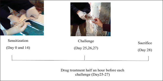
Figure 1. Protocol flow diagram of the study.
Lung Histopathology
The lungs were dissected, the animals were killed under chloroform anesthesia, and they were fixed in a 10% formalin solution for 24 hours to create cassettes. To ascertain the lung morphology and level of inflammation, samples were cut into 5 µm slices after tissue processing, placed on glass slides, and stained with eosin and hematoxylin. Lung inflamation parameters (alveoler septal infiltrates, perivascular inflitrates and combined bronchus associated lymphoid tissue hyperplasia and peribrochial
inflitrates) were scored as follows.
Score 0, for no inflamation
Score 1, for occasional cuffing with inflamatory cells (minimal)
Score 2, when most bronchus or vessels were surrounded by thin layer of 1-2 inflamatory cell layer (mild)
Score 3,when most bronchus or vessels were surrounded by thin layer of 3-4 inflamatory cell layer (moderate)
Score 4,when most bronchus or vessels were surrounded by thick layer more than 5 inflamatory cell layers (severe)
The total lung inflammation was calculated by the addition of alveolar septal, perivascular, combined bronchus associatedlymphoid tissue hyperplasia and peribronchial inflammation scores for each animal .21
Airway epithelial desquamation was scored according to the fallowing criteria
Score 0, normal intact airway epithelium.
Score 1, elongation and distortion of the cuboidal and columnar epithelial cells lining the airways.
Score 2, elongation associated with infolding of the epithelium and narrowing of airway lumen.
Score 3, loss of epithelial cells resulting in broken airways.22
Data Analysis:
SPSS 20 was used to enter and evaluate the data. The histology scores for epithelial alterations and inflammatory states were recorded, together with their frequencies and probabilities. Moreover, the results were compared between groups using the Kruskal-Wallis analysis of variance (ANOVA). The post hoc analysis was conducted using the Mann-Whitney U test.
RESULTS:
Table 2 presents the histological scores of lung inflammation and epithelial alterations in guinea pigs across different study groups. In negative group, no signs of perivascular, peribronchial, or alveolar septal infiltrates, nor bronchial epithelial changes, were observed. In contrast, the positive group exhibited moderate to severe perivascular, peribronchial, and alveolar septal infiltrates, along with epithelial alterations such as ciliated cell damage, mucous goblet cell hyperplasia, and/or squamous metaplasia. The DVA treated group showed these changes at minimal to moderate levels, while in the dexa treated group, they ranged from minimal to mild.
Table 2: Histological parameters analyzed for NC, PC, DVA and DEXA treated groups.
|
Total lung inflammation |
|||||||||||||||||
|
|
|
NC |
PC |
DVA |
DEXA |
|
|||||||||||
|
|
Lesion score |
n |
% |
n |
% |
n |
% |
n |
% |
|
|||||||
|
Perivascular infiltrates
|
Normal (0) |
6 |
100.0 |
0 |
0.0 |
0 |
0.0 |
1 |
16.6 |
|
|||||||
|
Minimal (1) |
0 |
0.0 |
0 |
0.0 |
1 |
16.6 |
4 |
66.6 |
|
||||||||
|
Mild (2) |
0 |
0.0 |
0 |
0.0 |
4 |
66.6 |
1 |
16.6 |
|
||||||||
|
Moderate(3) |
0 |
0.0 |
2 |
33.3 |
1 |
16.6 |
0 |
0.0 |
|
||||||||
|
Severe (4) |
0 |
0.0 |
4 |
66.6 |
0 |
0.0 |
0 |
0.0 |
|
||||||||
|
Peribronchial Infiltrates and BALT hyperplasia
|
Normal (0) |
6 |
100.0 |
0 |
0.0 |
0 |
0.0 |
1 |
16.6 |
|
|||||||
|
Minimal (1) |
0 |
0.0 |
0 |
0.0 |
3 |
50 |
5 |
83.3 |
|
||||||||
|
Mild (2) |
0 |
0.0 |
0 |
0.0 |
2 |
33.3 |
0 |
0.0 |
|
||||||||
|
Moderate (3) |
0 |
0.0 |
2 |
33.3 |
1 |
16.6 |
0 |
0.0 |
|
||||||||
|
Severe (4) |
0 |
0.0 |
4 |
66.6 |
0 |
0.0 |
0 |
0.0 |
|
||||||||
|
Alveolar septal infiltrates
|
Normal (0) |
6 |
100.0 |
0 |
0.0 |
0 |
0.0 |
0 |
0.0 |
|
|||||||
|
Minimal (1) |
0 |
0.0 |
0 |
0.0 |
1 |
16.6 |
2 |
33.3 |
|
||||||||
|
Mild (2) |
0 |
0.0 |
0 |
0.0 |
4 |
66.6 |
3 |
50 |
|
||||||||
|
Moderate(3) |
0 |
0.0 |
1 |
16.6 |
1 |
16.6 |
1 |
16.6 |
|
||||||||
|
Severe (4) |
0 |
0.0 |
5 |
83.3 |
0 |
0.0 |
0 |
0.0 |
|
||||||||
|
Airway epithelial desquamation |
|||||||||||||||||
|
Epithelial damage and narrowing
|
|
NC |
PC |
DVA |
DEXA |
|
|||||||||||
|
Lesion score |
n |
% |
n |
% |
n |
% |
n |
% |
|
||||||||
|
Normal (0) |
6 |
100.0 |
0 |
0.0 |
0 |
0.0 |
1 |
16.6 |
|
||||||||
|
Mild (1) |
0 |
0.0 |
0 |
0.0 |
2 |
33.3 |
4 |
66.6 |
|
||||||||
|
Moderate (2) |
0 |
0.0 |
5 |
83.3 |
3 |
50 |
1 |
16.6 |
|
||||||||
|
Severe (3) |
0 |
0.0 |
1 |
16.6 |
1 |
16.6 |
0 |
0.0 |
|
||||||||
|
|
|||||||||||
|
|
Figure 2a. Photomicrographs of (H & E stain, 400×). stained lung tissue samples from NC, PC, DVA and DEXA treated groups for assessment of lung inflammation in (a) bronchial, (b) peri bronchial, (c) alveolar septal, and (d) perivascular inflammation .
|
|
||||
|
|
Fig 2b:Photomicrographs of (H & E stain, 400×). stained lung tissue samples from NC, PC, DVA and DEXA treated groups for assessment of lung inflammation in perivascular area.
Overall histological scores for total lung inflammation and epithelial damage and narrowing were generated by taking means of individual lesion scores for each guinea pig of groups PC, DVA and DEXA respectively (Table 3a). NC group demonstrated no inflammation normal bronchial epithelium architecture (lesion score =0)
Table 3 a: Effect of deer velvet antler and dexametasone on total lung inflammation of ovalbumin sensitized guinea pigs (n=6).
|
Group |
Mean |
SD |
SE |
|
NC |
.000 |
.000 |
.000 |
|
PC |
3.71 |
0.44 |
0.18 |
|
DVA |
1.86 |
0.66 |
0.27 |
|
DEXA |
1.22 |
0.54 |
0.22 |
NC= negative control group, PC= positive control group, DVA=deer velvet antler and DEXA= dexamethasone treatment group.
Table 3b: Effect of deer velvet antler and dexametasone on bronchial epithelial damage of ovalbumin sensitized guinea pigs (n=6).
|
Group |
Mean |
SD |
SE |
|
NC |
.000 |
.000 |
.000 |
|
PC |
2.16 |
0.40 |
0.16 |
|
DVA |
1.83 |
0.75 |
0.30 |
|
DEXA |
1 |
0.63 |
0.25 |
NC= negative control group, PC= positive control group, DVA=deer velvet antler and DEXA= dexamethasone treatment group.
When comparison of histological parameters was performed among groups by using Kruskal Wallis ANOVA, it was found that the difference was significant for total lung inflammation and bronchial epithelial damage and narrowing, all with p values 0.000 and 0.001(Table 4).
Table 4: Comparison of histological parameters in four study groups.
|
|
Groups
|
n |
Mean Rank |
Kruskal Wallis ANOVA |
||
|
Chi-square |
Df |
P-value |
||||
|
Total lung inflammation |
NC |
6 |
3.50 |
20.53 |
3 |
0.000 |
|
PC |
6 |
21.4 |
|
|
|
|
|
DVA |
6 |
14.3 |
|
|
|
|
|
DEXA |
6 |
10.7 |
|
|
|
|
|
Total |
24 |
|
|
|
|
|
|
Epithelial damage and narrowing
|
NC |
6 |
4.0 |
17.46 |
3 |
0.001 |
|
PC |
6 |
18.9 |
|
|
|
|
|
DVA |
6 |
16.4 |
|
|
|
|
|
DEXA |
6 |
10.6 |
|
|
|
|
|
Total |
24 |
|
|
|
|
|
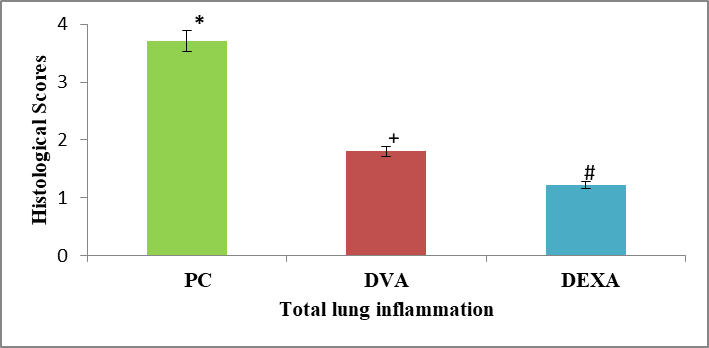
Fig 3 a: Effect of deer velvet antler and dexamethasone on total lung inflammation (Mean±SE) of ovalbumin sensitized guinea pigs (n=6). NC group demonstrated no inflammation (lesion score =0).
PC= positive control group, DVA= deer velvet antler and DEXA= dexamethasone treatment groups.
* p value ≤ 0.01, + p value ≤ 0.01 vs PC and #p value ≤ 0.01 vs PC
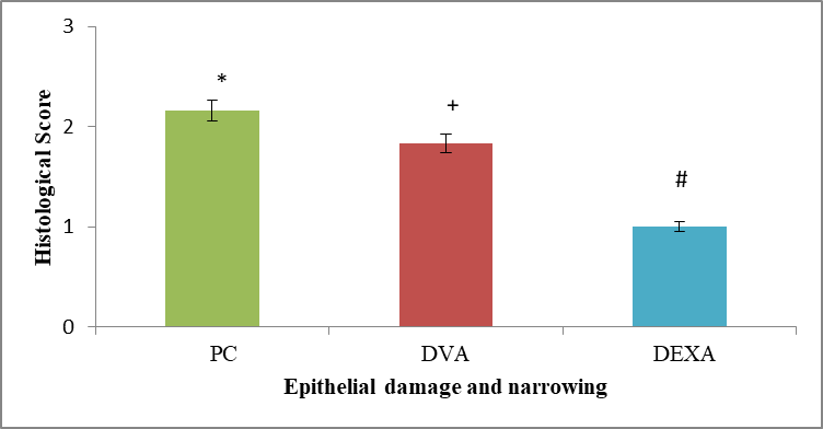
Fig 3 b: Effect of deer velvet antler and dexamethasone on epithelial damage and narrowing (Mean±SE) of ovalbumin sensitized guinea pigs (n=6). NC group demonstrated normal bronchial epithelium architecture (lesion score =0).
PC= positive control group, DVA= deer velvet antler and DEXA= dexamethasone treatment groups.
* p value ≤ 0.001, + Not Significant difference vs PC and # p value ≤ 0.05 vs PC
When pair wise comparison was evaluated for total lung inflammation by using Mann Whitney U Test, it was observed that group PC, DVA and DEXA had significantly increased total lung inflammation as compared to group NC with p values 0.002 in each case. DVA treated group had shown reduced total lung inflammation when compared with PC group with p value 0.004. Whereas DEXA treated group had significantly low inflammation as compared to PC group with p value 0.003. DVA treated group showed less reduction in inflammation score as compared to
DEXA group but difference was insignificant with p value 0.08. (Table 15)
When pair wise comparison was evaluated for airway epithelial damage and narrowing by using Mann Whitney U test, it was observed that group PC, DVA and DEXA had significantly enhanced airway epithelial damage and narrowing as compared to NC with p values 0.001, 0.002 and 0.006 respectively. DVA treated group had insignificantly reduced airway epithelial damage and narrowing when compared with PC group with p value 0.338. DEXA treated group reduced airway epithelial damage and narrowing when compared with PC group with p value 0.007. DVA treated group showed less reduction in airway epithelial damage and narrowing as compared to DEXA group but difference was insignificant with p value 0.067. (Table 5)
Table 5: Pair wise comparison of histological parameters in four study groups.
|
Histological parameter |
(I) group |
(J) group |
Mann Whitney U |
Z |
p-value |
|
Total lung inflammation |
NC |
PC |
0.000 |
- 3.14** |
0.002 |
|
DVA |
0.000 |
-3** |
0.002 |
||
|
DEXA |
0.000 |
-3.1** |
0.002 |
||
|
PC |
DVA |
0.500 |
-2.8** |
0.004 |
|
|
DEXA |
0.000 |
-2.9** |
0.003 |
||
|
DVA |
DEXA |
7.50 |
-1.7 |
0.08 |
|
|
Epithelial damage and narrowing
|
NC |
PC |
.000 |
-3.20*** |
0.001 |
|
DVA |
.000 |
-3.1** |
0.002 |
||
|
DEXA |
3.00 |
-2.73* |
0.006 |
||
|
PC |
DVA |
13 |
-0.95 |
0.338 |
|
|
DEXA |
2.50 |
-2.7* |
0.007 |
||
|
DVA |
DEXA |
7.50 |
-1.8 |
0.067 |
*** p value ≤ 0.001,** p value ≤ 0.01 and*p-value ≤ 0.05.
DISCUSSION :
Because conventional asthma treatments often have side effects and may not be effective for many patients, there is increasing interest in safe, affordable, and scientifically validated herbal alternatives.23 An experimental model of induced asthma was established in guinea pigs to evaluate the beneficial effects of DVA on airway inflammation. Histological examination of lung tissue revealed that DVA treatment produced an improvement in lung inflammation comparable to that of dexamethasone, although the reduction in epithelial damage was less pronounced.
The study included four groups, with a normal control group serving as the reference. The other three groups were sensitized using ovalbumin (OVA) and alum. Among these, the OVA group received distilled water as a placebo, the drug control group was administered the standard drug dexamethasone for comparison, and the experimental group was treated with DVA. Sensitization with OVA led to marked infiltration of inflammatory cells and epithelial alterations in the airway mucosa, a well-established method for inducing airway inflammation.24
In this study, histological analysis of lung tissues revealed that inflammatory cells infiltration and airway epithelial desquamation was significantly increased in the sensitized, non treated group (PC) when compared with NC with p values 0.01 and 0.001 respectively. These histopathological parameters were absent in the NC. These observations are consistent with previous study that have shown increased number of inflammatory cells in the lung tissue and enhanced bronchial epithelial destruction after sensitization and challenge with ovalbumin in murine model of allergic asthma,25 these observations are considered as a sign of successful sensitization.
Administration of DVA to guinea pigs markedly ameliorated airway inflammation, inflammatory cells were reduced in lung tissues compared with the PC group with p value 0.01 but bronchial epithelial injury was reduced to lesser extent with p value 0.33.
Kuo et al., 17 had shown that DVA administration in asthmatic murine model significantly reduced infiltration of inflammatory cells in the lung connective tissue with p value 0.001,this statistical difference with the present study is may be due to differences in duration of treatment.
Yang et al., studied the protective effects of herba houttuyniae aqueous extract against ova‐induced inflammation in asthmatic mice and observed that airway inflammation scores were declined as compared to sensitized group with p value 0.05.27
In contrast to previously published studies that demonstrated statistically significant reductions in total lung inflammation (p < 0.01), the findings of the present study did not reveal a significant difference between DVA and dexamethasone treatment groups.28,29
Different studies proposed multiple possible anti-inflammatory mechanisms of DVA as mentioned in the literature review. Deer velvet antler has been used for decades in the treatment of various diseases, but there is still no definitive information about the active biochemical component that performs the main role in various pharmacological activities. In one study, 3.2 kDa polypeptide was isolated from DVA extracts, and in vitro analysis determined that this component enhanced natural killer cell activity, and cell-mediated and humoral immunity. This polypeptide promoted the secretion of Th1 cytokines like IL-2, TNF-α, IL-12 and IFN-γ, and decreased the secretion of Th2 cytokines (IL-4 and IL-10). This polypeptide could be useful for TH2 mediated diseases like multiple allergies including asthma.16
In conclusion, both DVA and dexamethasone demonstrated comparable effects on total lung inflammation; however, DVA showed no significant impact on epithelial alterations. These findings suggest that DVA may serve as a potential therapeutic agent for reducing lung inflammation in experimental asthma models.
CONCLUSION :
DVA demonstrated an improvement in lung inflammation in an experimental model of allergic airway inflammation, showing effects comparable to those of dexamethasone. However, its ameliorative impact on alveolar and bronchial epithelial cells was less pronounced than that observed with dexamethasone. These findings suggest that DVA possesses promising anti-inflammatory potential for asthma management, warranting further investigation.
BIBLIOGRAPHY:
1. World Health Organization. Fact sheets: asthma. WHO. 2021. Available from: https://www.who.int/news-room/fact-sheets/detail/asthma
2. Asthma and Allergy Foundation of America. Asthma review. AAFA. 2021. Available from: https://www.aafa.org/asthma.aspx
3. Mattiuzzi C, Lippi G. Worldwide asthma epidemiology: insights from the global health data exchange database. Int Forum Allergy Rhinol. 2020;10(1):75–80. doi:10.1002/alr.22464
4. World Health Organization. Chronic respiratory diseases: asthma. WHO. 2020. Available from: https://www.who.int/news-room/q-a-detail/asthma
5. King GG, James A, Harkness L, Wark PA. Pathophysiology of severe asthma: we’ve only just started. Respirology. 2018;23(3):262–271. doi:10.1111/resp.13251
6. Reddel HK, Bacharier LB, Bateman ED, Brightling CE, Brusselle GG, Buhl R, et al. Global initiative for asthma strategy 2021: executive summary and rationale for key changes. Am J Respir Crit Care Med. 2022;205(1):17–35. doi:10.1164/rccm.202109-2205PP
7. Neffen H, Chahuàn M, Hernández DD, Vallejo-Perez E, Bolivar F, Sánchez MH, et al. Key factors associated with uncontrolled asthma: the asthma control in Latin America study. J Asthma. 2020;57(2):113–122.
8. Wu F, Li H, Jin L, et al. Deer antler base as a traditional Chinese medicine: a review of its traditional uses, chemistry and pharmacology. J Ethnopharmacol. 2013;145(2):403–415. doi:10.1016/j.jep.2012.12.008
9. Hung YK, Ho ST, Kuo CY, Chen MJ. In vitro effects of velvet antler water extracts on barrier integrity in Caco-2 cells. Int J Med Sci. 2021;18(8):1778.
10. Lee SH, Yang HW, Ding Y, Wang Y, Jeon YJ, Moon SH, et al. Anti-inflammatory effects of enzymatic hydrolysates of velvet antler in RAW 264.7 cells and zebrafish model. EXCLI J. 2015;14:1122–1132.
11. Kim HK, Kim MG, Leem KH. Comparison of the effect of velvet antler from different sections on longitudinal bone growth of adolescent rats. Evid Based Complement Alternat Med. 2016;2016:1927534. doi:10.1155/2016/1927534
12. Zang ZJ, Tang HF, Tuo Y, Xing WJ, Ji SY, Gao Y, Deng CH. Effects of velvet antler polypeptide on sexual behavior and testosterone synthesis in aging male mice. Asian J Androl. 2016;18(4):613–619.
13. Huo YS, Huo H, Zhang J. The contribution of deer velvet antler research to modern biological medicine. Chin J Integr Med. 2014;20(10):723–728. doi:10.1007/s11655-014-1808-7
14. Sun H, Xiao D, Liu W, Li X, Lin Z, Li Y, Ding Y. Well-known polypeptides of deer antler velvet with key actives: modern pharmacological advances. Naunyn Schmiedebergs Arch Pharmacol. 2024;397(1):15–31. doi:10.1007/s00210-023-02558-z
15. Kim YK, Kim KS, Chung KH, Kim JG, Kim KS, Lee YC, Chang YC, Kim CH. Inhibitory effects of deer antler aqua-acupuncture on type II collagen-induced arthritis in rats. Int Immunopharmacol. 2003;3(7):1001–1010.
16. Enhui Z, Xingxia L, Dandan L, Xuesong G, Shenyang G, Xiqing Y. Immunomodulatory effects of a 3.2 kDa polypeptide from velvet antler of Cervus nippon temminck. Int Immunopharmacol. 2013;16(2):210–213.
17. Kuo CY, Wang T, Dai TY, Wang CH, Chen KN, Chen YP, Chen MJ. Effect of the velvet antler of Formosan sambar deer on prevention of allergic airway response in mice. Evid Based Complement Alternat Med. 2012;2012:481318. doi:10.1155/2012/481318
18. Kwon Y, Sohn S, Lee G, Kim Y, Lee H, Shin M, Bae H. Electroacupuncture attenuates ovalbumin-induced allergic asthma via modulating CD4⁺CD25⁺ regulatory T cells. Evid Based Complement Alternat Med. 2012;2012:647308. doi:10.1155/2012/647308
19. Dai T, Wang C, Chen K, Huang I, Hong W, Wang S, Chen Y, Kuo C, Chen M. Anti-infective effects of velvet antler of Formosan sambar deer on Staphylococcus aureus-infected mice. Evid Based Complement Alternat Med. 2011;2011:534069.
20. Lowe AP, Thomas RS, Nials AT, Kidd EJ, Broadley KJ, Ford WR. Route of administration affects corticosteroid sensitivity of a combined ovalbumin and lipopolysaccharide asthma model in guinea pigs. J Pharmacol Exp Ther. 2017;362(2):327–337. doi:10.1124/jpet.117.241927.
21. Kumar M, Ahmad T, Sharma A, Mabalirajan U. Let-7 microRNA-mediated regulation of IL-13 and allergic airway inflammation. J Allergy Clin Immunol. 2011;128(5):1077-1085.
22. Nandedkar SD, Feroah TR, Hutchins W, Weihrauch D, Konduri KS, Wang G, Strunk RC, DeBaun MR, Hillery CA, Pritchard KA. Histopathology of experimentally induced asthma in a murine model of sickle cell disease. Blood. 2008;112(6):2529-2538.
23. Amaral-Machado L, Oliveira WN, Moreira-Oliveira SS, Pereira DT, Alencar EN, Tsapis N, et al. Use of natural products in asthma treatment. Evid Based Complement Alternat Med. 2020;2020:1021258. doi:10.1155/2020/1021258
24. Mincham KT, Scott NM, Lauzon-Joset JF, Leffler J, Stumbles PA, Holt PG, et al. Early life ovalbumin sensitization and aerosol challenge for induction of allergic airway inflammation in a BALB/c murine model. Bio Protoc. 2019;9(5):e3181. doi:10.21769/BioProtoc.3181
25. Vasconcelos LH, Silva MD, Costa AC, de Oliveira GA, de Souza IL, Queiroga FR, et al. A guinea pig model of airway smooth muscle hyperreactivity induced by chronic allergic lung inflammation: contribution of epithelium and oxidative stress. Front Pharmacol. 2019;9:1547. doi:10.3389/fphar.2018.01547
26. Sakat MS, Kilic K, Kandemir FM, Yildirim S, Sahin A, Kucukler S, et al. The ameliorative effect of berberine and coenzyme Q10 in an ovalbumin-induced allergic rhinitis model. Eur Arch Otorhinolaryngol. 2018;275(10):2495-2505. doi:10.1007/s00405-018-5104-3
27. Yang Y, Lai Q, Wang C, Zhou G. Protective effects of Herba Houttuyniae aqueous extract against OVA-induced airway hyperresponsiveness and inflammation in asthmatic mice. Evid Based Complement Alternat Med. 2022;2022:7609785. doi:10.1155/2022/7609785
28. Antwi AO, Obiri DD, Osafo N. Stigmasterol modulates allergic airway inflammation in a guinea pig model of ovalbumin-induced asthma. Mediators Inflamm. 2017;2017:2953930. doi:10.1155/2017/2953930
29. Antwi AO, Ekuadzi E, Boakye-Gyasi E, Agbettor ON, Opare CA, Atobrah R, et al. Pseudocedrela kotschyi limits airway inflammation in ovalbumin-induced asthma in guinea pigs. Sci Afr. 2021;14:e01023. doi:10.1016/j.sciaf.2021.e01023