VARIOUS RECONSTRUCTION OPTIONS AVAILABLE FOR POST TRAUMATIC MAXILLARY DEFECTS: A RETROSPECTIVE STUDY
- Shalini N , Undergraduate , Saveetha Dental College and Hospitals, Saveetha Institute of Medical and Technical Sciences, Saveetha University,Chennai, Tamil Nadu, India
- Kathiravan Selvarasu , Professor, Department of Oral and maxillofacial surgery, Saveetha Dental College and Hospital, Saveetha Institute of Medical and Technical Sciences Saveetha University, Chennai, Tamil Nadu , India.
- Kalyani P , Senior Lecturer, Department of Oral and maxillofacial surgery, Saveetha Dental College and Hospital, Saveetha Institute of Medical and Technical SciencesSaveetha University, Chennai, Tamil Nadu, India
Article Information:
Abstract:
Post-traumatic maxillary defects pose significant challenges due to their impact on function, esthetics, and quality of life. These defects commonly result from high-impact injuries, leading to oronasal communication and facial deformity. Various reconstruction options, including prosthetic and surgical approaches, are available depending on defect characteristics. However, variability in outcomes necessitates retrospective evaluation to guide optimal treatment selection.AIM - The aim of the study is to evaluate the various reconstruction options available for post traumatic maxillary defects.Materials and methods - A retrospective study was conducted using DIAS records from Saveetha Dental College. Patients who underwent reconstruction for post-traumatic maxillary defects were included and relevant data were collected. The type of reconstruction used was analyzed and compared using descriptive statistics.Result - A total of 22 patients with post-traumatic maxillary defects underwent reconstruction. Among the various techniques, the pectoralis major myocutaneous (PMMC) flap was the most commonly utilized modality. Other reconstruction options included fibula free flap, radial forearm free flap, and local flaps with lower frequency.Conclusion - PMMC flap remains the most commonly employed reconstruction option for post-traumatic maxillary defects. Its widespread use may be attributed to its reliability, versatility, and cost-effectiveness. However, selection of reconstruction modality should be individualized based on defect characteristics and patient factors.
Keywords:
Article :
INTRODUCTION :
The craniomaxillofacial skeleton has a variety of bone abnormalities, from minor periodontal defects to significant bone loss. These deficiencies are challenging to reconstruct due to their complex structure and can cause significant damage to the nearby structures, deformities, and restricted functioning. Periodontal disease, tooth loss, trauma, and infection are some of the factors that can cause bone defects. A variety of genetic and acquired conditions, including cleft lip and palate, congenital developmental malformations and trauma, jaw tumors, or tooth extraction, can cause jaw-bone defects. The maxilla and mandible make up the maxillofacial region. Jaw bone plays an important role in mastication, stability of the oral system and in facial appearance. Masticatory difficulties, aesthetic issues, and language impairment might result from the delayed healing or nonunion of jaw-bone abnormalities.
The putty, which is made from surgical-grade calcium sulfate hemihydrate, DBM, calcium sulfate dihydrate, and hydroxypropyl methyl cellulose, hardens about five to ten minutes after being mixed. To address these issues, calcium sulfate-based putties comprising DBM and CB were created. When used in hardened form or as an injectable putty containing DBM, calcium sulfate has been demonstrated in numerous animal and clinical investigations to be an efficient bone graft alternative material.
In the 1950s, bone cement was first used. Since then, the original polymethylmethacrylate (PMMA) cement has undergone numerous modifications, and substitutes have been created. Currently most suitable cements are PMMA based cements. Since plain PMMA has the most experience, it is the material of choice for this system's early development. An antibiotic-loaded PMMA cement can be chosen if there is a considerable risk of infection. However, alternatives to PMMA cements that offer benefits over PMMA cements include modified PMMA cements, composite resin cements, osteoinductive calcium phosphate compounds, and cementless fixation. The aim of the study is to evaluate injectable putty/bone cement for treating oral and maxillofacial bone defects.
MATERIALS AND METHODS:
This retrospective study was conducted using patient records retrieved from the DIAS database of Saveetha Dental College. The study included patients who had undergone reconstruction for post-traumatic maxillary defects, while those with defects of non-traumatic origin or incomplete clinical records were excluded. Data collected included patient demographics, etiology of trauma, type and extent of maxillary defect, and the reconstruction modality employed. All relevant information was systematically recorded and tabulated for analysis. The different reconstruction techniques were categorized and analyzed using descriptive statistics to determine their frequency and distribution. Ethical approval for the study was obtained from the institutional review board prior to data collection.
RESULTS::
A total of 22 patients with post-traumatic maxillary defects were included in the study, presenting with varying degrees of defect size, location, and complexity. The analysis of reconstruction modalities revealed that the pectoralis major myocutaneous (PMMC) flap was the most commonly utilized technique, accounting for the majority of cases.
Free vascularized flaps, including fibula free flap and radial forearm free flap, were employed in selected patients with larger and more complex defects requiring extensive bony and soft tissue reconstruction. In contrast, local flaps were primarily used in cases with smaller, less extensive defects where minimal tissue replacement was required.
The distribution pattern of reconstruction methods indicated a clear preference for PMMC flap across a wide range of clinical scenarios, owing to its reliability, ease of harvest, and relatively shorter operative time. Free flaps, although less frequently used, were preferred in cases demanding superior functional and esthetic outcomes, particularly in extensive defects. Overall, the choice of reconstruction modality appeared to be influenced by the size and extent of the defect, as well as the surgical requirements and patient-related factors.
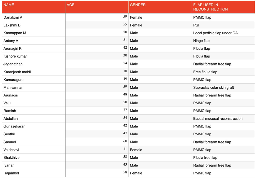
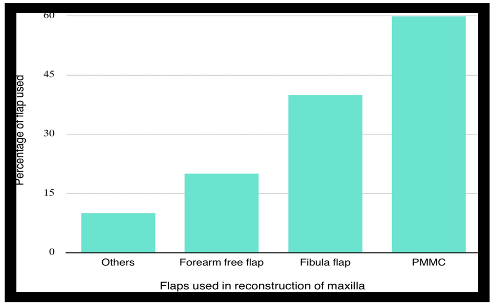
The distribution of reconstruction modalities showed that the pectoralis major myocutaneous (PMMC) flap was the most frequently used technique, accounting for approximately 55–60% of cases. Fibula free flap was the second most commonly employed method, contributing to around 35–40% of cases, while radial forearm free flap accounted for a smaller proportion (approximately 15–20%). Other reconstruction techniques, including local flaps, hinge flaps, and mucosal grafts, were used minimally.
The graphical representation further emphasizes the predominance of PMMC flap over other modalities, indicating a clear preference for this technique in clinical practice. Free vascularized flaps such as fibula and radial forearm flaps were selectively used in cases requiring more complex reconstruction, particularly where bony support or extensive tissue replacement was necessary. In contrast, local and less invasive techniques were limited to smaller defects.
DISCUSSION :
Reconstruction of post-traumatic maxillary defects remains a challenging aspect of maxillofacial rehabilitation due to the complex anatomy of the maxilla and its critical role in function and esthetics. The present study evaluated various reconstruction modalities and demonstrated a clear predominance of the pectoralis major myocutaneous (PMMC) flap as the most commonly utilized technique. This finding can be attributed to the reliability, ease of harvest, shorter operative time, and cost-effectiveness of the PMMC flap, making it a preferred choice, especially in resource-limited settings.
Free vascularized flaps, particularly the fibula free flap and radial forearm free flap, were used less frequently in this study. However, these techniques are considered superior in cases requiring extensive reconstruction, especially when both bony and soft tissue components need to be restored. The fibula free flap, in particular, provides adequate bone length and allows for future implant placement, thereby improving functional outcomes. Despite these advantages, the requirement for microsurgical expertise, longer operative time, and increased cost may limit their routine use.
In the present study, local flaps and other minor reconstructive procedures were utilized in a limited number of cases, primarily for smaller defects. This highlights the importance of defect size, location, and complexity in determining the appropriate reconstruction modality. The findings of this study are consistent with previous literature, which suggests that while advanced microvascular techniques are gaining popularity, PMMC flap continues to remain a dependable option in clinical practice.
Overall, the choice of reconstruction method should be individualized based on the extent of the defect, patient-related factors, and available surgical expertise. Although free flaps offer superior functional and esthetic outcomes in selected cases, PMMC flap remains a versatile and practical option for the management of post-traumatic maxillary defects.
CONCLUSION :
Within the limitations of this retrospective study, the pectoralis major myocutaneous (PMMC) flap was found to be the most commonly utilized reconstruction modality for post-traumatic maxillary defects. Its widespread use can be attributed to its reliability, versatility, and cost-effectiveness in managing a variety of defect sizes. Although free vascularized flaps such as fibula and radial forearm flaps provide superior functional and esthetic outcomes in extensive defects, their use is often limited by surgical complexity and resource requirements. Therefore, the selection of an appropriate reconstruction technique should be individualized based on the extent of the defect, patient factors, and available expertise to achieve optimal functional and esthetic rehabilitation.
BIBLIOGRAPHY:
1. Flores Garcia R, Vidriales Garcia V, Uribe Marquez SK, Valderrabano CPG. A novel technique for transportation osteogenesis in a severe maxillary post traumatic defect. J Stomatol Oral Maxillofac Surg. 2021 Sep;122(4):462–5.
2. Rajkumar K, Neelakandan RS, Devadoss P, Bandyopadhyay TK. Reconstruction of a Post Traumatic Anterior Maxillary Defect by Transport Distraction Osteogenesis. J Maxillofac Oral Surg. 2017 Mar;16(1):118–22.
3. Kudva A, Srikanth G, Singh A, Chitra A, Suryanarayan RK, Francis M. Reconstruction of Maxillary Defects Using Virtual Surgical Planning and Additive Manufacturing Technology: A Tertiary Care Centre Experience. J Maxillofac Oral Surg. 2024 Jun;23(3):644–52.
4. Mittal A, Ramanojam S, Khandelwal S, Valiulla MU. Rehabilitation of Post-traumatic Anterior Maxillary Osseous Deficit Using Iliac Onlay Bone Graft Combined With Dental Implants. Cureus. 2023 Apr;15(4):e37188.
5. Putri AAA, Muzakkie M. Complex soft tissue and facial bone reconstruction after a bear attack in Indonesia: a case report of definitive management following an 8-hour interfacility transfer. J Surg Case Rep. 2026 Mar;2026(3):rjag114.
6. Bhuyan MMI, Arefin MH, Sarkar MA, Hosna AU, Sultana G, Rahman MS, et al. Outcome of Temporalis Myofascial Flap in Maxillary Reconstruction. Mymensingh Med J. 2026 Apr;35(2):427–33.
7. Fiorellini J, Lin GH, Rocchietta I, Mojaver S, Aghaloo T, Ahn KM, et al. Consensus Report of Group 3 of the 1st Global Consensus for Clinical Guidelines for the Rehabilitation of the Edentulous Maxilla: Advanced Diagnostic Imaging, Augmentation Techniques, and Management of Complications. Clin Oral Implants Res. 2026 Feb;37 Suppl 30(Suppl 30):S68–80.
8. Pala K, Neugebauer J, Buser D, Brunello G, Chmielewski K, Chowdhary R, et al. Consensus Report of Group 2 of the 1st Global Consensus for Clinical Guidelines for the Rehabilitation of the Edentulous Maxilla: Zygomatic, Standard-Length, and Short Implant-Supported Prostheses. Clin Oral Implants Res. 2026 Feb;37 Suppl 30(Suppl 30):S49–67.
9. Ding Y, Wu X, Gao Y, Sun Y, Li Q, Song L. [A CBCT study on the inclination of the maxillary occlusal plane and related measurements in adolescent skeletal ClassⅠand ClassⅡpatients with normal vertical angles]. Shanghai Kou Qiang Yi Xue. 2026 Jan;35(1):83–7.