HETEROGENEITY IN CLINICAL MANIFESTATIONS OF JUVENILE NEPHRONOPHTHISIS: UNDERSTANDING THE ROLE OF ASSOCIATED GENES AND VARIANTS
- Shahida Perveen , Associate Professor Pediatric Nephrology, UCHS & CHL, Lahore
- Junaid Ahmed , Senior Registrar Department of Pediatric Nephrology, Children Hospital Faisalabad
- Saad Qayyum , Child Specialist Registrar Paediatric Medicine, UCHS & CHL, Lahore
- AMEEN ULLAH , HOD Pediatrics Department, Combined Military Hospital Kohat
- Ayesha Mansoor , Assistant Professor Paediatric Medicine, UCHS & CHL, Lahore
- SamreenAshraf , Assistant Professor Paediatric Medicine, UCHS & CHL, Lahore
- Usman Ghafoor , Registrar Paediatric Medicine, UCHS & CHL, Lahore
Article Information:
Abstract:
To evaluate the heterogeneity in clinical manifestations of juvenile nephronophthisis and to understand the role of associated genes and genetic variants in affected children.Study Design: Descriptive observational study.Place and Duration of the Study: Department of Pediatric Nephrology, University of Child Health Sciences, The Children’s Hospital Lahore, Pakistan, from September 2023 to August 2024.Methodology: A total of 25 children aged 5–15 years with clinical suspicion of juvenile nephronophthisis were included in the study. Demographic characteristics, anthropometric measurements, clinical symptoms, laboratory parameters, and requirement for dialysis were recorded. Radiological evaluation was performed using renal ultrasonography. Genetic analysis was carried out to identify associated gene mutations. Results: Among the 25 patients, 72% were male and 28% were female. The majority of children (55%) were aged 5–10 years, while 45% were between 11–15 years. Consanguinity was present in 76% of patients. The most common genetic mutation was NPHP1 detected in 68% of patients, followed by NPHP4 (12%) and NPHP5 (8%), while 12% showed no detectable mutation. Polyuria and polydipsia were present in all patients. Growth retardation was observed in 68%, night blindness in 56%, and hypertension in 8% of children. Renal ultrasonography showed shrunken kidneys with poor or absent corticomedullary differentiation in all patients, while renal cysts were identified in 24%. Conclusion: It is concluded that juvenile nephronophthisis demonstrates considerable heterogeneity in clinical presentation and genetic mutations. Nephronophthisis is a genetically diverse disorder involving multiple genes, with NPHP1 being the most common mutation identified in this study.
Keywords:
Article :
INTRODUCTION :
Juvenile nephronophthisis (JNPH) is a genetic kidney disease and among the most prevalent genetic mechanisms leading to chronic kidney disease and end-stage renal disease in children and adolescents [1]. Progressive tubulointerstitial fibrosis, corticomedullary cysts and slow loss of renal function are the features of the disease. The patients tend to have nonspecific symptoms clinically, including polyuria, polydipsia, nocturia, growth retardation, anemia, and impaired urinary concentrating ability [2]. Due to the subtle nature of these manifestations at an earlier age, the diagnosis happens late when in most cases, the damages to the kidney have already been very serious [3]. Consequently, a significant number of patients show up late in the illness when the renal failure has already developed significantly. Juvenile nephronophthisis is one of the diseases of the group called ciliopathies, which are caused by structural or functional defects of primary cilia [4]. Primary cilia are the microscopic hair-like organelles located on the surface of most epithelial cells and they have the critical role of cellular signaling pathways to regulate the tissue development, differentiation and homeostasis of cells. Primary cilia are mechanosensory in the kidney and they sense the movement of fluids in the renal tubules and control pathways of cell proliferation and differentiation [5]. These signalling pathways are disrupted by mutations in proteins of the cilia and cause anomalous development of tubules, fibrosis, and cysts of the kidney [6]. The high clinical heterogeneity is considered one of the most peculiar characteristics of juvenile nephronophthisis. Whereas, the development of isolated kidney disease is prevalent in many patients, some of them present with the syndromic forms of the disease where other organ systems are involved. Extra-renal appearance can consist of retinal degeneration, liver fibrosis, cerebellar malformations, skeletal defects and retarded development [7]. The existence of these systemic features also brings to light the multisystem characteristics of ciliopathies and the complexity of the biological mechanisms that contribute to the development of the disease. Well-known syndromic forms of nephronophthisis dysfunctions comprise Senior-Loken syndrome, Joubert syndrome, and Meckel-Gruber syndrome, each of which exhibits different levels of renal and extrarenal dysfunction [8]. The genetic variance that leads to the heterogeneity in juvenile nephronophthisis is highly linked to the genetic variability of the disorder. In the last twenty years, molecular genetics has contributed to the discovery of over 25 genes linked to nephronophthisis, also known as NPHP genes [9]. These genes encode proteins that are related to ciliary, intracellular transport, centrosome and signal transduction pathways. The defects in these genes interfere with the ciliary functioning and eventually lead to the disruption of the cellular homeostasis of the renal tubules [10]. Some of the commonest suspect genes include NPHP1, NPHP3, NPHP4, NPHP5, and NPHP6. The NPHP1 gene deletions are deemed as the most prevalent gene defect resulting in the juvenile nephronophthisis and normally lead to a case of isolated kidney disease that progresses to end-stage renal failure in adolescence [11]. Conversely, the other genes that have been implicated in mutations that cause syndromic forms of the disease are NPHP5 and NPHP6, which is accompanied by retinal degeneration and neurological defects. This difference in gene participation is a factor that can be attributed to the wide range of clinical presentations that are exhibited in different individuals that are affected [12-14].
Objective
To explore the clinical features and genetic abnormalities in a group of individuals affected by nephronophthisis-related ciliopathy.
METHODOLOGY:
This descriptive observational study conducted at the Department of Pediatric Nephrology, University of Child Health Sciences, The Children's Hospital Lahore, Pakistan, from September 2023 to August 2024. A total of 25 children with clinical suspicion of juvenile nephronophthisis were included in the study. The participants were between the ages of 5 and 15 years at the time of presentation. Children aged between 5 and 15 years presenting with clinical features suggestive of nephronophthisis were included in the study. Patients with symptoms such as polyuria, polydipsia, unexplained chronic kidney disease, growth retardation, or impaired urinary concentrating ability were considered eligible. Children with radiological findings suggestive of nephronophthisis, including increased renal echogenicity or corticomedullary cysts on renal ultrasound, were also included. Children diagnosed with chronic kidney disease secondary to other known etiologies such as congenital anomalies of the kidney and urinary tract, glomerulonephritis, obstructive uropathy, or systemic diseases affecting the kidneys were excluded from the study.
Data Collection
After obtaining institutional approval, data were collected from all eligible patients presenting to the pediatric nephrology department during the study period. The following detailed demographic details (age, gender, family history of renal disease) were taken. All patients were assessed in terms of anthropometric measurements of height, weight and body mass index to assess nutritional status and growth. To report clinical presentation symptoms and signs of juvenile nephronophthisis, clinical assessment was conducted. These were polyuria, polydipsia, fatigue, anemia, growth retardation and other such symptoms. The patients were also documented on the information about disease progression and the need to undergo renal replacement therapy such as dialysis. Lab tests were conducted to determine kidney functioning and related malfunctions. These examinations comprised serum creatinine, blood urea nitrogen, electrolyte levels, hemoglobin concentration and urinalysis. Further studies were conducted when clinically relevant, to assess the extent of involvement of kidney and other features of systems involved. Radiological evaluation was done by doing renal ultrasonography which established structural anomalies in keeping with nephronophthisis. It was found that there were increased renal echogenicity, decreased corticomedullary differentiation, and corticomedullary cysts. All the patients were subjected to genetic assessment on the basis of clinical suspicion of nephronophthisis and presence of typical characteristics in relation to known mutations of the NPHP genes. Molecular genetic testing could not be done in all of the cases due to the lack of availability of molecular diagnostic facilities. Rather, genetic associations had been assessed on the basis of clinical presentation, family history and previously known genotype-phenotype associations.
Data Analysis
Data were analyzed using SPSS v26.0. All collected data were compiled and analyzed to identify patterns in demographic characteristics, clinical manifestations, laboratory findings, and disease progression among the patients. Quantitative variables such as age and laboratory parameters were presented as mean ± standard deviation, while categorical variables such as gender, clinical symptoms, and dialysis requirement were presented as frequencies and percentages.
RESULTS:
Data were collected from 25 patients. The majority of patients were male, accounting for 18 (72%) cases, while females comprised 7 (28%). Regarding age distribution, 14 (55%) children were between 5–10 years of age and 11 (45%) were between 11–15 years. Consanguinity among parents was highly prevalent and was observed in 19 (76%) children, whereas 6 (24%) had no parental consanguinity. Genetic analysis revealed that the most common mutation was NPHP1, detected in 17 (68%) patients. Mutations in NPHP4 and NPHP5 were identified in 3 (12%) and 2 (8%) children, respectively, while no detectable mutation was found in 3 (12%) cases.
Table 1. Demographic Characteristics of Patients with Juvenile Nephronophthisis (n = 25)
|
Variable |
Category |
n (%) |
|
Gender |
Male |
18 (72%) |
|
Female |
7 (28%) |
|
|
Age Group |
5–10 years |
14 (55%) |
|
11–15 years |
11 (45%) |
|
|
Consanguinity |
Present |
19 (76%) |
|
Absent |
6 (24%) |
|
|
Gene Mutation |
NPHP1 |
17 (68%) |
|
NPHP4 |
3 (12%) |
|
|
NPHP5 |
2 (8%) |
|
|
No mutation detected |
3 (12%) |
Polyuria and polydipsia were universal symptoms and were reported in all 25 (100%) patients. Growth retardation was observed in 17 (68%) children, while 8 (32%) had normal growth parameters. Night blindness, indicating possible retinal involvement, was present in 14 (56%) patients and absent in 11 (44%). Hypertension was relatively uncommon and was detected in only 2 (8%) patients, whereas the remaining 23 (92%) children had normal blood pressure.
Table 2. Clinical Manifestations of Juvenile Nephronophthisis (n = 25)
|
Variable |
Category |
n (%) |
|
Polyuria |
Present |
25 (100%) |
|
Absent |
0 (0%) |
|
|
Polydipsia |
Present |
25 (100%) |
|
Absent |
0 (0%) |
|
|
Growth Retardation |
Present |
17 (68%) |
|
Absent |
8 (32%) |
|
|
Night Blindness |
Present |
14 (56%) |
|
Absent |
11 (44%) |
|
|
Hypertension |
Present |
2 (8%) |
|
Absent |
23 (92%) |
Renal ultrasonography revealed shrunken kidneys with poor or absent corticomedullary differentiation in all 25 (100%) patients, confirming the typical radiological features of nephronophthisis. Renal cysts were identified in 6 (24%) children, while 19 (76%) showed no cystic changes on ultrasound. Dialysis was required in 4 (16%) patients due to severe renal dysfunction, whereas 21 (84%) did not require dialysis at the time of evaluation. Syndromic associations were also noted, with Joubert syndrome present in 3 (12%) children and Senior–Løken syndrome identified in 5 (20%) patients.
Table 3. Radiological Findings and Disease Severity (n = 25)
|
Variable |
Category |
n (%) |
|
Shrunken Kidneys with Poor / Absent Corticomedullary Differentiation |
Present |
25 (100%) |
|
Absent |
0 (0%) |
|
|
Renal Cysts |
Present |
6 (24%) |
|
Absent |
19 (76%) |
|
|
Dialysis Requirement |
Yes |
4 (16%) |
|
No |
21 (84%) |
|
|
Joubert Syndrome |
Present |
3 (12%) |
|
Absent |
22 (88%) |
|
|
Senior–Løken Syndrome |
Present |
5 (20%) |
|
Absent |
20 (80%) |
Among patients with the NPHP1 mutation (n = 17), growth retardation was observed in 13 (76.5%) compared with 4 (50.0%) in patients with other or no mutations; however, this difference was not statistically significant (p = 0.182). Night blindness was present in 11 (64.7%) patients with NPHP1 mutation compared to 3 (37.5%) in the other mutation group (p = 0.213). Renal cysts were observed in 5 (29.4%) patients with NPHP1 mutation and in 1 (12.5%) patient with other mutations (p = 0.348). Dialysis requirement was seen in 3 (17.6%) patients with NPHP1 mutation and 1 (12.5%) patient with other mutations (p = 0.741). Syndromic variants were identified in 4 (23.5%) patients with NPHP1 mutation and in 4 (50.0%) patients with other mutations, although this difference was also not statistically significant (p = 0.184).
Table 4. Association of Genetic Mutations with Clinical Manifestations and Outcomes (n = 25)
|
Variable |
Category |
NPHP1 Mutation (n = 17) n (%) |
Other / No Mutation (n = 8) n (%) |
p-value |
|
Growth Retardation |
Present |
13 (76.5%) |
4 (50.0%) |
0.182 |
|
Absent |
4 (23.5%) |
4 (50.0%) |
||
|
Night Blindness |
Present |
11 (64.7%) |
3 (37.5%) |
0.213 |
|
Absent |
6 (35.3%) |
5 (62.5%) |
||
|
Renal Cysts on Ultrasound |
Present |
5 (29.4%) |
1 (12.5%) |
0.348 |
|
Absent |
12 (70.6%) |
7 (87.5%) |
||
|
Dialysis Requirement |
Yes |
3 (17.6%) |
1 (12.5%) |
0.741 |
|
No |
14 (82.4%) |
7 (87.5%) |
||
|
Syndromic Variant |
Present |
4 (23.5%) |
4 (50.0%) |
0.184 |
|
Absent |
13 (76.5%) |
4 (50.0%) |
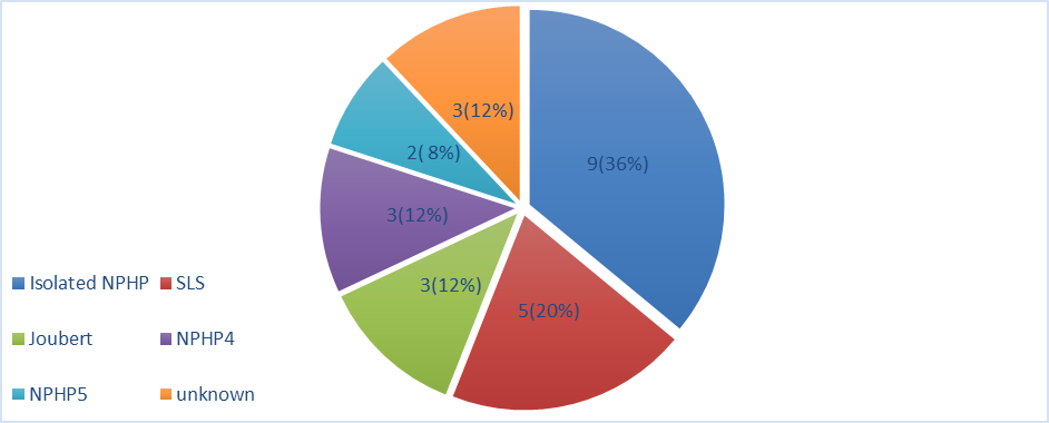
Figure 1: NEPHRONOPHTHISIS VARIANTS/SYNDROMES IN COHORT
Patients with NPHP1 mutation had a median age of 10 years (range 5.9–15), while those with NPHP4 and NPHP5 mutations had median ages of 8 years (range 6–10) and 7.5 years (range 7–8), respectively. Consanguinity was particularly frequent among patients with NPHP1 mutation (82.4%) and was present in all patients without a detected mutation. Family history of renal disease was most observed in the NPHP1 group (94.1%).
Table 5: Genotype–Phenotype Association in Children with Juvenile Nephronophthisis (n = 25)
|
Variable |
Not Known (n = 3) |
NPHP1 (n = 17) |
NPHP4 (n = 3) |
NPHP5 (n = 2) |
|
Age Median (Range), years |
12 (11–15) |
10 (5.9–15) |
8 (6–10) |
7.5 (7–8) |
|
Consanguinity |
3 (100%) |
14 (82.4%) |
0 (0%) |
0 (0%) |
|
Family History of Renal Disorder |
0 (0%) |
16 (94.1%) |
1 (33.3%) |
2 (100%) |
|
Polyuria |
3 (100%) |
17 (100%) |
3 (100%) |
2 (100%) |
|
Polydipsia |
3 (100%) |
16 (94.1%) |
3 (100%) |
2 (100%) |
|
Bed Wetting |
2 (66.7%) |
15 (88.2%) |
3 (100%) |
0 (0%) |
|
Growth Retardation / Stunting |
2 (66.7%) |
12 (70.6%) |
2 (66.7%) |
1 (50.0%) |
|
Hypertension |
0 (0%) |
2 (11.8%) |
0 (0%) |
0 (0%) |
|
Night Blindness |
0 (0%) |
2 (11.8%) |
0 (0%) |
1 (50.0%) |
|
Left Kidney Normal Size |
0 (0%) |
1 (5.9%) |
0 (0%) |
1 (50.0%) |
|
Left Kidney Shrunken |
3 (100%) |
16 (94.1%) |
3 (100%) |
1 (50.0%) |
|
Right Kidney Normal Size |
0 (0%) |
1 (5.9%) |
0 (0%) |
1 (50.0%) |
|
Right Kidney Shrunken |
3 (100%) |
16 (94.1%) |
3 (100%) |
1 (50.0%) |
DISCUSSION :
Juvenile nephronophthisis is an importance genetic heterogeneous ciliopathy and a significant cause of chronic kidney disease amongst kids and teenagers. The current paper considered clinical variations and genetic profiles of children suspected to have juvenile nephronophthisis. The results illustrate that there is a high heterogeneity in clinical manifestation, genetic mutations, and related phenotypical disorders which show the complexity of this disorder. In the current research, this was found to have a male preponderance in the sense that 72% of the patients were males and 28% were females. The same male dominance has been observed with a number of earlier studies carried out to examine child hereditary kidney disorders. Even though nephronophthisis is an autosomal recessive disease and theoretically both genders are equally affected, gender difference has been observed among clinical cohorts, perhaps because of the referral tendencies or limited number of studies [15].
The age distribution of the patients was that the largest number of children were aged between 5-10 years (55%), and 45% between 11-15 years. This observation is in line with the classic clinical history of juvenile nephronophthisis where signs and symptoms may manifest in childhood but may be undiagnosed until later as the kidney function starts to deteriorate. Past research has also indicated that a high number of patients would be diagnosed in late childhood or early adolescence stages when the progressive renal insufficiency would have become evident. In this study, consanguinity was found in 76 percent of the patients, which shows that the genetic factor of the disorder is high [16]. The high levels of consanguinity have been frequently documented in the studies that have been conducted in areas where consanguinity marriages are prevalent such as South Asia and the Middle East. Consanguinity enhances the risk of having autosomal recessive disorders like nephronophthisis and also leads to accumulation of genetic diseases in family. NPP1 was the most prevalent mutated gene in genetic analysis found in 68 percent of the patients [17]. The observation is consistent with past reports that have continuously cited deletions in NPHP1 as the most common genetic pathology in juvenile nephronophthisis. NPHP4 and NPHP5 were found to be mutated in 12 and 8 percent of patients respectively with 12 percent of the patients having no detectable mutation. The fact that not all patients have identifiable mutations could either be explained by the constraints of the genetic testing methods or the fact that the mutations are in genes that are not yet discovered [18].
The clinical signs presented in the paper are typical of nephronophthisis. All patients had polyuria and polydipsia, and this suggests that patients had low concentrating ability of urine because of tubular dysfunction. These symptoms are early warning signs of the disease and very often they are the first symptoms that are presented by the affected children. It was found that growth retardation occurred in 68% of the patients, which underscored the relationship between chronic kidney disease and nutritional status and total growth in children. Fifty six percent of the patients reported night blindness indicating that there was retinal involvement that is based on the ciliopathy-related disorders [19]. Senior-Loken syndrome is a syndromic disorder that is typically linked to retinal degeneration. The occurrence of night blindness in this study is relatively high, which is why it is possible to assume the existence of extra-renal manifestations in a considerable percentage of patients. The incidence of hypertension in children was only 8% and this is relatively low in comparison to other types of chronic kidney diseases. This observation is in line with the observation of tubulointerstitial nature of nephronophthisis, in which hypertension is usually manifested later in the course of the disease. Syndromic forms of nephronophthisis were also found in the study. Joubert syndrome was found in 12% of the patients and Senior-Loken syndrome was found in 20 percent of children. These syndromic associations also indicate the multisystem nature of the ciliopathies [20]. Joubert syndrome is mostly linked with neuronal disorders and cerebellar malformation, as compared to Senior-Loken syndrome which involves the combination of nephronophthisis and retinal degeneration [21]. There are several limitations in this study that ought to be taken into consideration during the interpretation of the findings. To begin with, the sample size was quite small (25% only), which may reduce the statistical power and extrapolation of the findings. Second, the research was done in one tertiary care facility and thus the results might not necessarily be applicable to the general population of children with juvenile nephronophthisis. Third, not all patients underwent comprehensive molecular genetic testing as there is low access to sophisticated testing centers hence could have under-detected some genetic mutations that cause the disease. Besides this, the study had a cross-sectional design, which restricted the possibility of evaluating the progression of disease over a long period, treatment failure and survival rates.
CONCLUSION :
It is concluded that juvenile nephronophthisis demonstrates marked heterogeneity in clinical presentation, genetic mutations, and associated syndromic manifestations among affected children. Nephronophthisis is a genetically diverse disorder with involvement of multiple genes, and the predominant genetic mutation observed in our study was NPHP1. The majority of patients exhibited early symptoms such as polyuria, polydipsia, and growth retardation, with a high prevalence of parental consanguinity supporting the autosomal recessive inheritance pattern of the disease. In addition to renal involvement, a considerable proportion of patients presented with extra-renal features including retinal abnormalities and syndromic conditions such as Joubert syndrome and Senior–Løken syndrome.
BIBLIOGRAPHY:
1. Zhong Z, Yan X, Fang Z, Dong Y, Tan J, Xie J, Hu L, Zhang S and Qin W (2022) Case Report: Adolescent-Onset Isolated Nephronophthisis Caused by a Novel Homozygous Inversin Mutation. Front. Genet. 13:847397. doi: 10.3389/fgene.2022.847397
2. König J, Kranz B, König S, Schlingmann KP, Titieni A, Tönshoff B, Habbig S, Pape L, Häffner K, Hansen M, Büscher A, Bald M, Billing H, Schild R, Walden U, Hampel T, Staude H, Riedl M, Gretz N, Lablans M, Bergmann C, Hildebrandt F, Omran H, Konrad M; Gesellschaft für Pädiatrische Nephrologie (GPN). Phenotypic Spectrum of Children with Nephronophthisis and Related Ciliopathies. Clin J Am Soc Nephrol. 2017 Dec 7;12(12):1974-1983. doi: 10.2215/CJN.01280217. Epub 2017 Nov 16. PMID: 29146700; PMCID: PMC5718263.
3. König JC, Karsay R, Gerß J, Schlingmann KP, Dahmer-Heath M, Telgmann AK, Kollmann S, Ariceta G, Gillion V, Bockenhauer D, Bertholet-Thomas A, Mastrangelo A, Boyer O, Lilien M, Decramer S, Schanstra JP, Pohl M, Schild R, Weber S, Hoefele J, Drube J, Cetiner M, Hansen M, Thumfart J, Tönshoff B, Habbig S, Liebau MC, Bald M, Bergmann C, Pennekamp P, Konrad M; NEOCYST consortium. Refining Kidney Survival in 383 Genetically Characterized Patients With Nephronophthisis. Kidney Int Rep. 2022 Jun 16;7(9):2016-2028. doi: 10.1016/j.ekir.2022.05.035. PMID: 36090483; PMCID: PMC9459005.
4. Tata A, Rocha G, Hureaux M, Serafin AS, Porée E, Menguy L, Goudin N, Cagnard N, Gréau L, Fila M, Briseño-Roa L, Annereau JP, Saunier S, Benmerah A. Prostaglandin Analogs and Eupatilin as Treatments for Nephronophthisis. Kidney Int Rep. 2025 May 2;10(8):2821-2835. doi: 10.1016/j.ekir.2025.04.060. PMID: 40814642; PMCID: PMC12347849.
5. Gupta S, Ozimek-Kulik JE, Phillips JK. Nephronophthisis-Pathobiology and Molecular Pathogenesis of a Rare Kidney Genetic Disease. Genes. 2021; 12(11):1762. https://doi.org/10.3390/genes12111762
6. Wolf MTF, Bonsib SM, Larsen CP, Hildebrandt F. Nephronophthisis: a pathological and genetic perspective. Pediatr Nephrol. 2024;39:1977–2000. doi:10.1007/s00467-023-06174-8
7. Benmerah A, Briseño-Roa L, Annereau JP, Saunier S. Repurposing small molecules for nephronophthisis and related renal ciliopathies. Kidney Int. 2023;104:245–253. doi:10.1016/j.kint.2023.04.027
8. Devlin L, Dhondurao Sudhindar P, Sayer JA. Renal ciliopathies: promising drug targets and prospects for clinical trials. Expert Opin Ther Targets. 2023;27:325–346. doi:10.1080/14728222.2023.2218616
9. Garcia H, Serafin AS, Silbermann F, et al. Agonists of prostaglandin E2 receptors as potential first-in-class treatment for nephronophthisis and related ciliopathies. Proc Natl Acad Sci U S A. 2022;119. doi:10.1073/pnas.2115960119
10. Garfa Traoré M, Roccio F, Miceli C, Ferri G, Parisot M, Cagnard N, Lhomme M, Dupont N, Benmerah A, Saunier S, Delous M. Fluid shear stress triggers cholesterol biosynthesis and uptake in inner medullary collecting duct cells, independently of nephrocystin-1 and nephrocystin-4. Front Mol Biosci. 2023 Oct 17;10:1254691. doi: 10.3389/fmolb.2023.1254691. PMID: 37916190; PMCID: PMC10616263.
11. Miri Karam Z, Gohari AK, Khabaz MJR, Yari A, Meybodi SME, Attari R, Torabi M, Vafaeie F, Moraddahande FM, Amiri S, Saeidi K. Identification of a Novel Deletion Variant (c.2999_3005delTGTGTGT/p.Asn1000SerfsTer4) in NPHP4 Associated With Nephronophthisis-4. J Clin Lab Anal. 2024 Jun;38(11-12):e25077. doi: 10.1002/jcla.25077. Epub 2024 Jun 19. PMID: 38895833; PMCID: PMC11252830.
12. Dahmer-Heath M, Optenhövel S, Hechler T, Konrad M, König J. Systematic review of outcomes reported in clinical research on nephronophthisis: how do they align with SONG Kids priorities? Pediatr Nephrol. 2026 Feb;41(2):339-351. doi: 10.1007/s00467-025-06912-0. Epub 2025 Aug 20. PMID: 40836044; PMCID: PMC12727818.
13. Knoers N, Antignac C, Bergmann C, Dahan K, Giglio S, Heidet L, Lipska-Ziętkiewicz BS, Noris M, Remuzzi G, Vargas-Poussou R, Schaefer F. Genetic testing in the diagnosis of chronic kidney disease: recommendations for clinical practice. Nephrol Dial Transplant. 2022 Jan 25;37(2):239-254. doi: 10.1093/ndt/gfab218. PMID: 34264297; PMCID: PMC8788237.
14. Ijaz A, Alfadhli F, Alharbi A, Khan YN, Alhawas YK, Hashmi JA, Wali A, Basit S. NPHP3 splice acceptor site variant is associated with infantile nephronophthisis and asphyxiating thoracic dystrophy; A rare combination. Eur J Med Genet. 2022 Oct;65(10):104578. doi: 10.1016/j.ejmg.2022.104578. Epub 2022 Aug 17. PMID: 35987473.
15. Yari A, Vafaeie F, Karam ZM, Hosseini M, Hashemzade H, Rahimi MS, Ehsanbakhsh A, Miri-Moghaddam E. Identification of a Novel ARSA Gene Mutation Through High-Throughput Molecular Diagnosis Method in Two Girls with Late Infantile Metachromatic Leukodystrophy. Neuromolecular Med. 2023 Dec;25(4):563-572. doi: 10.1007/s12017-023-08757-y. Epub 2023 Sep 8. PMID: 37682448.
16. Yari A, Etesam S, Zarifi S, Parvizpour S, Miri-Moghaddam E. Clinical Manifestations Associated with the Domain-Containing Protein 2 Gene Mutation in an Iranian Family with Spastic Paraplegia 54. Neurodegener Dis. 2022;22(3-4):139-150. doi: 10.1159/000530375. Epub 2023 Mar 28. PMID: 36977391.
17. Vafaeie F, Miri Karam Z, Yari A, Safarpour H, Kazemi T, Etesam S, Mohammadpour M, Miri-Moghaddam E. Clinical and genetic screening in a large Iranian family with Marfan syndrome: A case study. Health Sci Rep. 2023 Oct 23;6(10):e1647. doi: 10.1002/hsr2.1647. PMID: 37877128; PMCID: PMC10591539.
18. Modarage K, Malik SA and Goggolidou P (2022) Molecular Diagnostics of Ciliopathies and Insights Into Novel Developments in Diagnosing Rare Diseases. Br J Biomed Sci 79:10221. doi: 10.3389/bjbs.2021.10221
19. Kofotolios, I., Bonios, M. J., Adamopoulos, M., Mourouzis, I., Filippatos, G., Boletis, J. N., Marinaki, S., & Mavroidis, M. (2024). The Han:SPRD Rat: A Preclinical Model of Polycystic Kidney Disease. Biomedicines, 12(2), 362. https://doi.org/10.3390/biomedicines12020362
20. Zaikova NM, Magon VA. Clinical features of juvenile nephronophthisis in an adolescent girl. Urol Nephrol Open Access J. 2025;13(1):12–14. doi:10.15406/unoaj.2025.13.0036
21. Bhimma, R., Jembere, E. & Hariparshad, S. Case report of a child with nephronophthisis from South Africa. BMC Pediatr 24, 431 (2024). https://doi.org/10.1186/s12887-024-04872-2