EFFECTIVENESS AND COMPLICATIONS OF PCNL IN PEDIATRIC PATIENTS WITH RENAL STONES THAT FAILED ESWL OR F.URS.
- Sami Ul Haq , Department of Urology, Timergara Teaching Hospital, Dir Lower, Pakistan
- Ghufran Ullah , Department of Urology, Timergara Teaching Hospital, Dir Lower, Pakistan
- Aziz Ul Wahab , Department of Urology, Timergara Teaching Hospital, Dir Lower, Pakistan
- Irshad Khan , Assistant Professor Zeb Medical and Dental College Timergara Dir L.:
- Irshad Ahmad , Consultant Urologist, Regional Headquarter Hospital, Skardu
Article Information:
Abstract:
Pediatric nephrolithiasis is an increasing concern and often requires complete stone clearance due to high recurrence risk. Managing large renal stones (>2 cm), lower calyceal stones (>1 cm), and stones refractory to Extracorporeal Shock Wave Lithotripsy (ESWL) or flexible ureteroscopy (F.URS) remains challenging. Percutaneous nephrolithotomy (PCNL) is a key minimally invasive option for these cases. Objective: To evaluate the effectiveness and complications of PCNL in pediatric patients with large renal stones (>2 cm), lower calyceal stones (>1 cm), and smaller stones (<2 cm) that failed ESWL or F.URS. Methods: This prospective study was conducted from August 2024 to February 2025 at the Urology Unit, District Head Quarter Hospital Dir Lower Timergara and Regional Headquarter Hospital, Skardu. A total of 72 (36 of each center) children (<18 years) with eligible renal stones underwent PCNL under general anesthesia. Preoperative evaluation included clinical assessment, laboratory tests, and imaging (X-ray KUB, ultrasonography, and non-contrast CT when required). Stone fragmentation was performed using Holmium:YAG laser, pneumatic lithoclast, or ultrasonic lithotripter depending on stone characteristics. Postoperative outcomes and complications were recorded, including bleeding, hematuria, fever, perforation, residual stones, hemoglobin drop, transfusion need, and hospital stay. Results: The mean age was 10.5 ± 3.5 years; 69.4% were male. Mean stone size was 2.4 ± 0.5 cm, and 70.8% of stones were radiopaque. Mean operative time was 85.3 ± 16.7 minutes, and hospital stay was 2–3 days. Overall, 87.5% of patients were stone-free or had insignificant residual fragments (≤4 mm), while 12.5% had residual stones. Intraoperative complications included bleeding (12.5%) and calyceal perforation (13.9%). Postoperatively, hematuria occurred in 12.5% and fever in 16.6%. Blood transfusion was required in 4.2%, with a mean hemoglobin drop of 1.1 ± 0.6 g/dL. No colon injury or urine leakage was reported. Conclusion: PCNL is a safe and effective treatment for pediatric renal stones, particularly in large, lower calyceal, or treatment-resistant cases. It achieves a high stone-free rate with acceptable complication rates and a short hospital stay, even in resource-limited settings. Long-term follow-up and metabolic evaluation are recommended to reduce recurrence.
Keywords:
Article :
INTRODUCTION :
The prevalence of urolithiasis in pediatric patients is approximately 1%. However, all children diagnosed with urolithiasis are considered at high risk for stone recurrence, making complete stone clearance an essential goal of treatment. The choice of management is individualized and depends on the patient’s age, stone size, number and location, clinical condition, and urinary tract anatomy. Pediatric stone disease is often complex and may be associated with underlying metabolic disorders or structural abnormalities of the urinary system. ¹
Over recent decades, the management of renal stones has shifted from open surgery to minimally invasive techniques. Current treatment options include extracorporeal shock wave lithotripsy (ESWL), flexible ureteroscopy (FURS), and percutaneous nephrolithotomy (PCNL). According to the European Association of Urology (EAU) and the American Urological Association (AUA) guidelines, PCNL is the preferred treatment for renal stones larger than 2 cm.²
Pediatric PCNL was first reported in 1985 and has since enabled effective management of children with large stone burdens or in cases where ESWL is contraindicated or unlikely to succeed. PCNL in children has been performed using both adult and pediatric instruments. The rationale for miniaturized instruments (“mini-perc”) is to reduce morbidity while maintaining high stone clearance rates. Despite this, stone-free rates and complication profiles vary depending on the technique used and the surgeon’s experience. ² The objective of this study was to evaluate the safety and effectiveness of PCNL as a treatment modality for pediatric patients with renal calculi.
METHODOLOGY:
This prospective study was conducted from August 2024 to February 2025 at the Urology Unit, District Head Quarter Hospital Dir Lower Timergara and Regional Headquarter Hospital, Skardu. A total of 72 (36 of each center) children (<18 years) with renal stones, comprising large stones (>2 cm), lower calyceal stones (>1 cm), or smaller stones (<2 cm) that had failed previous treatment such as extracorporeal shock wave lithotripsy (ESWL) or flexible ureteroscopy (F.URS); ethical approval was obtained and informed consent was taken from parents/guardians, while all patients underwent preoperative evaluation including medical history, clinical examination, laboratory investigations, and imaging (X-ray KUB, ultrasonography, and non-contrast CT urography when indicated); PCNL was performed under general anesthesia using a standardized approach starting with urethro-cystoscopy to identify the ureteric orifice, followed by guidewire placement and insertion of an open-tip ureteric catheter, then repositioning for calyceal puncture and tract access, with stone fragmentation achieved using Holmium:YAG laser, pneumatic lithoclast, or ultrasonic lithotripter based on stone characteristics and instrument availability; postoperative care included monitoring for complications, pain control with IV NSAIDs, and antibiotics, with nephrostomy tube removal after 2–3 days in uncomplicated cases and DJ stent removal at 4 weeks in patients who were stone-free or had clinically insignificant residual fragments (≤4 mm); data were analyzed using SPSS version 20.0 to assess outcomes and complications, demonstrating a high success rate with 87.5% of patients being stone-free or having minimal residual stones.
RESULTS:
Across 72 pediatric patients, the mean age was 10.5 ± 3.5 years, including 4 (5.6%) preschool children (3–6 years), 26 (36.1%) school-age children (6–12 years), and 42 (58.3%) adolescents (12–18 years), with 50 males (69.4%) and 22 females (30.6%); stones were predominantly right-sided in 60 (83.3%) and left-sided in 12 (16.7%), with single stones in 48 (66.7%) and multiple stones in 24 (33.3%), and the stone location was renal pelvis in 24 (33.3%), upper calyx in 9 (12.5%), middle calyx in 9 (12.5%), lower calyx in 9 (12.5%), pelvis + lower calyx in 9 (12.5%), upper + middle calyx in 6 (8.3%), and middle + lower calyx in 6 (8.3%); regarding radiology and burden, 51 (70.8%) stones were radiopaque and 21 (29.2%) were radiolucent, the mean stone size was 2.4 ± 0.5 cm, and median density was HU 850.0 (range 550.0–1130.0); operative outcomes showed a mean operative time of 85.3 ± 16.7 minutes, with PCNL performed using a 20 Fr nephroscope in 42 (58.3%), 9.5 Fr semi-rigid URS in 20 (27.8%), and 12 Fr MIP-M system in 10 (13.9%), and access was achieved using a single tract in 56 (77.8%) versus multiple tracts in 16 (22.2%), with tract sites including lower calyx in 39 (54.2%), middle calyx in 16 (22.2%), lower + middle calyx in 6 (8.3%), and lower + upper calyx in 4 (5.6%); tract dilation was done with Amplatz dilators in 42 (58.3%), single-step dilatation in 21 (29.2%), and telescoping metal dilators in 9 (12.5%), while lithotripsy modality used was Holmium:YAG laser in 30 (41.7%), pneumatic in 24 (33.3%), and ultrasonic in 18 (25%); postoperative drainage/stenting included open-tip ureteral catheter + nephrostomy in 21 (29.2%), DJ + nephrostomy in 39 (54.2%), and DJ without nephrostomy in 12 (16.7%); effectiveness was high with 63 (87.5%) patients having no residual stones, while 9 (12.5%) had residuals, specifically pelvic residual stone 0.9 cm in 3 (4.2%), upper calyx residual stone 1.5 cm in 3 (4.2%), and lower calyceal residual stone 1.6 cm in 3 (4.2%); intraoperative complications included bleeding in 9 (12.5%) and calyceal perforation in 10 (13.9%), and postoperative complications included hematuria in 9 (12.5%) (classified as mild in 6 (8.3%) and severe in 3 (4.2%)) and fever in 12 (16.6%), with low-grade fever (37.5–38.5°C) in 6 (8.3%) and high-grade fever (>38.5°C) in 6 (8.3%); blood transfusion was required in 3 (4.2%) while 69 (95.8%) required none, and importantly colon injury was 0/72 (0%) and urine leakage was 0/72 (0%); recovery was short with a mean hemoglobin drop of 1.1 ± 0.6 g/dL and hospital stay of 2 days in 57 (79.2%) and 3 days in 15 (20.8%), confirming a strong overall outcome profile with high clearance and low serious complication rates.
Table: Demographics, Stone Characteristics, Operative Details, Outcomes, and Complications
|
Domain |
Variable |
Result |
|
Patient Demographics |
Total patients |
72 |
|
Age (years), mean ± SD |
10.5 ± 3.5 |
|
|
Age group: Preschool (3–6) |
4 (5.6%) |
|
|
Age group: School-age (6–12) |
26 (36.1%) |
|
|
Age group: Adolescent (12–18) |
42 (58.3%) |
|
|
Sex: Male |
50 (69.4%) |
|
|
Sex: Female |
22 (30.6%) |
|
|
Stone Characteristics |
Stone side: Right |
60 (83.3%) |
|
Stone side: Left |
12 (16.7%) |
|
|
Multiplicity: Single |
48 (66.7%) |
|
|
Multiplicity: Multiple |
24 (33.3%) |
|
|
Stone site: Pelvis |
24 (33.3%) |
|
|
Stone site: Upper calyx |
9 (12.5%) |
|
|
Stone site: Middle calyx |
9 (12.5%) |
|
|
Stone site: Lower calyx |
9 (12.5%) |
|
|
Stone site: Pelvis + Lower calyx |
9 (12.5%) |
|
|
Stone site: Upper + Middle calyx |
6 (8.3%) |
|
|
Stone site: Middle + Lower calyx |
6 (8.3%) |
|
|
Opacity: Radiopaque |
51 (70.8%) |
|
|
Opacity: Radiolucent |
21 (29.2%) |
|
|
Stone size (cm), mean ± SD |
2.4 ± 0.5 |
|
|
HU (median, range) |
850.0 (550.0–1130.0) |
|
|
Operative Data |
Operative time (min), mean ± SD |
85.3 ± 16.7 |
|
Type of PCNL: 20 Fr nephroscope |
42 (58.3%) |
|
|
Type of PCNL: 9.5 Fr semi-rigid URS |
20 (27.8%) |
|
|
Type of PCNL: 12 Fr MIP-M system |
10 (13.9%) |
|
|
Tract number: Single |
56 (77.8%) |
|
|
Tract number: Multiple |
16 (22.2%) |
|
|
Tract site: Lower calyx |
39 (54.2%) |
|
|
Tract site: Middle calyx |
16 (22.2%) |
|
|
Tract site: Lower + Middle calyx |
6 (8.3%) |
|
|
Tract site: Lower + Upper calyx |
4 (5.6%) |
|
|
Dilatation: Amplatz dilators |
42 (58.3%) |
|
|
Dilatation: Single-step dilatation |
21 (29.2%) |
|
|
Dilatation: Telescoping metal dilators |
9 (12.5%) |
|
|
Lithotripsy: Holmium:YAG laser |
30 (41.7%) |
|
|
Lithotripsy: Pneumatic |
24 (33.3%) |
|
|
Lithotripsy: Ultrasonic |
18 (25.0%) |
|
|
Stents/drainage: Open-tip ureteral catheter + nephrostomy |
21 (29.2%) |
|
|
Stents/drainage: DJ + nephrostomy |
39 (54.2%) |
|
|
Stents/drainage: DJ without nephrostomy |
12 (16.7%) |
|
|
Outcomes / Residuals |
Stone-free / no residuals |
63 (87.5%) |
|
Residual stones present |
9 (12.5%) |
|
|
Pelvic residual stone 0.9 cm |
3 (4.2%) |
|
|
Upper calyx residual stone 1.5 cm |
3 (4.2%) |
|
|
Lower calyceal residual stone 1.6 cm |
3 (4.2%) |
|
|
Complications |
Intraoperative bleeding |
9 (12.5%) |
|
Calyceal perforation |
10 (13.9%) |
|
|
Hematuria (overall) |
9 (12.5%) |
|
|
Hematuria: Mild |
6 (8.3%) |
|
|
Hematuria: Severe |
3 (4.2%) |
|
|
Fever (overall) |
12 (16.6%) |
|
|
Fever: Low grade (37.5–38.5°C) |
6 (8.3%) |
|
|
Fever: High grade (>38.5°C) |
6 (8.3%) |
|
|
Blood transfusion required |
3 (4.2%) |
|
|
Colon injury |
0 (0%) |
|
|
Urine leakage |
0 (0%) |
|
|
Recovery / Hospital Stay |
Hb drop (g/dL), mean ± SD |
1.1 ± 0.6 |
|
Hospital stay: 2 days |
57 (79.2%) |
|
|
Hospital stay: 3 days |
15 (20.8%) |
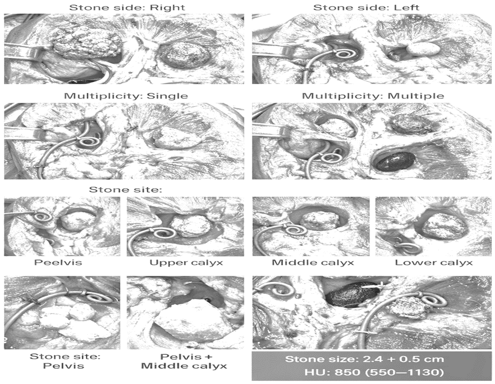
Figure 1: Combined Demographic, Stone Characteristics, Operative Details, Outcomes, Complications, and Recovery Data of Pediatric PCNL (n = 72)
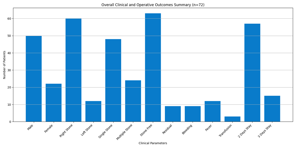
Figure Mean age was 10.5 ± 3.5 years (preschool 5.6%, school-age 36.1%, adolescents 58.3%) with male 69.4% and female 30.6%; stones were mainly right-sided (83.3%), single (66.7%), commonly in pelvis (33.3%), mostly radiopaque (70.8%), with size 2.4 ± 0.5 cm and HU 850 (550–1130).
PCNL showed strong outcomes: mean operative time 85.3 ± 16.7 min, stone-free rate 87.5%, residual stones 12.5%, complications included bleeding 12.5%, perforation 13.9%, hematuria 12.5%, fever 16.6%, transfusion 4.2%, with no colon injury/urine leak, Hb drop 1.1 ± 0.6 g/dL, and hospital stay 2 days (79.2%) or 3 days (20.8%).
DISCUSSION:
Kidney stones, although traditionally thought to be uncommon in pediatric populations, have become an increasingly significant health concern, particularly in developing nations. These conditions are often associated with anatomical and metabolic abnormalities, as well as infections, which can lead to higher recurrence rates. Such challenges highlight the need for more efficient and minimally invasive treatments in this demographic [3, 4]. In the present study, treatment methods were guided by the stone burden, patient age, and the availability of instruments. For patients with larger stone burdens, older children required dilatation up to 24 Fr. In contrast, younger children had dilatation limited to 16 Fr, depending on the instrument available, including the 9.5 Fr semi-rigid ureteroscope or the 12 Fr MIP-M system. Previous studies have suggested that dilating to 24 Fr or 26 Fr in pediatric patients does not significantly increase morbidity. Traxer et al. [5] noted that using smaller access sizes does not yield better outcomes in terms of renal scarring, as observed in animal models. While some studies suggest a reduction in complication rates with smaller instruments, Bilen et al. [7] found no significant difference in complication rates when comparing devices of varying sizes. On the other hand, other
studies have shown that smaller nephroscopes may help reduce morbidity and mortality rates [6]. Zeren et al. [8] demonstrated a correlation between intraoperative bleeding and factors such as stone burden, surgical time, and sheath size. The dilatation methods in our study included Amplatz dilators in the majority of cases, with a smaller proportion using single-step or telescoping metal dilators. Salerno et al. [9] reported that Amplatz dilators were the preferred choice in all cases of PCNL, while El-Nahas et al. [10] noted that coaxial telescopic dilators were commonly used in pediatric PCNL procedures. Holmium: YAG laser lithotripsy was the primary method of stone fragmentation in our study, with an average power of 30 W applied. Ultrasonic lithotripsy and pneumatic lithotripsy were used less frequently. Bujons et al. [11] employed a high-power Ho: YAG laser for treating difficult stones in pediatric patients, while Jou et al. [12] found that adjusting the power to 30 W improved efficiency. In a similar study, El-Nahas et al. [10] employed ultrasonic, pneumatic, and Ho: YAG lasers to fragment stones. Postoperative complications were managed with DJ stents and open-tip ureteric catheters. Rashid et al. [14] used DJ stents in 86% of cases, while Mahmood et al. [15] recommended routine DJ insertion. In our study, 62.5% of patients received DJ stents postoperatively, which aligns with the findings of other studies. Bleeding was a minor complication in our study, with only mild bleeding observed in 3 patients, and no cases required the procedure to be abandoned. Desai et al. [16] and Zeren et al. [17] have shown that bleeding is associated with factors such as tract size, stone load, and operative time. Gunes et al. [18] suggested that complications are more common in children under 7 years old when adult-sized equipment is used. According to Aron et al. [19], bleeding in PCNL is primarily due to torque applied to the nephroscope, and they recommend using multiple tracts instead of a single tract to minimize complications. Postoperative fever was observed in 4 patients (16.7%), with three experiencing low-grade fever and one experiencing high-grade fever. Similar findings were reported by Caione et al. [20], Mahmood et al. [13], and Sebaey et al. [21]. Hypothermia was absent in our patients, likely due to the regulated operating room temperature and the use of warmed saline for irrigation. Previous studies have confirmed the absence of hypothermia in similar settings [6, 22]. Urine leakage and colon injury were not observed in our study. Previous studies, such as those by El-Nahas et al. [10], Aldaqadossi et al. [22], Öztürk et al. [23], and Moussavi-Bahar et al. [24], have reported rare occurrences of urine leakage and colon injury in pediatric percutaneous nephrolithotomy (PCNL). Regarding the stone-free rate (SFR), our study demonstrated an 87.5% SFR after the initial percutaneous nephrolithotomy (PCNL) session, with only three cases of residual stones. Other studies, including those by Badway et al. [22] and Mahmood et al. [13], have reported success rates (SFRs) ranging from 84% to 94%, indicating the overall effectiveness of PCNL.
Limitations: The study's limitations include a relatively small sample size and a short follow-up period. Additionally, variability in the types of nephroscopes, lithotripters, and dilators used may have influenced the outcomes.
CONCLUSION:
PCNL is a safe and effective method for treating large renal stones in children. Tailoring the tract dilation technique based on stone burden, patient age, degree of hydronephrosis, and available instruments is crucial. While miniaturization of instruments is important, optimization of techniques plays a more significant role in ensuring better outcomes.
Acknowledgement
The authors would like to thank all the participants for their valuable contribution to this study. There are no conflicts of interest to disclose, and no external funding was received for this research.
Authors' Contributions:
Sami Ul Haq: Contributed to study design, data collection, and manuscript writing.
Ghufran Ullah: Assisted in data collection and manuscript editing.
Aziz Ul Wahab: Contributed to data analysis and manuscript preparation.
Irshad Khan & Irshad Ahmad: Provided overall guidance, contributed to study design, data analysis, and manuscript writing.
The authors acknowledge the assistance of the entire urology department Timergara Teaching Hospital, Dir Lower, and RHQ Skardu for their support throughout the study.
BIBLIOGRAPHY:
1. Straub M, Gschwend J, Zorn C. Pediatric urolithiasis: the current surgical management. Pediatr Nephrol. 2010; 25:1239-44.
2. Rizvi SAH, Sultan S, Zafar MN, Ahmed B, Aba Umer S, Naqvi SAA, et al. Pediatric urolithiasis in emerging economies. Int Surg J. 2016; 36:705-12.
3. Rizvi S, Naqvi S, Hussain Z, Hashmi A, Hussain M, Zafar M, et al. Management of pediatric urolithiasis in Pakistan: experience with 1,440 children. Urol J. 2003; 169:634-7.
4. Samad L, Aquil S, Zaidi Z. Paediatric percutaneous nephrolithotomy: setting new frontiers. BJU Int. 2006; 97:359-63.
5. Traxer O, Smith TG, Pearle MS, Corwin TS, Saboorian H, Cadeddu JA, et al. Renal parenchymal injury after standard and mini percutaneous nephrolithotomy. J Urol. 2001; 165:1693-5.
6. Unsal A, Resorlu B, Kara C, Bozkurt OF, Ozyuvali E. Safety and efficacy of percutaneous nephrolithotomy in infants, preschool age, and older children with different sizes of instruments. Urol J. 2010; 76:247-52.
7. Bilen C, Kocak B, Kitirci G, Ozkaya O, Sarikaya S. Percutaneous nephrolithotomy in children: Lessons learned in 5 years at a single institution. Urol J. 2007; 177:1867-71.
8. Ganpule AP, Mishra S, Desai MR. Percutaneous nephrolithotomy for pediatric urolithiasis. Indian J Urol. 2010; 26:549-54.
9. Salerno A, Nappo SG, Matarazzo E, De Dominicis M, Caione P. Treatment of pediatric renal stones in a western country: a changing pattern. J Pediatr Surg. 2013; 48:835-9.
10. El-Nahas AR, Shokeir AA, El-Kenawy MR, Shoma AM, Eraky I, El-Assmy AM, et al. Safety and efficacy of supracostal percutaneous nephrolithotomy in pediatric patients. Urol J. 2008; 180:676-80.
11. Bujons A, Millán F, Centeno C, Emiliani E, Martín FS, Angerri O, et al. Mini-percutaneous nephrolithotomy with high-power holmium YAG laser in pediatric patients with staghorn and complex calculi. J Pediatr Urol. 2016; 12:253-5.
12. Jou YC, Shen JH, Cheng MC, Lin CT, Chen PC. Percutaneous nephrolithotomy with holmium: yttrium-aluminum-garnet laser and fiber guider-report of 349 cases. Urol J. 2005; 65:454-8.
13. Mahmood SN, Aziz BO, Tawfeeq HM, Fakhralddin SS. Mini–versus standard percutaneous nephrolithotomy for treatment of pediatric renal stones: is smaller enough? J Pediatr Urol. 2019; 15:664-9.
14. Rashid AO, Amin SH, Kadum A, Abed M, Mohammed SK, Buchholz N, et al. Mini-percutaneous nephrolithotomy for complex staghorn stones in children. Urol Int. 2019; 102:356-9.
15. Mahmood SN, Falah B, Ahmed C, Fakhralddin S, Tawfeeq H. Is mini percutaneous nephrolithotomy a game changer for the treatment of renal stones in children? Eur Urol Open Sci. 2022; 37:45-9.
16. Desai M. Endoscopic management of stones in children. Curr Opin Urol. 2005; 15:107-12.
17. Zeren S, Satar N, Bayazit Y, Bayazit AK, Payasli K, Özkeçeli R, et al. Percutaneous nephrolithotomy in the management of pediatric renal calculi. J Endourol. 2002; 16:75-8.
18. Gunes A, Ugras MY, Yilmaz U, Baydinc C, Soylu A. Percutaneous nephrolithotomy for pediatric stone disease: Our experience with adult-sized equipment. Scand J Urol. 2003; 37:477-81.
19. Aron M, Yadav R, Goel R, Kolla SB, Gautam G, Hemal AK, et al. Multi-tract percutaneous nephrolithotomy for large complete staghorn calculi. Urologia internationalis. 2005; 75:327-32.
20. Caione P, Collura G, Innocenzi M, De Dominicis M, Gerocarni Nappo S, Capozza N, et al. Percutaneous Endoscopic Treatment for Urinary Stones in Pediatric Patients: Where We Are Now. Transl Pediatr. 2016; 5:26674.
21. Sebaey A, Abdelaal A, Elshaer A, Alazaby H, Kadeel W, Soliman T, et al. Modified tubeless minimally invasive percutaneous nephrolithotomy for management of renal stones in children: A single-center experience. Arab J Urol. 2019; 17:285-91.
22. Smaldone MC, Docimo SG, Ost MC. Contemporary surgical management of pediatric urolithiasis. Urol Clin North Am. 2010; 37:253-67.
23. Öztürk H. Treatment of colonic injury during percutaneous nephrolithotomy. Rev Urol. 2015; 17:194201.
24. Mousavi-Bahar SH, Mehrabi S, Moslemi MK. Percutaneous nephrolithotomy complications in 671 consecutive patients: a single-center experience. Urol J. 2011; 8:271-6.
25. Brodie KE, Lane VA, Lee TWJ, Roberts JP, Raghavan A, Hughes D, et al. Outcomes following ‘mini’ percutaneous nephrolithotomy for renal calculi in children. A single-centre study. J Pediatr Urol. 2015; 11:120-5.