ASSOCIATION BETWEEN PRE-OPERATIVE HBA1C LEVELS AND EARLY POST-OPERATIVE CORNEAL EDEMA AFTER CATARACT SURGERY
- Gowtham P. S , Junior Resident, Department of Ophthalmology, Saveetha Institute of Medical and Technical Sciences (SIMATS), Chennai, Tamil Nadu.
- Veni priya , Professor, Department of Ophthalmology, Saveetha Institute of Medical and Technical Sciences (SIMATS), Chennai, Tamil Nadu.
- Sindhiya , Senior Resident, Department of Pharmacology, Sri Muthukumaran Medical College Hospital and Research Institute ,Chennai , Tamil Nadu
Article Information:
Abstract:
Diabetes mellitus increases the risk of postoperative corneal edema following cataract surgery by impairing the corneal endothelium's ability to function. Despite the prevalence of many cataract surgeries , little is known about the relationship between postoperative corneal edema and long-term glycemic management after cataract surgery. Therefore this study was conducted to determine the relationship between the HBA1C levels and corneal edema on day 1 & day 7.AIM: To assess the relationship between the postoperative corneal edema in diabetic patients undergoing cataract surgery with respect to preoperative HbA1c levels.METHODS: In this prospective observational study , diabetic patients undergoing cataract surgery at a tertiary care facility. Patients were grouped according to glycemic management, and preoperative HbA1c levels were documented. Slit-lamp biomicroscopy was used to measure postoperative corneal edema, which was graded on days 1 and 7. Chi-square test and Mc Nemar test was used for the correlation between ocular edema and HbA1c groups. p < 0.05 is regarded as statistically significant.RESULTS: On the first postoperative day, a higher incidence and severity of ocular edema were linked to higher HbA1c levels. By postoperative day 7, patients with inadequate glucose control also had delayed corneal edema resolution. HbA1c levels were found to be positively correlated with the degree of ocular edema.CONCLUSION: Lower rate of resolution of corneal edema after cataract surgery is linked to poor preoperative glycemic management, as demonstrated by raised HbA1c values. For diabetic patients having cataract surgery, preoperative HbA1c testing may aid in risk assessment, patient education, and perioperative care optimization.
Keywords:
Article :
INTRODUCTION :
Chronic glucose exposure is reflected in HbA1c. The cornea's endothelium, which is crucial for preserving corneal deturgescence, is especially affected by structural and functional changes brought on by persistently high HbA1c.(1) The corneal endothelium undergoes structural and functional alterations as a result of chronic hyperglycemia, which is shown by higher HbA1c values. Endothelial cell loss, increased polymegathism, and pleomorphism result from persistent non-enzymatic glycation of endothelial cell proteins, which lowers the endothelium's functional reserve. Further jeopardizing endothelial integrity is the buildup of advanced glycation end products, which thickens Descemet's membrane and raises oxidative stress.Endothelial pump dysfunction is also linked to elevated HbA1c (2). Because of mitochondrial dysfunction and reduced ATP availability, hyperglycemia lowers Na+/K+-ATPase activity, which leads to ineffective fluid transfer from the corneal stroma to the anterior chamber. The cornea is more prone to edema, particularly after surgical stress, due to diabetes-related changes in stromal extracellular matrix and higher baseline corneal thickness(3).
During small incision cataract surgery, endothelial trauma and transient increases in intraocular pressure place additional stress on an already compromised endothelium in poorly controlled diabetics(4). Delayed endothelial cell migration and repair in these patients further prolong recovery. Consequently, patients with higher HbA1c levels exhibit a greater incidence, increased severity, and delayed resolution of postoperative corneal edema compared to those with good glycemic control.The effect of long-term glycemic control on postoperative corneal outcomes has not been sufficiently studied, despite the high prevalence of diabetes among patients undergoing cataract surgery(5). The majority of current research concentrates on phacoemulsification, although there is little data assessing short incision cataract surgery (SICS), which is still often carried out in underdeveloped nations(6).
Prior studies have mostly used central corneal thickness measures or specular microscopy to measure corneal endothelial alterations, but they have not linked these results to clinically noticeable corneal edema and its gradual clearance(7). Furthermore, it is unclear how preoperative HbA1c levels relate to the intensity and length of early postoperative ocular edema. Few studies have assessed HbA1c as a prognostic marker for postoperative corneal healing in diabetics having straightforward SICS, and there is a paucity of uniform HbA1c classification in prior research(8). As a result, HbA1c's clinical value in preoperative risk assessment, patient counseling, and surgical planning for corneal outcomes is still underutilized.
AIM
To evaluate the relationship between pre-operative HbA1c levels and the frequency and degree of corneal edema after cataract surgery.
OBJECTIVES:
• To compare patients with varying HbA1c levels in terms of ocular edema incidence
• termine the degree of ocular edema on days 1 and 7 following surgery
METHOLOGY:
This is a Prospective observational analytical study conducted at the Department of Ophthalmology, tertiary care teaching hospital for a period of 2 months. The study was started after the approval from the Institutional Ethical Committee. The Study Population included Adult patients with diabetes mellitus undergoing surgery. Informed consent was obtained from all participants before the start of the study. Patients of age ≥ 40 years, Known diabetic patients, well controlled hypertensive patients and not associated with ocular complications. Undergoing uncomplicated SICS, Pre-operative HbA1c available (within last 3 months) were included in the study. Patients with Pre-existing corneal pathology (dystrophy, degeneration, scarring),Previous intraocular surgery, Uncontrolled hypertension (e.g., BP persistently >160/100 mmHg), Hypertensive retinopathy with ocular complications, History of hypertensive ocular emergencies, Intraoperative complications (PCR, vitreous loss),Uveitis or glaucoma, Other systemic conditions affecting corneal health, long-term corticosteroid therapy, Topical Antiglaucoma Medications, Drugs Causing Ocular Surface or Corneal Toxicity (Amiodarone, Chloroquine / Hydroxychloroquine,Tamoxifen).
Sample Size: The sample size was calculated using the formula n = 4pq/d². Assuming a 50% prevalence of postoperative corneal edema among diabetic patients undergoing SICS and allowing a 20% margin of error, the calculated sample size was 100 eyes. Considering feasibility and practical constraints, a sample size ranging from 60 to 100 eyes was planned for the study. The patients were classified by Grouping Based on HbA1c where - Group A: HbA1c < 7% (good control),Group B: HbA1c 7–8.9% (moderate control), Group C: HbA1c ≥ 9% (poor control). Pre-operatively, HbA1c levels of the patients was taken. Patients with controlled diabetic profile and hypertension was posted for the SICS. Post-operatively, slit lamp examination was done on day 1 and day 7 to grade the corneal edema. Assessment of corneal edema done using the using slit lamp and were graded based on the following categories:
• Grade 0 – No edema
• Grade 1 – Mild (Descemet folds)
• Grade 2 – Moderate (stromal haze)
• Grade 3 – Severe (epithelial + stromal edema)
Primary outcome was based on the presence and grade of Corneal edema on the first surgical day .Secondary outcome was Persistence of edema on day 7.
Statistical Analysis: Using the Chi-square test, the relationship between HbA1c levels and the existence of ocular edema was evaluated independently on postoperative Days 1 and 7. The distribution of ocular edema across the three HbA1c groups at each time point was compared using this assay. A paired analysis was necessary to assess changes in corneal edema status over time within the same patients. Consequently, the presence or absence of ocular edema between Day 1 and Day 7 was compared using McNemar's test. Statistical significance was defined as a p-value of less than 0.05
RESULTS:
A comparison of corneal edema during the postoperative phase after cataract surgery was studied. With evaluations conducted on Day 1 and Day 7, the results are discussed in connection to glycemic control, cataract type, and surgical method. Corneal edema's distribution, intensity, and changes over time are emphasized. To assess correlations and changes over time, the proper statistical tests were used. These findings shed light on the variables affecting the development and maintenance of ocular edema following surgery.
The study revealed that higher incidence and severity of corneal edema in patients with poor glycemic control (HbA1c ≥ 9%) and delayed resolution of edema. Chi-square test was used to examine the relationship between HbA1c levels and ocular edema on the postoperative day 1 & 7. On the postoperative day 1, study revealed a Chi-square value (χ²) of 25.76. With a p-value of around 0.0000025 (p < 0.001), this result was highly statistically significant. These results show a significant correlation between ocular edema on the first day following surgery and elevated HbA1c levels. On postoperative Day 7, the study produced a Chi-square value (χ²) of 31.26. With a p-value of around 0.00000016 (p < 0.001), this link was determined to be highly statistically significant. According to these results, higher HbA1c is highly associated with persistent corneal edema on day and 1 & 7, which show that the prevalence of corneal edema rose dramatically with rising HbA1c levels. McNemar’s test was used to compare the presence of corneal edema between postoperative Day 1 and Day 7 within each HbA1c group where no new-onset corneal edema was developed after Day 1.McNemar’s test revealed no statistically significant change in the presence of corneal edema between postoperative Day 1 and Day 7 across all HbA1c categories (p > 0.05). While a numerical reduction in corneal edema was observed in patients with HbA1c ≤7 and 7.1–9, the difference did not reach statistical significance. Patients with HbA1c >9 demonstrated persistent corneal edema with no improvement over time.
On Day 1 and Day 7 following surgery, the distribution of ocular edema differed according on the kind of cataract. Corneal edema was most common in patients with posterior subcapsular cataract (PSCC) on both days, indicating increased corneal stress following surgery. Between Day 1 and Day 7, cortical (CC) and mature cataracts (MC) showed a moderate decrease in edema, suggesting a slow healing. On the other hand, ocular edema was modest in hypermature cataracts (HMC), perhaps because of the smaller sample size. For all cataract types, ocular edema generally decreased from Day 1 to Day 7.(Table 1)
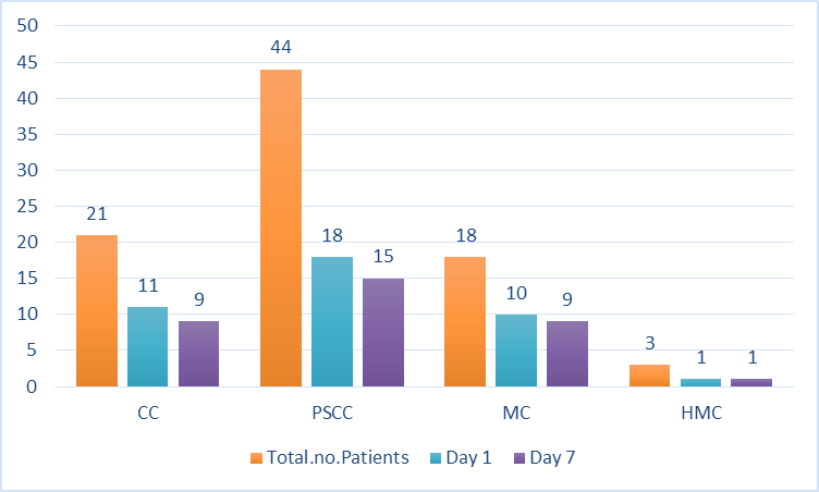
Table 1: Comparision Of Corneal Edema Based On Types Of Cataract On Day 1 & Day 7
Higher HbA1c levels on Day 1 and Day 7 were associated with increased corneal edema severity. By Day 7, patients with HbA1c <7 had improved from their mild, mostly lower-grade edema. On the other hand, there was minimal decrease over time in the number of Grade 2 and Grade 3 edema cases among patients with HbA1c >9. In general, more severe and long-lasting ocular edema was linked to poor glycemic control.(Table 2)
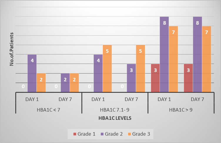
Table 2: Distribution of Corneal Edema Grades Across Different HbA1c Levels on Postoperative Days 1 and 7
Corneal edema was most common in patients with combination CC and PSCC on both Day 1 and Day 7. By Day 7, edema had slightly decreased in the majority of mixed cataract types. Slower resolution was suggested by the CC & MC group's lack of change over time. Corneal edema was minor in PSCC and HMC instances, probably because there were fewer individuals. Combinations including PSCC were often linked to increased ocular edema following surgery.(Table 3)
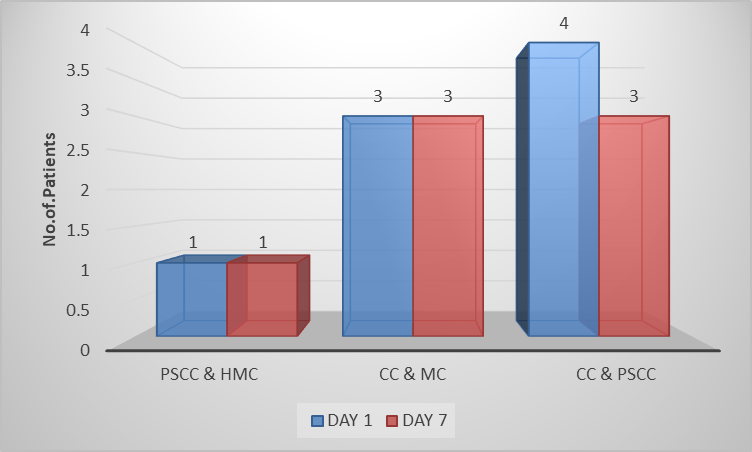
Table 3: Patient Distribution by Diagnosis: Day 1 vs Day 7
XFrom Day 1 to Day 7, both surgical methods demonstrated a decrease in corneal edema. Corneal edema in the SICS group decreased gradually after surgery, from 54% on Day 1 to 46% on Day 7. A more noticeable decrease was seen in the phacoemulsification group, where edema dropped from 62% on Day 1 to 38% on Day 7. In comparison to SICS, phacoemulsification was generally linked to a quicker clearance of corneal edema (Fig.1 & 2).
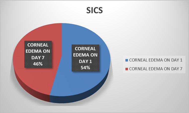
Figure1:Proportion of Corneal Edema in SICS Patients on Day 1 and Day 7”
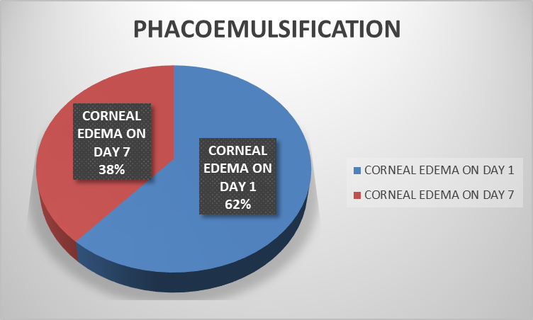
Figure2:Proportion of Corneal Edema in Patients on Day 1 and Day 7”
Overall the findings demonstrates that individuals with higher HbA1c levels—especially those with HbA1c >9—had more frequent and prolonged postoperative ocular edema. Compared to other cataract types, those undergoing SICS and those with posterior subcapsular components had a greater burden of edema. While corneal edema decreased from Day 1 to Day 7, poorly managed diabetics had a slower rate of resolution. These results emphasize how surgical variables and glucose condition affect early postoperative corneal healing.
DISCUSSION :
The current study assessed the relationship between the postoperative corneal edema and glycemic control, cataract features, and surgical technique. The results show a strong correlation between the occurrence and durability of ocular edema on postoperative Days 1 and 7 and elevated HbA1c levels. Corneal edema was much more common in patients with poor glycemic control (HbA1c >9), and it resolved extremely slowly over time, indicating decreased corneal endothelium regeneration in uncontrolled diabetes.
This observation is in line with findings from recent clinical research conducted in India, which indicates that patients with diabetes, particularly those with poor glycemic control, are more likely to develop than non-diabetics to experience postoperative complications, such as corneal edema, after cataract surgery, and that poor control has a negative impact on both visual recovery and complication rates(9).
Diabetes-related endothelial dysfunction can account for the observed correlation between higher HbA1c and ocular edema. Corneal deturgescence is known to be compromised by chronic hyperglycemia's effects on corneal endothelial cell density, Na+/K+-ATPase pump function, and oxidative stress. Even in the early postoperative phase, individuals with greater HbA1c levels may experience delayed clearance of edema due to these pathophysiological alterations.
In this study, surgical corneal outcomes were also influenced by cataract morphology. On both Day 1 and Day 7, posterior subcapsular cataracts were linked to a higher prevalence of corneal edema, especially when paired with cortical cataracts. These cataract varieties frequently necessitate more phaco energy and surgical manipulation, which increases endothelial stress and postoperative edema. Conversely, there were fewer incidences of edema in hypermature cataracts, which is probably due to the smaller sample size rather than a protective effect.
Additionally, studies have shown similar trends of increased endothelial vulnerability in diabetic eyes following cataract extraction, even though the majority of earlier Indian research has concentrated on overall complication profiles and endothelial changes rather than HbA1c-stratified corneal edema specifically. Our observation of prolonged edema with rising HbA1c is supported by comparative investigations showing increased corneal endothelial stress and delayed recovery following surgery in diabetics.(10)
McNemar's test revealed that although ocular edema improved in individuals with good to moderate glycemic control from Day 1 to Day 7, the change was not statistically significant across specific HbA1c categories. This implies that endothelium healing in diabetes patients may need a longer follow-up period to show substantial clinical resolution, even though numerical improvement does occur.
The study emphasizes how crucial thorough surgical planning and preoperative metabolic management are for diabetic individuals having cataract surgery. The risk and duration of postoperative corneal edema may be decreased by strict glycemic control, choosing the right surgical technique, and minimizing endothelial stress. The long-term effects of glycemic status on corneal recovery following cataract surgery would be further clarified by future research using bigger sample numbers, longer follow-up, and endothelial cell count monitoring.
Our study's trend supports these more general conclusions, demonstrating the critical role metabolic regulation plays in postoperative tissue response and healing. Tight glycemic control prior to surgery may lessen early postoperative corneal edema and improve overall recovery, according to our combined data, highlighting the importance of careful preoperative evaluation and perioperative optimization in diabetic cataract patients.
CONCLUSION :
This study shows that people with diabetes who have cataract surgery are more likely to experience ocular edema after the procedure. HbA1c can serve as a simple pre-operative predictor for corneal recovery. In order to reduce this risk, optimal preoperative glycemic management is essential. Effective surgical planning requires a thorough evaluation of the kind and density of cataracts. Intraoperative corneal stress can be decreased by using the right surgical approach. Corneal healing is enhanced by careful perioperative management, which includes monitoring and postoperative care. Timely interventions are possible when high-risk patients are identified early. These results support evidence-based methods and are consistent with current Indian research.For improved surgical results, systemic and ocular factors must be integrated in future to improve patient safety and practices,
BIBLIOGRAPHY:
1. Ladea L, Zemba M, Calancea MI, Călțaru MV, Dragosloveanu CD, Coroleucă R, Catrina EL, Brezean I, Dinu V. Corneal epithelial changes in diabetic patients: A review. International Journal of Molecular Sciences. 2024 Mar 19;25(6):3471.
2. Chen C, Zhang B, Xue J, Li Z, Dou S, Chen H, Wang Q, Qu M, Wang H, Zhang Y, Wan L. Pathogenic role of endoplasmic reticulum stress in diabetic corneal endothelial dysfunction. Investigative ophthalmology & visual science. 2022 Mar 2;63(3):4-.
3. Ghenciu LA, Hațegan OA, Bolintineanu SL, Dănilă AI, Faur AC, Prodan-Bărbulescu C, Stoicescu ER, Iacob R, Șișu AM. Immune-Mediated Ocular Surface Disease in Diabetes Mellitus—Clinical Perspectives and Treatment: A Narrative Review. Biomedicines. 2024 Jun 12;12(6):1303.
4. Yang C, An Q, Zhou H, Ge H. Research progress on the impact of cataract surgery on corneal endothelial cells. Advances in Ophthalmology Practice and Research. 2024 Nov 1;4(4):194-201.
5. Boroojeny AB, Nunney I, Dhatariya KK. The association between glycaemic control, renal function and post-operative ophthalmic complications in people with diabetes undergoing cataract surgery—a single-centre retrospective analysis. Diabetes Therapy. 2022 Apr;13(4):723-32.
6. Das S, Shergill SS. Cataract Surgery, Their Postoperative Analysis, and a Comparison of Phacoemulsification and Manual Small-Incision Cataract Surgery at a Tertiary Care Hospital: A Retrospective Study. Medical Journal of Dr. DY Patil Vidyapeeth. 2023 Jan 1;16(1):80-4.
7. Feizi S. Corneal endothelial cell dysfunction: etiologies and management. Therapeutic advances in ophthalmology. 2018 Dec;10:2515841418815802.
8. Ahmed SZ. Endothelial Cell Changes After Small Incision Cataract Surgery in Diabetic and Non Diabetic Patients–A Cohort Study (Master's thesis, Rajiv Gandhi University of Health Sciences (India)).
9. Uddin MF. Evaluating Surgical Outcomes of Cataract Surgery in Patients with Diabetes. Barind Medical College J. [Internet]. 2025 Dec. 15 [cited 2026 Jan. 23];11(2):34-9. Available from: https://bmcj.org/index.php/bmcj/article/view/228
10. Chaurasia RK, Khasnavis A, Mittal J. Comparison of corneal endothelial changes following phacoemulsification in diabetic and non-diabetic patients. Indian J Ophthalmol. 2022 Apr;70(4):1208-1213. doi: 10.4103/ijo.IJO_1955_21. PMID: 35326017; PMCID: PMC9240528.