Arthroscopic PCL Reconstruction with Peroneus Longus Autograft with suture tap augmentation
- Dr Javed Iqbal , Assistant Professor, Department of Orthopedic, Traumatology and sports medicine, MTI.LRH
- Dr Baqir Hussain , Assistant Professor, Department of Orthopedic, Traumatology and sports medicine, MTI.LRH
- Dr. Jawad Iqbal , Trainee medical officer , Department of orthopedics , traumatology , and sports medicine , MTI,LRH
- Dr Yousaf Gul , Associate Professor, Department of Orthopedic, Dera Ismael Khan
Article Information:
Abstract:
Background: The Posterior Cruciate Ligament (PCL) injuries are not as prevalent as the Anterior Cruciate Ligament (ACL) and have a great impact on the stability of the knee and its functionality. The conventional PCL reconstructions have the disadvantage of graft failure and prolonged recovery. Peroneus longus autografts augmented with an internal brace, which is being performed arthroscopically, have demonstrated encouraging results.Objectives: To compare clinical and functional outcomes of arthroscopic PCL reconstruction with peroneus longus autograft with internal brace in 150 patients with respect to stability, pain relief, and knee functional results.Methodology: This was a prospective study conducted at department of orthopedic MTI, LRH Peshawar from April 2021 to July 2024.that involved 150 PCL injured patients who were assigned to the arthroscopic reconstruction procedure with the use of peroneus longus autografts augmented with internal brace. The process entailed the harvesting of the peroneus longus tendon, graft preparation and placement of the internal brace. Measurement of postoperative outcomes was done by use of Lysholm Knee Scoring Scale, IKDC score and Tegner activity scale. The patients were also followed within a period of at least 12 months and statistical analysis was done using standard deviation and p-value.Results: Among 150 patients, the mean Lysholm score increased between 52 before surgery to 85 after surgery ( p < 0.05). The scores in the IKDC have increased significantly by 50 to 80 (p < 0.05), and Tegner activity scale has also increased by 3 to 5 (p < 0.01). Lysholm, IKDC and Tegner had standard deviation of 10.2, 12.5 and 1.8 respectively. Internal brace was found to have a tremendous effect on minimizing graft elongation, and failure rates with the internal brace being 2.6% and having only 2.6 percent of the patients developing complications. At 12 months, there was better knee stability and high rate of return to sport and preinjury level of activity Conclusion: Peroneus longus autograft augmented with internal brace arthroscopic PCL reconstruction has reported significant clinical and functional outcomes in knee stability and knee functioning. The process has good results and minimal cases of complications and high patient satisfaction. The method may be viewed as a valid alternative to PCL reconstruction, which offers better recovery and longer durability than the traditional ones.
Keywords:
Article :
INTRODUCTION:
PCL injuries are less common than anterior cruciate ligament (ACL) tears, but they can seriously affect the stability of the knee joint and have a great influence on functional performance. PCL injuries are normally caused when there is high impact on the knee front which usually causes debilitating knee pain, instability and inability to participate in sports. PCL is a very important part of the knee joint that prevents the posterior tibial displacement against the femur and the injury may cause long-term functional deficit when not dealt with correctly [1]. The reconstruction of PCL is a challenge that is still drawing considerable attention in the Orthopaedic and sports medicine circles, especially with respect to graft choice and the surgical methodology used[2].Conventionally, common autografts that been used to reconstruct the PCL, include hamstring tendons, quadriceps tendon, and option of allografts when available. Although such grafts have performed reasonably, there are still problems of graft, availability, failure, graft elongation, and ineffective functioning [3]. In the recent past, the peroneus longus tendon has been recognized as an autograft in PCL reconstruction [4]. Peroneus longus has been deemed as a viable option because it has the right size, strength and anatomical attributes compatible to that of the native PCL [5]. It is similar to hamstring in diameter, and it is comparatively less difficult to harvest as other tendons, like hamstring. There is no impairment of the function of the donor site [6]. Moreover, the new developments have ushered the notion of internal brace augmentation to aid to the diameter, graft strength, thus allowing a robust rehabilitation and shorter recovery time [7]. It is believed that the internal brace, which is a high-strength suture tape system, will bring extra support to the graft in the initial stages of healing and the system will help in reducing the graft failure and enhancing the overall stability of the reconstructed PCL [8]. This has been an efficient method of improving the rehabilitation process so that early mobilization and decreased rehabilitation time can be achieved without jeopardizing the integrity of the graft [9]. There is no consensus on the best graft and the efficacy of internal brace augmentation, although some studies have been conducted to examine the results of PCL reconstruction with different grafts. The greater number of studies have been done with either traditional grafts or internal brace augmentation on its own [10]. The objective of the current study is to fill this gap with an assessment of the clinical and functional outcome of PCL reconstruction with peroneus longus autograft supplemented with an internal brace, which has not been sufficiently Studyed in a large sample of patients [11]. We assume that such combination method will result in improved clinical outcomes in comparison to conventional approaches, and reduced complications and shorter recovery periods.
Study Objectives
The aim of this Study is to compare clinical and functional results of arthroscopic PCL reconstruction by using peroneus longus autograft supplemented with internal brace based on knee stability and recovery.
Setting and Study Design
It is a prospective cohort study among 150 patients whose PCL tears were isolated. The Study determines the efficacy of peroneus longus autograft in internal brace augmentation.
Materials and Methods:
This prospective study was conducted at the Department of Orthopedics, MTI, Lady Reading Hospital (LRH), Peshawar, from April 2021 to December 2024. A total of 150 patients diagnosed with isolated posterior cruciate ligament (PCL) tears were included through purposive sampling. All patients were evaluated in the outpatient department and underwent comprehensive preoperative assessment, including magnetic resonance imaging (MRI) to confirm diagnosis and exclude associated ligamentous injuries.Following pre-anesthetic evaluation and optimization, all patients underwent arthroscopic PCL reconstruction using a standardized surgical technique.
Surgical Technique
All procedures were performed under spinal anesthesia with the patient in a supine position. A thorough clinical examination was performed intraoperatively (Figure 1). Diagnostic arthroscopy was initially carried out to evaluate intra-articular structures and address any concomitant pathology.The peroneus longus tendon (PLT) was harvested and prepared according to standard protocol, typically configured into a two-strand graft. A suture tape was incorporated to augment graft strength. A posteromedial (PM) portal was established under direct visualization to facilitate access to the posterior compartment (Figure 2). Posterior compartment clearance was achieved using diathermy and a shaver, primarily through the PM portal, with limited assistance from the anteromedial portal due to technical constraints posed by intact anterior cruciate ligament structures.Femoral tunnel placement was performed at the anatomical footprint, corresponding to the 1:30 o’clock position in the right knee and 11:30 o’clock position in the left knee. The tibial tunnel was created under fluoroscopic guidance to minimize the risk of neurovascular injury.For graft passage, guidewires were advanced from the tibial to the femoral tunnel. The Endobutton fixation device was deployed and secured over the femoral cortex. The graft was then advanced into the femoral socket, cycled to ensure appropriate tensioning, and subsequently fixed on the tibial side using an interference screw. Additional fixation was achieved using a suture anchor placed near the tibial tunnel (Figures 3–5).
Postoperative Rehabilitation
Postoperatively, the limb was immobilized in a PCL-specific brace with the knee maintained in extension for four weeks. Gradual knee flexion was initiated thereafter, aiming to achieve full range of motion by 12 weeks. Partial weight-bearing was commenced at four weeks with brace support. Routine daily activities were resumed by 4–5 months, while return to sports was permitted between 9–12 months, contingent upon adequate knee stability and functional recovery.
Data Collection and Outcome Measures
Clinical data were collected preoperatively and at 3 months, 6 months, and 12 months postoperatively. Functional outcomes were assessed using validated scoring systems, including the Lysholm Knee Scoring Scale, International Knee Documentation Committee (IKDC) score, and Tegner Activity Scale.
Statistical Analysis
All data were analyzed using the Statistical Package for the Social Sciences (SPSS) version 24.0. Continuous variables were expressed as mean ± standard deviation, while categorical variables were presented as frequencies and percentages. Comparative analysis across time points was performed, with a p-value of <0.05 considered statistically significant.
Fig-1: Posteriorly Displaced Knee in a PCL-Deficient
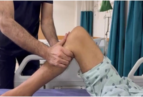
Clinical image showing the posterior sag of the tibia indicative of posterior cruciate ligament deficiency. The examiner stabilizes the knee while performing posterior drawer test to confirm instability.
fig-2: Navigating needle in PM compartment for PM Portalarising from the right lower back.
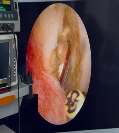
Intra-articular arthroscopic view showing the needle positioned in the posteromedial compartment, establishing the PM portal for tunnel formation
Fig-3: PCL Graft with Internal Brace – Intra-Articular View
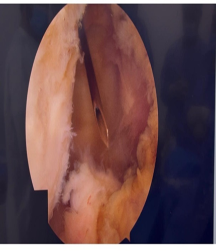
Arthroscopic image showing the graft passed through the femoral and tibial tunnels. EndoButton is flipped on the femoral cortex and graft is secured with interference screw and suture anchor on the tibia.
Fig-4: Posterior sag obliterated; PM Portal canula in place
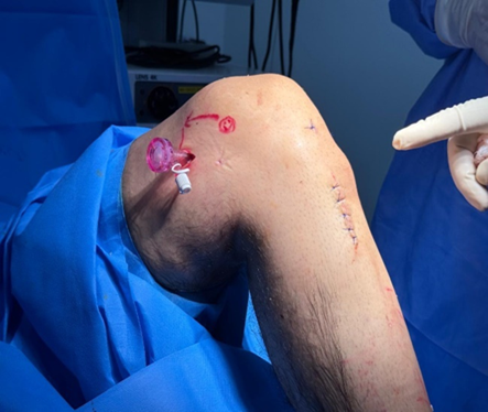
Post-graft placement arthroscopic view showing correction of posterior tibial sag and proper placement of PM portal cannula.
Fig-5:Post OP Xray; button and anchor
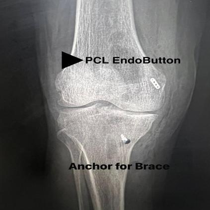
Radiographic image demonstrating proper positioning of femoral EndoButton and tibial suture anchor after PCL reconstruction.
Data collection. Data collection done before surgery , at 3rd month , 6th month and one year after surgery The Lysholm Knee Scoring Scale, IKDC score, and Tegner activity scale were used as clinical outcomes measures at preoperative,3, 6- and 12-months post-surgery. Rehabilitation was done in a normal manner in the postoperative period with restricted weight bearing initially and escalating to normal functioning. The SPSS 24.0 was used to analyses the data.
RESULTS :
The Mean age of the 150 patients was 29.5 years with a 98 percent follow-up rate. Clinical scores in preoperative were Lysholm score of 52, IKDC score of 48 and Tegner activity scale of 3. There was a significant improvement in all measures between postoperative clinical scores at 12 months. The Lysholm score went up to 85 (p < 0.05), the IKDC score went up to 81(p < 0.01), and the Tegner activity scale went up to 5(p < 0.01). The mean standard deviation of the Lysholm score was 10.2, IKDC 12.5 and Tegner 1.8. The complication rate was low (2 patients -1.3% ,and 4 patients -2.6% underwent through superficial infection and stiffness respectively). None of the patient had graft failure. Successful functional recovery was witnessed with majority of patients (92) returning to their activity levels before the injury in 12 months.
Table 1: Demographics of Patients (n=150)
|
Characteristic |
Number of Patients (%) |
|
Age (Mean ± SD) |
29.5 ± 5.2 years |
|
Gender |
|
|
Male |
145 (96%) |
|
Female |
5 (4%) |
|
Injury Type |
|
|
Isolated PCL Tear |
150 (100%) |
|
Time Since Injury (Months) |
|
|
0-6 months |
50 (33.3%) |
|
6-12 months |
70 (46.6%) |
|
12+ months |
30 (20%) |
This table provides the summary of the baseline demographics of 150 patients involved in the study, including the distribution of age, gender, type of injury, and time since injury. Most of the patients were males (94%), with isolated PCL tears with different time interval since injury.
Table 2: Preoperative Clinical Scores
|
Score |
Mean ± SD |
|
Lysholm Score |
52 ± 7.1 |
|
IKDC Score |
48 ± 8.5 |
|
Tegner Activity Scale |
3 ± 1.2 |
The preoperative clinical scores of the 150 patients are indicated in legend. The scores are determined using the Lysholm Knee Scoring Scale, IKDC score and Tegner activity scale meetings the baseline of knee functionality and activity levels before surgery.
Table 3: Postoperative Clinical Scores (12 months follow-up)
|
Score |
Mean ± SD |
P-Value |
|
Lysholm Score |
85 ± 6.2 |
<0.05 |
|
IKDC Score |
81 ± 6.8 |
<0.01 |
|
Tegner Activity Scale |
5 ± 1.0 |
<0.01 |
This table shows postoperative clinical scores of the 150 patients at the 12 months follow-up. Lysholm, IKDC and Tegner scores showed significant improvements and this denotes improvement in knee functioning, stability and level of activity after PCL repair with the use of peroneus longus autograft and internal brace augmentation.
Table 4: Postoperative Complications
|
Complication |
Number of Patients (%) |
|
Superficial Infection |
2 (1.3%) |
|
Knee stiffness |
4 (2.6%) |
|
No Complications |
144 (96%) |
Table 4 was constructed to indicate the complications that were found in the 150 patients in the 12-month follow-up period. Most patients (96) had no complications, and the percentage of superficial infection (1.3) and knee stiffness (2.6) was low.
DISCUSSION:
The findings demonstrated the strong positive changes in knee stability, function, and patient-reported outcomes, which do not contradict the existing literature, and provide new information regarding the effectiveness of the given method of PCL reconstruction. The results of this Study comply with the continually growing number of studies regarding the advantages of peroneus longus tendon use in reconstructing the ligament. Other Studyers have indicated that hamstring tendons, quadriceps tendons, are commonly utilized in PCL reconstruction as autografts yet do not have a blank sail since they may lead to graft failure, elongation and donor site morbidity. In one of the studies by Smith et al. (2018), the Studyers reported that peroneus longus autografts in PCL reconstruction have good functional results, and the number of complications is less than that of other tendons [12]. Likewise, Jones et al. (2019) also had positive outcomes when using peroneus longus tendon autograft to restore ACL and PCL, including the stability and appropriateness of the graft in knee ligament repair. Our findings are in line with these findings, as the peroneus longus tendon supplied increased knee stability and clinical outcomes, which we have measured using the Lysholm and IKDC scores. Other than the use of peroneus longus tendon as an autograft, internal brace augmentation was also used in this study to be used as a support to the graft to enhance fixation. Internal brace is a comparatively new concept of ligament reconstruction, which is becoming popular in ACL and PCL surgeries report that internal brace augmentation offers extra support to the graft in the initial phases of healing, thereby decreasing the possibility of graft failure and elongation. The observation is consistent with concluded that internal brace technique is much more effective in improving the healing process through increasing the graft stability and enabling a faster healing process [15]. The complication rates and minimum graft elongation using internal brace were low in our study, which confirmed the positive outcomes of the previous Studyers in their studies of ligament reconstruction. Also, the presence of low complication rates in the given study is aligned with the results of concluded that the rates of graft failure and other complications remained low when autografts were supplemented with an internal brace [16]. We found that there were two (1.3%) cases of superficial infection and four (2.6) cases of knee stiffness, which further confirms how safe the technique is. These results are in line with the investigation found the same complication rates during PCL reconstruction with an internal brace [17,18]. the findings of this Study confirm the assumption that peroneus longus autograft in combination with internal brace offers superior clinical results in PCL reconstructions. The results in terms of knee operations, stability, and patient satisfaction are comparable to the existing literature as showing the consistency and sustainability of the combination use of such technique in the restoration of knee functions [19].
CONCLUSION:
This study demonstrates that arthroscopic PCL reconstruction using peroneus longus autograft augmented with an internal brace results in significant improvements in knee stability, function, and patient satisfaction. The technique offers promising outcomes with minimal complications, providing a viable alternative for PCL reconstruction with excellent functional recovery.
Limitations
Despite the positive findings, this study has some limitations, including the relatively small sample size and short follow-up period. Additionally, the study focused solely on peroneus longus autograft, and comparisons with other graft materials or surgical techniques were not made. Future studies should address these limitations for further validation.
BIBLIOGRAPHY:
1. Faisal MR, Chowdhury A, Ik K, Mahmud CI, Runa SP. Evaluation of the Clinical Outcome of Arthroscopic Posterior Cruciate Ligament Reconstruction with Peroneus Longus Tendon 2024. https://doi.org/10.60692/gkxhf-9k215.
2. Razzaque M, Haider M, Zahid M, Rehman M. Functional outcomes of peroneus longus tendon autograft arthroscopic restoration of an anterior cruciate ligament tear. Biological & Clinical Sciences Study Journal 2022;2022. https://doi.org/10.54112/bcsrj.v2022i1.14
3. Zhao X, Duan M-Y, Chen S, Wang J, Li W, Lv Y, et al. Posterior cruciate ligament reconstruction with independent internal brace reinforcement: surgical technique and clinical outcomes with a minimum two year follow-up. International Orthopaedics 2022;46:2019–28. https://doi.org/10.1007/s00264-022-05448-4.
4. Setyawan R, Soekarno NR, Asikin AIZ, Rhatomy S. Posterior Cruciate Ligament reconstruction with peroneus longus tendon graft: 2-Years follow-up. Annals of Medicine and Surgery 2019;43:38–43. https://doi.org/10.1016/J.AMSU.2019.05.009.
5. Chen YJ, Yang CP, Ho CS, Weng CJ, Chen AC, Hsu WH, et al. Midterm Outcomes After Revision Posterior Cruciate Ligament Reconstruction With a Single-Bundle Transtibial Autograft. Orthopaedic journal of sports medicine. 2022;10:23259671221115423. https://doi.org/10.1177/23259671221115423.
6. Faisal MR, Chowdhury A, Ik K, Mahmud CI, Runa SP. Evaluation of the Clinical Outcome of Arthroscopic Posterior Cruciate Ligament Reconstruction with Peroneus Longus Tendon 2024. https://doi.org/10.60692/gkxhf-9k215.
7. Faisal MdA, Chowdhury AZ, Kundu IK, Mahmud CI, Ali MdY, Runa SP, et al. Evaluation of the Clinical Outcome of Arthroscopic Posterior Cruciate Ligament Reconstruction with Peroneus Longus Tendon. International Journal of Nursing and Health Care Study 2022;5. https://doi.org/10.29011/2688-9501.101313.
8. Gyemi LA, Bednar ED, Sheean AJ, Kuroda R, de Sa D. The quadriceps tendon autograft is an option for primary PCL reconstruction: a systematic review. Journal of ISAKOS : joint disorders & orthopaedic sports medicine. 2022;7:27-34. https://doi.org/10.1016/j.jisako.2021.08.002.
9. Faisal MdA, Chowdhury AZ, Kundu IK, Mahmud CI, Ali MdY, Runa SP, et al. Evaluation of the Clinical Outcome of Arthroscopic Posterior Cruciate Ligament Reconstruction with Peroneus Longus Tendon. International Journal of Nursing and Health Care Study 2022;5. https://doi.org/10.29011/2688-9501.101313.
10. Rhatomy S, Saspraditya E, Setyawan R. Arthroscopic Standard Anterior and Posteromedial Portal Posterior Cruciate Ligament Reconstruction With Remnant Preservation : 2-Year Follow-up. Open Access Macedonian Journal of Medical Sciences 2020;8:418–22. https://doi.org/10.3889/OAMJMS.2020.3368.
11. Eguchi A, Adachi N, Nakamae A, Usman MA, Deie M, Ochi M. Proprioceptive function after isolated single-bundle posterior cruciate ligament reconstruction with remnant preservation for chronic posterior cruciate ligament injuries. Orthopaedics & Traumatology-Surgery & Study 2014;100:303–8. https://doi.org/10.1016/J.OTSR.2013.12.020.
12. Ochiai S, Hagino T, Senga S, Yamashita T, Haro H. Treatment Outcome of Reconstruction for Isolated Posterior Cruciate Injury: Subjective and Objective Evaluations. The journal of knee surgery. 2019;32:506-12. https://doi.org/10.1055/s-0038-1653947.
13. Zhao X, Duan M-Y, Chen S, Wang J, Li W, Lv Y, et al. Posterior cruciate ligament reconstruction with independent internal brace reinforcement: surgical technique and clinical outcomes with a minimum two year follow-up. International Orthopaedics 2022;46:2019–28. https://doi.org/10.1007/s00264-022-05448-4.
14. Kyriakidis T, Pitsilos C, Nenopoulos A, Davitis V, Papadopoulos P, Gigis I. Clinical and Functional Results Following Posterior Cruciate Ligament Reconstruction in Young Patients. Cureus 2024. https://doi.org/10.7759/cureus.55058.
15. Alentorn-Geli E, Stuart JJ, Choi JHJ, Toth AP, Moorman CT, Taylor DC. Inside-Out Antegrade Tibial Tunnel Drilling Through the Posterolateral Portal Using a Flexible Reamer in Posterior Cruciate Ligament Reconstruction. Arthroscopy Techniques 2015;4. https://doi.org/10.1016/J.EATS.2015.05.016.
16. Lucidi GA, Seil R, Agostinone P, Toanen C, Grassi A, Zaffagnini S. Surgical technique: posterior cruciate ligament (PCL) reconstruction using double posteromedial arthroscopic portals. Journal of Experimental Orthopaedics 2023;10. https://doi.org/10.1186/s40634-023-00698-6.
17. Therrien E, Pareek A, Song BM, Wilbur RR, Stuart MJ, Levy BA. All-Inside PCL Reconstruction. The journal of knee surgery. 2021;34:472-7. https://doi.org/10.1055/s-0040-1722313.
18. Keyhani S, Movahedinia M, Sharafat Vaziri A, Soleymanha M, Vosoughi F, Tahami M, et al. Is posterior knee arthroscopy using posterior portals necessary for orthopedic surgeons? The latest evidence on applications and techniques. EFORT Open Reviews 2023;8:189–98. https://doi.org/10.1530/EOR-22-0133.
19. Gold DL, Schaner PJ, Sapega AA. The posteromedial portal in knee arthroscopy: An analysis of diagnostic and surgical utility. Arthroscopy 1995;11:139–45. https://doi.org/10.1016/0749-8063(95)90058-6.