Titanium-reinforced expanded polytetrafluoroethylene membrane in vertical ridge augmentation—A Systematic Review
- Dr Snigdha S K , Department of Periodontics, KLE VK Institute of Dental Sciences, Belagavi, India
- Dr Vinayak Kumbhojkar , Dean and Professor, Department of Periodontics, KLE VK Institute of Dental Sciences, Belagavi, India
Article Information:
Abstract:
This systematic review assessed the effectiveness of titanium-reinforced expanded polytetrafluoroethylene (e-PTFE) membranes in vertical ridge augmentation (VRA). A comprehensive literature search was performed in PubMed, Scopus, ScienceDirect, and Google Scholar for studies published between 2006 and 2024. Six studies met the inclusion criteria, comprising randomised controlled trials and prospective clinical investigations with follow-ups ranging from 6 months to 5 years. The included studies evaluated vertical bone gain, implant survival, membrane exposure, and complications. Titanium-reinforced e-PTFE membranes consistently achieved significant vertical bone augmentation, with mean bone gains exceeding 4 mm in most studies. Their mechanical strength and rigidity provided superior space maintenance and stability, supporting predictable bone regeneration and high implant survival rates (>90%). However, membrane exposure was a common complication, reported in 15–40% of cases, often leading to partial loss of grafted bone. The requirement for a secondary procedure to remove the non-resorbable membrane was another drawback. Despite variations in study design, graft materials, and follow-up periods, the overall evidence supports the efficacy of titanium-reinforced e-PTFE membranes in achieving reliable vertical ridge augmentation. Careful case selection, meticulous surgical technique, and stringent plaque control are essential to minimise complications and optimise outcomes. Further long-term randomised controlled trials with standardised protocols are necessary to confirm these findings and establish clinical guidelines for their optimal use in vertical ridge reconstruction.
Keywords:
Article :
INTRODUCTION:
Over the past decade, vertical ridge augmentation techniques have evolved significantly. Guided bone regeneration (GBR) has proven predictable in short- and long-term studies. Simion et al first demonstrated vertical bone augmentation in atrophic ridges using a titanium-reinforced e-PTFE membrane in 1994, with subsequent studies confirming that particulated autogenous bone graft enhances regeneration potential.¹,²
Autogenous bone remains the "gold standard" for GBR-based vertical ridge augmentation due to its biocompatibility, osteogenic and osteoinductive properties, and space-maintaining capacity, despite being technically demanding to harvest. In the early 1990s, Mellonig et al proposed demineralised freeze-dried bone allograft (DFDBA) as an alternative.³ Simion et al later confirmed beneficial effects of DFDBA under membranes,⁴ though its osteoinductive reliability remains controversial.⁵,⁶ A malleable allogeneic bone matrix (Regenaform) was introduced to address this inconsistency, combining assayed DFDBA with cortico-cancellous chips in a thermoplastic collagen carrier, tested for osteoinductivity per lot.⁷,⁸,⁹
Bone deficiency remains the primary limitation for implant-prosthetic rehabilitation. While short/ultra-short implants may suffice in mild atrophy, GBR is indicated where insufficient bone volume precludes implant placement or where functional and esthetic demands require augmentation. GBR relies on barrier membranes to exclude soft tissue cells and allow osteoprogenitor colonization.¹⁰,¹¹ Non-resorbable PTFE membranes offer durable cell exclusion and, with titanium reinforcement, space maintenance in non-contained defects. Collagen membranes, though biocompatible, require titanium support to prevent collapse in severe defects. Multiple studies confirm GBR efficacy for vertical augmentation,¹²,¹³,¹⁴ and recent systematic reviews support it as the most predictable approach.¹⁵ However, randomized clinical trials on peri-implant bone levels, crestal bone loss, and soft tissue outcomes remain scarce, warranting a comprehensive systematic review.
RESULTS:
The present systematic review was conducted to assess whether the titanium-reinforced polytetrafluoroethylene membrane is efficacious in vertical ridge augmentation. The screening process was undertaken in three steps that included screening of titles, followed by screening of abstracts and finally screening of full text for inclusion in the review. The characteristics of the studies included in the systematic review are presented in the tables below.”
“Table no.1- Details of the studies included in the systematic review
|
Study Id |
Author |
Year |
Study design |
Sample size |
|
1 |
Park JW et al |
2006 |
A Report of Four Cases |
n=4 patients |
|
2 |
Merli M et al |
2007 |
A Preliminary Report of a Blinded Randomized Controlled Clinical Trial |
n=22 |
|
3 |
Fontana F et al |
2008 |
A Prospective Pilot Study |
n=25 implants (10 edentulous areas, 5 patients) |
|
4 |
Siciliano VL et al |
2011 |
A 12-Month Randomized Controlled Clinical Trial |
n=40 patients |
|
5 |
Ronda M et al |
2013 |
a prospective randomized controlled clinical trial |
n=23 patients (78 implants) |
|
6 |
Cucchi A et al |
2020 |
1-year results of a randomized clinical trial. |
n=40 patients |
Table 1 represents eight studies included in the systematic review as per the pre-defined eligibility criteria. All studies evaluated whether the titanium-reinforced expanded polytetrafluoroethylene membrane is efficacious in vertical ridge augmentation. With respect to publication year, the studies were published from 2006 to 2020. Regarding the study design, from the included studies, 4 studies were randomised controlled studies/trials, 1 study was a prospective study, and the remaining study was a case report. The sample size across the studies ranged from as less as 4 to a maximum of 40 patients. In the case report, 4 cases were analysed; in the randomised controlled studies, the majority of them were single-blinded.
Table 2- Details of the study participants, intervention, and comparator of the studies included in the systematic review
|
Sr. no |
1 |
2 |
3 |
4 |
5 |
6 |
|
Author |
Park JW et al |
Merli M et al |
Fontana F et al |
Siciliano VL et al |
Ronda M et al |
Cucchi A et al |
|
Population |
patients who received bone augmentation surgery because of inadequate alveolar ridge widths for implant placement |
partially edentulous patients requiring bone augmentation |
patients with bilateral posterior mandibular partial edentulism. Patients were treated with a split-mouth design approach |
patients with deep, non-contained intrabony defects (i.e., with a ‡80% 1-wall component and a residual 2- to 3-wall compo nent inthemostapicalpart) |
patients requiring bone augmentation with guided bone regeneration (GBR) procedures for placing implants in atrophic posterior mandibles (available bone height |
patients with vertical defects were enrolled and treated according to the study protocol. |
|
Interventions/treatments used (Test group) |
bioactive glass particles of a narrow size range with GBR procedure and titanium-reinforced ex |
resorbable collagen barriers supported by osteosynthesis plates (test) |
test group (titanium-reinforced e-PTFE membrane and allogeneic bone matrix) |
an enamel matrix derivative(EMD) |
A composite bone graft (50% autologous bone) d-PTFE membrane (test) |
reinforced-PTFE membranes (group-A) |
|
Exposure/Comparator (Control group) |
NA |
nonresorbable titanium-reinforced e- polytetrafluoroethylene (e-PTFE) barrier (control) |
control group (titanium-reinforced e-PTFE membrane and autogenous bone chips) |
guided tissue regeneration(GTR) |
50% mineralized bone allograft : e-PTFE membrane (control) |
titanium-meshes plus collagen membranes (group-B). |
|
Follow-up period |
6 months, 18 months |
5 years |
5 months, 1 year and 3 years |
At baseline and after 12 months |
6 months |
at baseline and after 1 year |
|
Primary outcomes |
area of newly formed bone (NB%, area of newly formed mineralized bone expressed as a percentage of the total de fect area) and remaining graft particle area |
amount of vertically regenerated bone measured intrasurgically, and biologic complications |
clinical parameters including the amount of vertically regenerated bone (DSB) and biologic complications were recorded. Histomorphometric analysis and the bone-implant contact percentage were performed |
clinical parameters including probing depths (PDs) and clinical at tachment levels (CAL) were recorded. |
healing, vertical defects |
peri-implant-bone-levels (PBL), interproximal bone-peaks (IBP), pocket-probing-depth (PPD), bleeding-on-probing (BoP), plaque-index(mPI), gingival-index(mGI), keratinized-tissue-thickness/width (tKT and wKT), and fornix-depth (FD). |
|
Secondary outcome, Any aditional outcomes |
NR |
NR |
NR |
NR |
NR |
NR |
|
Results |
The augmented sites showed clinically significant increases in alveolar ridge width. Implant stability was achieved by using long implants that engaged with the lateral or apical native bony wall. |
There was no statistically significant difference in bone gain between the 2 procedures |
Verti cal bone regeneration was evident in both groups since all the samples demonstrated trabecular bone with different degrees of maturation and mineralization in the regenerated area |
The mean CAL gain at sites treated with GTR was significantly greater |
The normalized data (percentage changes against baseline) did not show any statistically significant difference between test and control groups (P = NS). |
After 1 year, implants showed a change of PBL from 0.12 to 0.76 mm, with marginal bone loss of 0.67 and 0.61 mm for group A and B, respectively, without significant differences |
|
Conclusion |
the increase in the width of the alveolar ridge induced by bioactive glass was sufficient for placement of an implant combined with a titanium reinforced e-PTFE barrier membrane |
Both techniques were effective in augmenting bone; however, both were associated with complications. Clinicians and patients must care- fully weigh risks and benefits when considering the use of vertical guided bone regeneration |
it appears that the behavior of the allogeneic bone matrix is similar to that of autogenous bone chips when used for vertical ridge augmentation by means of guided bone regeneration techniques. Both grafts demonstrated analogous histologic characteristics |
Although the outcomes of open-flap debride ment alone were not investigated, the application of EMD alone appeared to yield less PD reduction and CAL gain com pared to GTRtherapyinthetreatmentofdeep, non-contained intrabony defects |
both d-PTFE and e-PTFE membranes showed identical clinical results in the treatment of vertical bone defects around implants, using the GBR technique |
The results indicate that GBR treatment with titanium-meshes plus collagen membranes (Group B) compared to reinforced-PTFE membranes does not appear to be inferior or superior in terms of PBL change |
NA: Not applicable/available, NR – Not reported.
Table 2 represents study characteristics with respect to sample, intervention, comparator, outcome, results and conclusion. The age of the patients in the included studies ranged from 30 years to 78 years. The mean age across the studies was 45.43 years. The population in the studies included the patients with bilateral posterior mandibular partial edentulousness, patients with deep, non-contained intrabony defects and those with vertical defects.
In the studies included in this review, the Interventions/treatments used (Test group) consisted of titanium-reinforced e-PTFE membrane and allogeneic bone matrix; in some studies, bioactive glass particles of a narrow size range with GBR procedure and titanium-reinforced expanded polytetrafluoroethylene (TR e-PTFE) membranes were used.
The Exposure/Comparator (Control group) was used in only randomised controlled studies and prospective studies; whereas for case reports, there was no control group. The comparator/control group consisted of nonresorbable titanium-reinforced e-polytetrafluoroethylene (e-PTFE) barrier or just the guided tissue regeneration(GTR). In the majority of the studies, the follow-up period ranged from baseline to up to a maximum of 5 years, and the majority of the studies had a follow-up of 12 months.
The primary clinical outcomes in all of the included studies were the amount of vertically regenerated bone measured intrasurgically and biologic complications. The clinical parameters, including the amount of vertically regenerated bone (DSB) and biologic complications, were recorded. Histomorphometric analysis and the bone-implant contact percentage were performed. There were no secondary outcomes reported across studies. Quantitative data were presented in the form of different outcomes and presented as mean and standard deviation. The majority of the study results showed that Vertical bone regeneration was evident in both groups since all the samples demonstrated trabecular bone with different degrees of maturation and mineralisation in the regenerated area. A few studies did not showstatistically significant differences between the 2 groups. The conclusion reported across studies was that both techniques were effective in augmenting bone; however, both were associated with complications. Clinicians and patients must carefully weigh risks and benefits when considering the use of vertical guided bone regeneration. Furthermore, both dense-PTFE and expanded-PTFE membranes showed identical clinical results in the treatment of vertical bone defects around implants, using the GBR technique.
RISK OF BIAS
The risk of bias was assessed for the 6 included studies. The RoB of 4 studies was done via the Cochrane RoB2 tool for randomised controlled studies. For one prospective study, a Newcastle Ottawa scale was used to determine the risk of bias, and for the remaining case report, the JBI’s critical appraisal tool was used.
A) Risk of Bias for Randomised Controlled studies
The RoB2 tool revealed that the majority of the studies (2) had low risk of bias, followed by 2 studies which had some concerns for missing outcome data domain. (Figure 2a and Figure 2b). The summary plot showed that about 50% low risk of bias was noted and about 50% some concerns for a missing outcome data domain in 2 studies. Thus, it can be interpreted that the overall low risk of bias is noted across all the studies.
Figure 2a: Risk of bias using RoB-2 tool
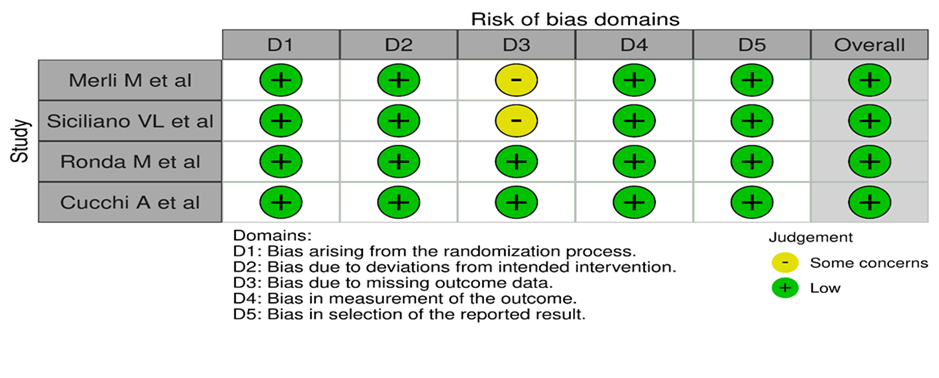
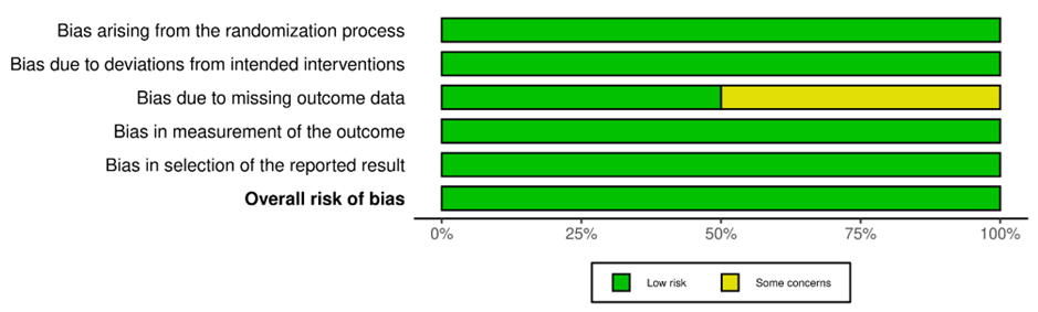
Figure 2b: RoB2 “Summary Plot” distribution of risk of bias among the studies
A) Risk of Bias for Prospective Studies
Only 1 study included in the systematic review was assessed for quality assessment by using the Newcastle Ottawa scale. The scale showed a score of 6/9, the main issues being a lack of adequate diagnosis, definition of controls, and lack of adjustment for potential confounders (Table 3).
Table 3- Study Quality as Assessed by the Newcastle Ottawa Scale as Judged by the 2 Reviewers Who Performed Data Extraction
|
S. No. |
Authors |
Year |
Selection (Maximum, 4 Asterisks) |
Comparability (Maximum, 2 Asterisks) |
Exposure/Intervention (Maximum, 3 Asterisks) |
|
|
Fontana F et al |
2008 |
*** |
** |
* |
C) Risk of Bias for Case Reports
Risk of bias assessment of one case report was performed. A JBI critical appraisal checklist for Case reports was used.
Table 4 – Critical appraisal of Case reports (JBI appraisal tool)
|
Study ID |
Author |
Were patient’s demographic characteristics clearly described? |
Was the patient’s history clearly described and presented as a timeline |
Was the current clinical condition of the patient on presentation clearly described? |
Were diagnostic tests or assessment methods and the results clearly described? |
Was the intervention(s) or treatment procedure(s) clearly described? |
Was the post-intervention clinical condition clearly described? |
Were adverse events (harms) or unanticipated events identified and described? |
Does the case report provide takeaway lessons? |
|
1 |
Park JW et al |
Yes |
Yes |
Yes |
Yes |
Yes |
Yes |
Unclear |
Yes |
The above table shows the critical appraisal of the case report included in the review. It has been found that the case report showed the majority of the items of appraisal as yes; whereas only an unclear outcome was reported for adverse events (harms) or unanticipated events not identified and described. Thus, when the overall quality assessment or Risk of bias is done, it can be interpreted that the case report shows a low risk of bias.
DISCUSSION:
This systematic review evaluated the clinical efficacy and limitations of titanium-reinforced e-PTFE membranes in vertical ridge augmentation (VRA).
Findings consistently demonstrated substantial vertical bone gain, often exceeding 4 mm, attributable to the membrane's structural rigidity enabling space maintenance and collapse resistance under soft tissue pressure.¹⁷A key limitation across studies was the high membrane exposure rate (15–40%), which risks graft instability and infection. However, minor exposures managed with local care and antibiotics did not always compromise regenerative outcomes.¹⁸,¹⁹ Prolonged healing periods (6–9 months) and mandatory second-stage membrane removal add to patient morbidity, unlike resorbable membranes, which lack the structural properties required for vertical augmentation.²⁰ Despite these concerns, implant survival rates in augmented sites generally exceeded 90%, reinforcing their value in selected cases.²¹,²²
Compared to other GBR approaches—titanium mesh, resorbable membranes, or autogenous block grafts—titanium-reinforced e-PTFE membranes showed comparable or superior bone gain, consistent with previous systematic reviews (e.g., Chiapasco et al., 2009; Aghaloo & Moy, 2007). High exposure rates align with findings by Urban et al. and Bartee, attributing this to membrane rigidity and sharp edges.²3,²4,²5 Surgical precision, particularly tension-free primary closure, remains critical to minimising complications, consistent with Urban et al. (2016).²6,²7
Discrepancies regarding the impact of membrane exposure on graft loss likely reflect differences in post-operative protocols. Variability in grafting materials, membrane retrieval timing (4–9 months), and study designs limits direct comparisons. Unlike RCT-exclusive reviews, this review incorporated both RCTs and well-documented case series, broadening real-world applicability but increasing heterogeneity.²8,29,30 The predominance of case series over RCTs reduces overall evidence quality, and standardisation in outcome reporting remains necessary.³1
The review addresses a clinically relevant question with a systematic, multi-database search strategy. Inclusion of both RCTs and observational studies enhances generalizability. Multiple parameters—bone gain, exposure rates, complication profiles, and implant success—were critically analysed, offering balanced interpretation and direct clinical applicability while identifying gaps for future research.Heterogeneity among studies, non-randomised designs, small sample sizes, and variable outcome measures precluded comprehensive meta-analysis. Limited long-term follow-up restricts insights into bone stability and implant durability.
Titanium-reinforced e-PTFE membranes are effective for VRA where space maintenance is critical. Clinicians must balance vertical bone gain benefits against exposure risk and retrieval requirements. Meticulous technique, tension-free closure, and patient compliance are essential.Well-designed RCTs with standardised protocols and long-term follow-up are needed. Comparative studies with newer materials or techniques will help clarify optimal strategies for complex bone augmentation.
CONCLUSION :
Within the limitations of this systematic review, it can be highlighted that titanium-reinforced e-PTFE membranes are effective in achieving significant vertical bone gain in patients undergoing vertical ridge augmentation (VRA). The inherent rigidity and space-maintaining properties of these membranes contribute to predictable regenerative outcomes, supporting their use in complex implant site development. However, the review also underscores notable limitations, particularly the high rate of membrane exposure and the need for a second-stage surgery for membrane removal. Despite these complications, implant survival rates in regenerated sites remain high, indicating the clinical viability of this approach when performed with proper surgical technique and patient compliance.
While current evidence supports the efficacy of titanium-reinforced e-PTFE membranes in VRA, the heterogeneity of study designs, materials used, and follow-up durations calls for caution in generalising results. Further well-designed randomised controlled trials with standardised protocols and long-term outcomes are essential to validate these findings and optimise treatment strategies. In conclusion, titanium-reinforced e-PTFE membranes represent a valuable tool in vertical ridge augmentation, particularly in cases where space maintenance is critical. Careful case selection, meticulous surgical execution, and proactive complication management are essential to maximise their clinical success.
REFERENCES:
1. Merli M, Migani M, Bernardelli F, Esposito M.Vertical bone augmentation with dental implant placement: Efficacy and complications associated with 2 different techniques.A retrospective cohort study. Int J Oral Maxillofac Implants 2006;21:600–606. 24.
2. Merli M, Migani M, Esposito M.Vertical ridge augmentation with autogenous bone grafts: Resorbable barriers supported by osteosynthetic plates versus titanium-reinforced barriers. A preliminary report of a blinded, randomised controlled clinical trial. Int J Oral Maxillofac Implants 2007;22:373–382.
3. Simion M, Fontana F, Rasperini G, Maiorana C.Vertical ridge augmentation by expanded-polytetrafluoroethylene membrane and a combination of intraoral autogenous bone graft and deproteinized anorganic bovine bone (Bio Oss).Clin Oral Implants Res 2007;18:620–629.
4. Esposito M, Grusovin MG, Worthington HV, Coulthard P. Interventions for replacing missing teeth: Bone augmentation techniques for dental implant treatment.Cochrane Database of Systematic Reviews 2006, Issue 1.
5. Dahlin C, Simion M, Nanmark U, Sennerby L. Histological morphology of the e-PTFE/tissue interface in humans subjected to guided bone regeneration in conjunction with oral implant treatment.Clin Oral Implants Res 1998;9:100–106.
6. Schenk RK, Buser D, Hardwick WR, Dahlin C. Healing pattern of bone regeneration in membrane-protected defects: A histologic study in the canine mandible.Int J Oral Maxillofac Implants 1994;9:13–29.
7. Lazzara RJ, Testori T, Trisi P, Porter SS, Weinstein RL.A human histologic analysis of osseotite and machined surfaces using implants with 2 opposing surfaces.Int J Periodontics Restorative Dent 1999;19:117–129
8. Aghaloo, T.L. & Moy, P.K. (2008). Which hard tissue augmentation techniques are the most successful in furnishing bony support for implant placement? International Journal of Oral & Maxillofacial Implants 22:49–
9. Barber, H.D., Lignelli, J., Smith, B.M., & Bartee, B.K. (2007). Using a dense PTFE membrane without primary closure to achieve bone and tissue regeneration. Journal of Oral and Maxillofacial Surgery 65: 748–752.
10. Barboza, E.P., Stutz, B., Ferreira, V.F. & Carvalho, W. (2010). Guided bone regeneration using nonexpanded polytetrafluoroethylene membranes in preparation for dental implant placements. A report of 420 cases. Implant Dentistry 19: 2–7.
11. Bartee, B.K. (1998). Evaluation of a new polytetrafluoroethylene guided tissue regeneration membrane in healing extraction sites. Compendium of Continuing Education in Dentistry 19: 1256–1264.
12. Bartee, B.K. (2001) Extraction site reconstruction for alveolar ridge preservation. Part 2: membrane-assisted surgical technique. Journal of Oral Implantology 27: 194–197.
13. Becmeur, F., Geiss, S., Laustriat, S., Bientz, J., Marcellin, L. & Sauvage, P. (1990) History of Teflon. European Urology 17: 299–300.
14. Buser, D., Weber, H.P., Brägger, U. & Balsiger, C. (1991) Tissue integration of one-stage ITI implants. 3-year results of a longitudinal study with Hollow Cylinder and Hollow-Screw implants. International Journal of Oral and Maxillofacial Implants 6: 405–412.
15. Carpio, L., Loza, J., Lynch, S. & Genco, R. (2000). Guided bone regeneration around endosseous implants with anorganic bovine bone mineral. A randomised controlled trial comparing bioabsorbable versus non-resorbable barriers. Journal of Periodontology 71: 1743–1749.
16. Dahlin, C., Linde, A., Gottlow, J. & Nyman, S. (1988) Healing of bone defects by guided tissue regeneration. Plastic and Reconstructive Surgery 81: 672–676.
17. Fontana F et al. Clinical and Histologic Evaluation of Allogeneic Bone Matrix Versus Autogenous Bone Chips Associated with Titanium-Reinforced e-PTFE Membrane for Vertical Ridge Augmentation: A Prospective Pilot Study INT J ORAL MAXILLOFAC IMPLANTS 2008;23:1003–1012
18. Ronda M, Rebaudi A, Torelli L, Stacchi C. Expanded vs. dense polytetrafluoroethylene membranes in vertical ridge augmentation around dental implants: a prospective randomised controlled clinical trial. Clin. Oral Impl. Res. 25, 2014, 859–866
19. Lamb, J.W., 3rd, Greenwell, H., Drisko, C., Hender son, R.D., Scheetz, J.P. & Rebitski, G. (2001) A comparison of porous and non-porous Teflon membranes plus demineralised freeze-dried bone allograft in the treatment of class II buccal/lingual furcation defects: a clinical reentry study. Journal of Periodontology 72: 1580–1587.
20. Llambes, F., Silvestre, F.J. & Caffesse, R. (2007) Vertical guided bone regeneration with bioabsorbable barriers. Journal of Periodontology 78: 2036–2042.
21. Lundgren, A., Lundgren, D. & Taylor, A. (1998). Influence of barrier occlusiveness on guided bone augmentation. An experimental study in the rat. Clinical Oral Implants Research 9: 251–260.
22. Merrill, E.W. (1987). Distinctions and correspondences among surfaces containing blood. Annals of the New York Academy of Sciences 516: 196–203.
23. Parma-Benfenati, S., Tinti, C., Albrektsson, T. & Johansson, C. (1999) Histologic evaluation of guided vertical ridge augmentation around implants in humans. International Journal of Periodontics and Restorative Dentistry 19: 424–437.
24. Polimeni, G., Koo, K.T., Qahash, M., Xiropaidis, A.V., Albandar, J.M. & Wikesj€o, U.M. (2004) Prognostic factors for alveolar regeneration: effect of tissue occlusion on alveolar bone regeneration with guided tissue regeneration. Journal of Clinical Periodontology 31: 730–735.
25. Preti, G., Martinasso, G., Peirone, B., Navone, R., Manzella, C., Muzio, G., Russo, C., Canuto, R.A. &Schierano, G. (2007) Cytokines and growth factors involved in the osseointegration of oral titanium implants positioned using piezoelectric bone surgery versus a drill technique: a pilot study in minipigs. Journal of Periodontology 78: 716–722.
26. Proussaefs, P., Lozada, J., Kleinman, A., Rohrer, M.D. & McMillan, P.J. (2003) The use of titanium mesh in conjunction with autogenous bone graft and inorganic bovine bone mineral (Bio-Oss) for localised alveolar ridge augmentation: a human study. International Journal of Periodontics and Restorative Dentistry 23: 185–195.
27. Rebaudi, A., Trisi, P., Cella, R. & Cecchini, G. (2010) Preoperative evaluation of bone quality and bone density using a novel CT/microCT-based hard-normal-soft classification system. International Journal of Oral and Maxillofacial Implants 25:75 85.
28. Parashis A, Tsiklakis K. Clinical and radiographic findings following application of enamel matrix derivative in the treatment of intrabony defects. A series of case reports. J Clin Periodontol 2000;27:705-713.
29. Silvestri M, Ricci G, Rasperini G, Sartori S, Cattaneo V. Comparison of treatments of infrabony defects with enamel matrix derivative, guided tissue regeneration with a nonresorbable membrane and Widman modified flap. A pilot study. J Clin Periodontol 2000;27: 603-610. 43.
30. Matuliene G, Pjetursson BE, Salvi GE, et al. Influence of residual pockets on progression of periodontitis and tooth loss: Results after 11 years of maintenance. J Clin Periodontol 2008;35:685-695.
31. Nishimura, I., Shimizu, Y. & Ooya, K. (2004) Effects of cortical bone perforation on experimental guided bone regeneration. Clinical Oral Implants Research 15: 293–300.