Green synthesis of silver nanoparticles (AgNPs) by using Adiantum venustum leaf’s and stem extract and its antibacterial assay
- Muhammad Abbas , Centre for Biotechnology and Microbiology, University of Swat, Swat, Pakistan, Department of Molecular Biology, Uskudar University, Istanbul, Turkey
- Said Azmat Ali , Centre for Biotechnology and Microbiology, University of Swat, Swat, Pakistan
- Efrah Mansoor , Islamic International Dental College, Riphah International University, Islamabad, Pakistan
- Ezza Mansoor , Islamic International Dental College, Riphah International University, Islamabad, Pakistan
- Naveed Alam , Department of Microbiology, Abdul Wali Khan University Mardan, Pakistan
- Aziz Ullah , Department of Microbiology, Abdul Wali Khan University Mardan, Pakistan
- Sadiq Ahmad , Centre for Biotechnology and Microbiology, University of Swat, Swat, Pakistan
- Muhammad Nazir Uddin , Department of Biotechnology, Genetics, Forensic and Microbiology, Faculty of Life Sciences, University of Swat, Swat 19130, Pakistan
- Dilawaiz , Department of Zoology, Quaid-i-Azam University Islamabad, Pakistan
- Syed Hamza Abbas , Department of Microbiology, Faculty of Biological Sciences, Quaid-i-Azam University, Islamabad, Pakistan
Article Information:
Abstract:
Abstract: Apart from huge interest in using biological control agents, nanoparticles are gaining popularity for their use as alternatives to chemical control agents against plant pathogens. Biologically synthesized silver nanoparticles are of particular interest due to their eco-friendly nature, generation of minimal hazards caused by products during synthesis and lesser toxicity of silver. In this study, we have optimized protocol for synthesis of antibacterial silver nanoparticles by using Adiantum venustum leaves and stem powder. The synthesized nanoparticles with ideal characteristics were used in different concentrations alone and in combination with plant extracts to inhibit Erwinia caratovora, Ralstonia solanacearum and Xanthomonas campestris. 2Mm stock solution of uncentrifuged AgNPs were used in different concentration by serial diluting the stock solution into 1mg/ml, 500µg/ml, 100µg/ml and 50µg/ml respectively. The above-mentioned concentrations showed different zones of inhibition against the used pathogens. In high throughout assays used to inhibit these plant pathogens, uncentrifuged nanoparticles showed highest activity against bacterial pathogens, suggesting the role of nanoparticles as delivery vehicles for antimicrobial secondary metabolites. This study can be exploited in plant assays using the same formulations of nanoparticles and tested microorganisms in controlled environment.
Keywords:
Article :
INTRODUCTION:
Nanotechnology is now making a mounting logic of anticipation in the field of science particularly biomedical and biotechnological devices. Nanoparticles encompass totally new and advanced goods created on exact characters such as shape, size and distribution. The need and scope of nanotechnology is on its high level because there is speedy emergence of antimicrobial resistance (AMR) which has emerged by different types of factors such as misuse and overuse of antimicrobials. Nanotechnology is helping to improve and develop medicines, transport, energy, food and science related to the biosphere and other. Nanotechnology is the only solution to the present days antimicrobial resistance to develop modified target drugs, in the form of nanoparticles, of different types of salt which will show broad spectrum activity. With the expression and development of microbial resistance in the many antibiotics there is adjustment and the discovery of new antimicrobial objectives. The term Nano: Greek-"Nanos" signifying "tiny/dwarf", refers to1 x 10−9 part of a substance. Nanoparticle is consequently characterized as a minuscule molecule of material with extent under 1 micron in one of the three possible extents. Nanoparticles which range between 1 nm to 100nm, and they are interestingly different merchaise than similar materials at unrivaled scale (Buzeaetal.,2007). Antimicrobial agents are synthetic or natural chemical substance shaving a static (inhibitory) or cidal (killing) action against microorganisms. Antimicrobial agents are synthetic or natural chemical substance shaving a static (inhibitory) or cidal (killing) action against microorganisms. It is stated that the antimicrobial mechanism of nanomaterial is because of their bigger surface area that enable straight interference with the microorganisms in different manners like penetrating the cell envelop, disturbance surface carriers, oxidizing cell parts, or produce secondary items for example melted substantial metal particles or oversensitive oxygen species (ROS) that increase the chances of cell killing (Li et al., 2008). Nanomaterials are well-known to restrict with cell chemicals and DNA or connect directly and make holes in the surface of microorganism, increasing permeability, leading to cell death. (Holt and Poet, 2005). Adiantum venustum, a maidenhair, is a species of fern in the genus Adiantum belongs to the family Petridaceae. The habitat of this plant is the upper local regions of swat Madayn Pakistan. It is also known as black Hasranj in India. The height of A. venustum is 15-25cm tall and 0.9cm wide. The roots of A. venustum are rhizomatous and the whole plant makes a stead spreading mate. The scientific kingdom of this plant is plantae and the class is polypodiopsida. A. venustum is used in the treatment of headache, cold, hydrophobia and Inflammation of the chest. It is also used against viral diseases as an antiviral and against bacterial disease as antibacterial. A. venustum extract can be used for the treatment of diabetes liver problems and for the treatment of diuretics as it helps on the stimulation of antidiuretic chromones. A. venustum can be used for the treatment of cancer because of its ethanolic extract from the stem and leaves composed of terpenoids, phytosterols, flavonoids and saponins and some other substances that control cancer and show anticancer properties. A. venustum was also used in experiments on mice by injecting the extract of this fern into mice which shows sedation, muscles relaxation and hypnosis in mice
MATERIALS AND METHODS:
Plant Collection
A. venustum was collected from upper local region of Swat Madayan. After plant Collection, the plants were dried at room temperature. After drying stem and leaves were separated from each other and then converted into powdered form with the help of electrical grander. A. venustum leaves were then recycled for further amalgamation of AgNPs.
Preparation of plant leafs extract
Plant extract of A, venustum was prepared by boiling of 100 t0 1000mg of the dried leaves powder of A. venustum in 10ml of distilled water for 5 to 8 minutes till the formation of surface layer. The aqueous extract was cooled till room temperature. After the cooling down, the extract was filtered one time through a porous fabric and two times through Whatman filter papers. The extracts were then stored at 4 degrees centigrade for further analysis.
Silver nitrate solution preparation
Silver nitrate (AgNO3) solution was prepared by taking 0.17g of AgNO3 in 100 ml of distilled water in a bottle followed by gentle shaking to dissolve AgNO3 in water properly. Aluminum foil was wrapped around the bottle to prevent reaction of AgNO3 with light because AgNO3 is extremely sensitive to light. The extract of AgNO3 is then stored for further analysis.
Table1: Assessment of different plant extract and AgNO3 concentration for AgNPs synthesis
|
AgNO3 Mm |
Plant extract (mg/ml) |
AgNO3 + Plant extract |
|||
|
8 |
8 |
8+8 |
8+6 |
4+8 |
2+8 |
|
6 |
6 |
8+6 |
6+6 |
4+6 |
2+6 |
|
4 |
4 |
8+4 |
6+4 |
4+4 |
2+4 |
|
2 |
2 |
8+2 |
6+2 |
4+2 |
2+2 |
Green synthesis of AgNPs using leaf extract
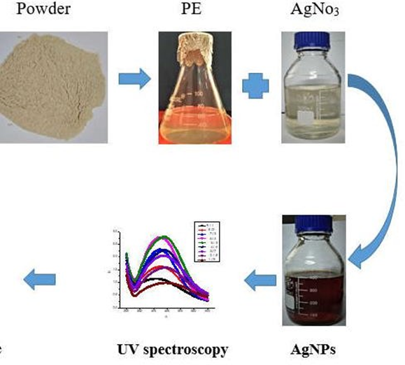
To synthesize AgNps, AgNO3 (1Mm) and plant extract (2mg ml-1) were assorted in dissimilar ratios. Briefly, 700μl of plant extract was mixed with 300μl of AgNO3 (7:3 ratio). The following mixtures were prepared by increasing plant extract and decreasing AgNO3 volumes by 100 μl until the final ratio of 7:3 was reached. Besides, appropriate concentrations of plant extract and AgNO3 for optimal AgNPs synthesis were resolute in series of these reactions, containing mixtures of equal volumes of two-fold serial dilutions of AgNO3(20mM) and plant extract (100 to 0.79 mg ml-1). These mixtures are either prepared in 1.5-ml Eppen dorf tubes by mixing 500μl of each reactant or in 96-well microtiter plates by combining 100μl of each reactant. The permitted reactions to headway at room temperature for diverse time periods.
Table.2.2: Synthesis formulation of biosynthesized nanoparticles at different ratios
|
Plant extract(2mg/ml) + AgNO3 1mM |
(v/v) ratios of Plant extract and AgNO3 |
|
900 µl + 100 µl |
9:1 |
|
800 µl + 200 µl |
8:2 |
|
700 µl + 300 µl |
7:3 |
|
600 µl + 400 µl |
6:4 |
|
500 µl + 500 µl |
5:5 |
|
400 µl + 600 µl |
4:6 |
|
300 µl + 700 µl |
3:7 |
|
200 µl + 800 µl |
2:8 |
|
100 µl + 900 µl |
1:9 |
Separation of Green synthesized Nanoparticles
Unreacted plant extract and AgNO3 were detached by washing and pelleting (AgNPs) as continues. 1ml mixture reaction was centrifuged at 13,000×g at room temperature for 18minutes. Useless Supernatants were discarded and the re suspended of AgNP pellet in 1 ml distilled water by centrifugation at 13,000×g for 18 minutes. This procedure was continual four times. The subsequent AgNPs from centrifugation of five times were suspended in distilled water and used for description
Antibacterial activity
The inhibition assay was conducted by two-fold dilution of AgNPs. Comparison of plant extract with centrifuged and non-centrifuged nanoparticles was performed against five gram-negative Phyto pathogens. First, nutrient broth was prepared and autoclaved at 121℃ for 15 minute and cooled it on room temperature. The nutrient broth was then poured to test tube at a volume of 9ml, each bacterial culture is inoculated in a test tube and incubate it on different optimal temperature for 24 hours. Next, for the evaluation of antimicrobial action of AgNPs, nutrient agar media was prepared by adding 7-gram nutrient agar and 8-gram technical agar in 250 ml distilled water and then autoclaved the suspension for 45 minutes at 90-degree temperature. The medium was then transferred onto sterilized petri dishes and kept in the closed plastic bags at 6-degree temperature each bacterial culture was then spread with the help of spreader on the petri plates and wells of standardized size according to the concentration of Nano particles solution were then made in the petri plates. The plates were then placed in incubator at 37 degrees for 24 and 24 hours and zone of inhibitions were recorded. The AgNPs from centrifugation weighed on balance which was about 0.82mg. The nanoparticles were then completely dissolved in 40 ml distilled water, and the solution was diluted further to obtained 2Mm, 4Mm, 6Mm, 8Mm and 10Mm stock solutions.
RESULTS:
Visual examination of the reaction mixture
After 24 hours, the plant-mediated production of AgNPs was first demonstrated graphically, as varied ratios of plant extract (9:1) and AgNO3 (1:9) were mixed, resulting in a colour change to brownie. Different ratios of colour shifts revealed distinct surface plasmon resonance Peak of green-synthesized AgNps.
UV-vis spectroscopy
The characterizations of green manufactured AgNPs were conceded out by using UV- spectrophotometer. The combination of above different proportions of plant extract and AgNO3 were inspected by UV-spectroscopy which showed a speed up in UV-Vis spectrum above 400, and increased to most noticeable range, from 400 to 450nm, using different plant extract and AgNO3 ratios
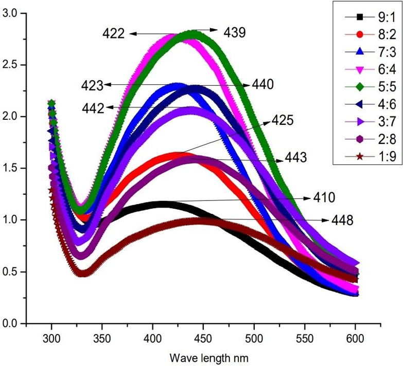
Fig.3.1 Different ratios UV absorbance spectrum of 1 mM AgNO3 and 2mg/ml of A. venustum
leaf and stem extract.
|
EXTRACT (µl): |
1000 |
900 |
800 |
700 |
600 |
500 |
400 |
300 |
200 |
100 |
0 |
|
AgNo3 (µl): |
0 |
100 |
200 |
300 |
400 |
500 |
600 |
700 |
800 |
900 |
1000 |

Figure2: Accurate ratios of AgNO3 and A. venustum leaf and stem extracts
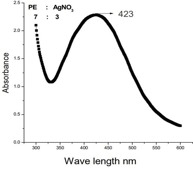
Fig.3 Accurate UV spectrum peak of plant extract and silver nitrate at (7:3) ratio
Antibacterial analysis
The AgNPs synthesized, assessed NPs with plant extract and filtrate of plant extract from the leaf and stem of A. venustum was verified for bacterial inhibition assay in opposing concentrations. Every concentration of conduct showed us divergences influence of bacterial reticence.
Antibacterial inhibition against Pseudomonas aeruginosa
Different nanoparticles concentration from 2Mm stock solution were tested against P. aeruginosa and the plates stayed incubated for 24 and 28 hours for 37 degrees. After 24 and 28 hours, the zone of inhibitions were noted. At 1mg/ml the zone of inhibition was 14 nm. At concentration 500µg/ml the zone of inhibitions was 12 nm. Further at 100µg/ml, the zone of inhibition was 7nm and at 50µg/ml, the nanoparticles showed 5nm zone of inhibition respectively.
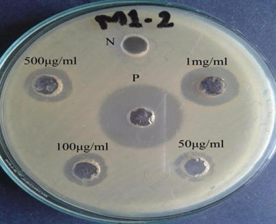
Fig4 Zones of inhibition of 2mM Ag-NPs against P. aeruginosa
Table1: Effectiveness of Ag nanoparticles against P. aeruginosa
|
S. No. |
Molar Solution |
Inhibition Zones (mm) |
||||
|
Positive Control |
50µg/ml |
100µg/ml |
500µg/ml |
1mg/ml |
||
|
1 |
2 Mm |
21 |
5 |
7 |
12 |
14 |
Antibacterial inhibition against E. acaratovora
Different concentrations of nanoparticles from 2Mm stock solution were tested against E. caratovora and the plates stayed incubated for 24 and 28 hours for 37 degrees. After 24 and 28 hours, the zone of inhibitions were noted. At 1mg/ml the zone of inhibition was 17 nm. At concentration 500µg/ml the zone of inhibitions was 14 nm. Further at 100µg/ml, the zone of inhibition was 13 and at 50µg/ml, the nanoparticles showed 10 nm zone of inhibition respectively.
Table2:Effectiveness of Ag nanoparticles against E. caratovora
|
S. No |
Molar Solution |
Inhibition Zones (mm) |
||||
|
Positive control |
50µg/ml |
100µg/ml |
500µg/ml |
1mg/ml |
||
|
1 |
2Mm |
18 |
10 |
12 |
14 |
17 |
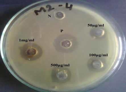
Fig5: Zones of inhibition of 4mM AgNps against E. caratovora
Antibacterial inhibition against X. campestris
Different concentrations of nanoparticles from 2Mm stock solution were tested against X. campestris and the plates stayed incubated for 24 and 28 hours for 37 degrees. After 24 and 28 hours, the zone of inhibitions were noted. At 1mg/ml the zone of inhibition was14 nm. At concentration 500µg/ml the zone of inhibitions was 13 nm. Further at 100µg/ml, the zone of inhibition was 11 and at 50µg/ml, the nanoparticles showed 9 nm zone of inhibition respectively.
Table3: Effectiveness of silver nanoparticles against X. campetris
|
S. No |
MolarSolution |
Inhibition Zones (mm) |
||||
|
Positive Control |
50 µg/ml |
100 µg/ml |
500 µg/ml |
1mg/ml |
||
|
1 |
2 Mm |
16 |
9 |
11 |
13 |
14 |
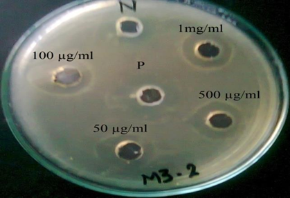
Fig6: Zone of inhibition of 6mM silver nanoparticles against X. campestris
Antibacterial inhibition against X. oryzae
Different concentrations of nanoparticles from 2Mm stock solution were tested against X. oryzae and the plates stayed incubated for 24 and 28 hours for 37 degrees. After 24 and 28 hours, the zone of inhibitions were noted. At 1mg/ml the zone of inhibition was 10 nm. At concentration 500µg/ml the zone of inhibitions was 8 nm. Further at 100µg/ml, the zone of inhibition was 7 and at 50µg/ml, the nanoparticles showed 5 nm zone of inhibition respectively.
Table4 :Effectiveness of silver nanoparticles against X. oryzae
|
S. No |
MolarSolution |
Inhibition Zones (mm) |
||||
|
Positive Control |
50µg/ml |
100µg/ml |
500µg/ml |
1mg/ml |
||
|
1 |
2 mM |
11 |
5 |
7 |
8 |
10 |
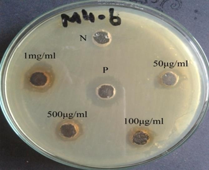
Fig7 Zone of inhibition of silver nanoparticles against X. oryzae
Antibacterial inhibition against Ralstonia Solanacearum
Different concentrations of nanoparticles from 2Mm stock solution were tested against R. Solanacearum and the plates were incubated for 24 and 28 hours for 37 degrees. After 24 and 28 hours, the zone of inhibitions were noted. At 1mg/ml the zone of inhibition was 9 nm. At concentration 500µg/ml the zone of inhibitions was 6 nm. Further at 100µg/ml, the zone of inhibition was 4 and at 50µg/ml, the nanoparticles showed 2.5 nm zone of inhibition respectively.
Table5: Effectiveness of silver nanoparticles against R. solanacearum
|
S. No |
Molar Solution |
Inhibition Zones (mm) |
||||
|
Positive Control |
50µg/ml |
100µg/ml |
500µg/ml |
1mg/ml |
||
|
1 |
2 mM |
20 |
2.5 |
4 |
6 |
9 |
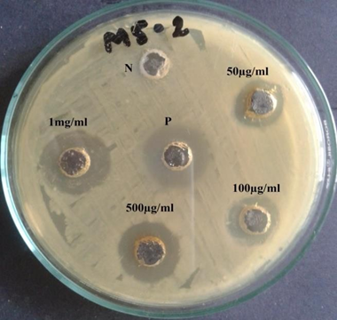
Fig8: Zone of inhibition of 10mM Ag nanoparticles against R. solanaceuru
DISCUSSION :
Presently, Due to the environmentally friendly nature, to produce AgNPs using plant materials attended a lot of attention and it has been chosen for traditional chemical approaches (Bose &Chatterjee, 2016). The production of AgNPs from medicinal plant extracts were used in a variety of applications that are advantageous to men. Because of its nontoxic qualities and less adverse effects, it has a wide range of clinical and biomedical uses (Marin et al., 2015). As a result, they may be useful in drug delivery systems, like anti-tumor, anticancer, antimicrobial agents and anti-inflammatory (Suri et al., 2007). The current study, silver nanoparticle was manufactured from AgNO3 solution with the use of A. venustum leafs and stem aqueous extract, under daylight the minimum reaction time of reaction mixture was 8 minutes. According to the work of (Garibo e al., 2020), within 8 minutes the change of Ag ion was confirmed to be a silver nanoparticle. Visually the color of the reaction mixture was converted from watery to coffee color. Likewise, findings were reported by (Vidhu et al., 2011) and (Bhuyar et al., 2020). This shifting of color of the reaction-mixture is responsible for superficial Plasmon resonance peak (SPR) (Shahverdi et al., 2007; Kanmani and Lim 2013). SPR peaks were examined by UV spectrophotometer, the absorption spectra of the obtained AgNPs peaked at 423nm of the solution, this result is moderately nearer to the results shown by (Ezealisiji et al., 2017; Shivananda et al., 2016) ) who observed 420nm and 422nm peaks, respectively. With close comparison of plant extracts the surface Plasmon resonance was attained in 423nm which is close comparison with the results of Cyperus conglomerates (Al-Nuairi et al.,2020); Zingiberofficinalerhizome (Mathew et al. 2018). The synthesized AgNPs from leaf and stem extract of A. venustum during our experiment shows significant antibacterial effect against the tested bacteria. Extensively it is reported That silver ion and silver composites materials exhibited extreme toxicity against bacteria (Uz-Zaman et al., 2020). Because of the presence of well-formed surface, these particles allow greater interaction with environment andshowed high potency of antibacterial inhibition (Choi et al., 2008). In recent study, the bacterial inhibition of silver nanoparticles, uncentrifuged nanoparticle and plant extract were used against three-gram negative phytopathogens. After 24 hours the bacterial effect of dis similar mixture of different concentration were obtained. The judgement of AgNPs with uncentrifuged nanoparticles, AgNPs showed high potency (Uz-Zaman et al., 2020) of antibacterial assay, additionally the assessment of un centrifuge filtrate plant extract with nano particles, nanoparticles which are uncentrifuged showed us great antibacterial activity against three phytopathogenic bacteria in different concentrations. The recent studies and the results were compared (Hernández‐Díaz et al., 2021). Our analyses demonstrated that AgNPs generation using plant extract can be done in a moderate, simple, and economical manner, and that green synthesized AgNPs is suitable for the novel antibacterial substance’s development
CONCLUSION :
In this study we successfully produce Ag nanoparticles from the leaf and stem extract of A. venustum. The biosynthesis of AgNPs depends on the deliberation of AgNO3 and plant extract. The minimum concentration of plant extract and AgNO3 showed narrow and low absorbance of peak. With increasing the concentration of plant extracts the peak became broader and large sized nanoparticles were formed. Furthermore, in our analysis dissimilar proportions of AgNO3 and plant extract were used to confirm narrow peak and low absorbance of peak which indicated small sized and constant nanoparticle. The synthesized nanoparticle was confirmed visually and by UV- spectroscopy. In this investigation, the obtained AgNPs were used for antibacterial potential against five bacteria strains, the results revealed remarkable antibacterial activity at different concentrations
REFERNCES:
1. Akbar, S., Tauseef, I., Subhan, F., Sultana, N., Khan, I., Ahmed, U. and Haleem, K.S., 2020. An overview of the plant-mediated synthesis of zinc oxide nanoparticles and their antimicrobial potential. Inorganic andNano-Metal Chemistry, 50(4), pp.257-271
2. Abbas S, Nasreen S, Haroon A, Ashraf MA, 2020. Synhesis of Silver and Copper Nanoparticles from Plants and Application as Adsorbents for Naphthalene decontamination. Saudi Journal of Biological Sciences 27, 1016-23
3. Abou El-Nour KM, Eftaiha AA, Al-Warthan A, Ammar RA, 2010. Synthesis and applications of silver nanoparticles. Arabian journal of chemistry 3, 135-40
4. Ahmad N, Sharma S, 2012. Green synthesis of silver nanoparticles using extracts of Ananascomosus.
5. Ahmed S, Ahmad M, Swami BL, Ikram S, 2016a. A review on plants extract mediated synthesis of silver nanoparticles for antimicrobial applications: a green expertise. Journal of advanced research 7, 17-28
6. Ahmed S, Ikram S, 2015. Silver nanoparticles: one pot green synthesis using Terminalia arjuna extract for biological application. J NanomedNanotechnol6, 309.
7. Ahmed S, Saifullah, Ahmad M, Swami BL, Ikram S, 2016b. Green synthesis of silver nanoparticles using Azadirachtaindica aqueous leaf extract. Journal of radiationresearch and applied sciences 9, 1-7
8. Ajitha B, Reddy YaK, Reddy PS, 2014. Biogenic nano-scale silver particles by Tephrosiapurpurea leaf extract and their inborn antimicrobial activity. SpectrochimicaActa Part A: Molecular and Biomolecular Spectroscopy 121, 164-72
9. Al-Nuairi AG, Mosa KA, Mohammad MG, El-Keblawy A, Soliman S, Alawadhi H, 2020. Biosynthesis, characterization, and evaluation of the cytotoxic effects of biologically synthesized AgNPs from Cyperusconglomeratus root extracts on breast cancer cell line MCF-7. Biological trace element research 194,560-9
10. Alfuraydi AA, Devanesan S, Al-Ansari M, Alsalhi MS, Ranjitsingh AJ, 2019. Ecofriendly green synthesis of AgNPs from the sesame oil cake and its potential anticancer and antimicrobial activities. Journal of Photochemistry and Photobiology B: Biology 192, 83-9
11. Ali M, Kim B, Belfield KD, Norman D, Brennan M, Ali GS, 2016. Green synthesis and characterization of silver nanoparticles using Artemisia absinthium aqueous extract—a comprehensive study. Materials Science and Engineering: C 58, 359-65.
12. Almadiy AA, Nenaah GE, 2018. Ecofriendly synthesis of silver nanoparticles using potato steroidal alkaloids and their activity against phytopathogenic fungi. Brazilian Archives of Biology and Technology 61
13. Anju T, Parvathy S, Veettil MV, et al., 2021. Green synthesis of silver nanoparticles from Aloe vera leaf extract and its antimicrobial activity. Materials Today: Proceedings
14. Aravind M, Ahmad A, Ahmad I, et al., 2021. Critical green routing synthesis of silver NPs using jasmine flower extract for biological activities and photo catalytical degradation of methylene blue. Journal of Environmental Chemical Engineering 9,104877
15. Benakashani F, Allafchian A, Jalali S, 2016. Biosynthesis of silver nanoparticles using Capparisspinosa L. leaf extract and their antibacterial activity. Karbala International Journal of Modern Science 2, 251-8.
16. Bhakya S, Muthukrishnan S, Sukumaran M, et al., 2016. Antimicrobial, antioxidant and anticancer activity of biogenic silver nanoparticles–an experimental report. RSCadvances 6, 81436-46.
17. Bhat M, Chakraborty B, Kumar RS, et al., 2021. Biogenic synthesis, characterization and antimicrobial activity of Ixorabrachypoda (DC) leaf extract mediated silver nanoparticles. Journal of King Saud University-Science 33, 101296
18. Bhuyar P, Rahim MHA, Sundararaju S, Ramaraj R, Maniam GP, Govindan N, 2020. Synthesis of silver nanoparticles using marine macro algae Padina sp. and its antibacterial activity towards pathogenic bacteria. Beni-Suef University Journal ofBasic and Applied Sciences 9, 1-15
19. Bose D, Chatterjee S, 2016. Biogenic synthesis of silver nanoparticles using guava (Psidiumguajava) leaf extract and its antibacterial activity against Pseudomonas aeruginosa. Applied Nanoscience 6, 895-901.
20. Carson L, Bandara S, Joseph M, et al., 2020. Green synthesis of silver nanoparticles with antimicrobial properties using Phyla dulcis plant extract. Foodborne pathogensand disease 17,504-11.
21. Chauhan HK, Bisht AK, Bhatt ID, Bhatt A, Gallacher D, Santo A, 2018. Population change of Trillium govanianum (Melanthiaceae) amid altered indigenous harvesting practices in the Indian Himalayas. Journal of ethnopharmacology213, 302-10
22. Cheng H-J, Wang H, Zhang J-Z, 2020. Phytofabrication of silver nanoparticles using three flower extracts and their antibacterial activities against pathogen Ralstonia solanacearum strain YY06 of bacterial wilt. Frontiers in microbiology 11, 2110.
23. Chidurala SC, Kalagadda VR, Tambur P, 2016. Antimicrobial activity of pure Cu nano particles synthesized by surfactant varied chemical reduction method. Environmental Nanotechnology, Monitoring & Management 6, 88-94.
24. Da Silva SL, Figueredo PM, Yano T, 2006. Antibacterial and antifungal activities ofvolatile oils from ZanthoxylumRhoifolium. Leaves. Pharmaceutical Biology 44, 657-9.
25. Dadashpour M, Firouzi-Amandi A, Pourhassan-Moghaddam M, et al., 2018. Biomimetic synthesis of silver nanoparticles using Matricariachamomilla extract and their potential anticancer activity against human lung cancer cells. Materials Scienceand Engineering: C 92,902-12.
26. Dangi S, Gupta A, Gupta DK, Singh S, Parajuli N, 2020. Green synthesis of silver nanoparticles using aqueous root extract of Berberisasiatica and evaluation of their antibacterial activity. Chemical Data Collections 28, 100411
27. Dutta T, Ghosh NN, Das M, Adhikary R, Mandal V, Chattopadhyay AP, 2020. Green synthesis of antibacterial and antifungal silver nanoparticles using Citrus limetta peel extract: Experimental and theoretical studies. Journal of Environmental ChemicalEngineering 8,104019.
28. Dwivedi AD, Gopal K, 2010. Biosynthesis of silver and gold nanoparticles using Chenopodium album leaf extract. Colloids and Surfaces A: Physicochemical andEngineering Aspects 369, 2733.
29. El-Desouky N, Shoueir KR, El- Mehasseb I, El-Kemary M, 2021. Bio-inspired greenmanufacturing of plasmonic silver nanoparticles/Degussa using Banana Waste Peduncles: Photocatalytic, antimicrobial, and cytotoxicity evaluation. Journal of materials research and technology 10, 671-86.
30. Esmaile F, Koohestani H, Abdollah-Pour H, 2020. Characterization and antibacterial activity of silver nanoparticles green synthesized using Ziziphoraclinopodioides extract. Environmental Nanotechnology, Monitoring & Management 14, 100303.
31. Franci G, Falanga A, Galdiero S, et al., 2015. Silver nanoparticles as potential antibacterial agents. Molecules 20, 8856-74.
32. Garibo D, Borbón-Nuñez HA, De León JND, et al., 2020. Green synthesis of silver nanoparticles using Lysilomaacapulcensis exhibit high-antimicrobial activity. Scientific reports 10, 1-11
33. Gopinath V, Velusamy P, 2013. Extracellular biosynthesis of silver nanoparticles using Bacillus sp. GP-23 and evaluation of their antifungal activity towards Fusarium oxysporum. SpectrochimicaActa Part A: Molecular and Biomolecular Spectroscopy106, 170-4
34. Helmlinger J, Sengstock C, Groß-Heitfeld C, et al., 2016. Silver nanoparticles with different size and shape: equal cytotoxicity, but different antibacterial effects. RSCadvances 6, 18490-501
35. Hernández‐Díaz JA, Garza‐García JJ, Zamudio‐Ojeda A, León‐Morales JM López‐Velázquez JC, García‐Morales S, 2021. Plant‐mediated synthesis of nanoparticles and their antimicrobial activity against phytopathogens. Journal of theScience of Food and Agriculture 101, 1270-87.
36. Hong X, Wen J, Xiong X, Hu Y, 2016. Shape effect on the antibacterial activity of silver nanoparticles synthesized via a microwave-assisted method. Environmentalscience and pollution research 23, 4489-97.