HISTOPATHOLOGICAL SPECTRUM OF SKIN LESIONS IN A TERTIARY CARE HOSPITAL: A CROSS-SECTIONAL STUDY
- Vyshakhi Sukumaran , Post Graduate , Department of Pathology, Shimoga Institute of Medical Sciences, Shivamogga, India
- Pradeep Kumar L , Assistant Professor, Department of Pathology, Shimoga Institute of Medical Sciences, Shivamogga, India
- Shobana Ahalisha Kalaivanan , Post Graduate, Shimoga Institute of Medical Sciences, Shivamogga, India
Article Information:
Abstract:
Skin diseases encompass a wide range of pathological entities with overlapping clinical features. Histopathological examination remains the gold standard for definitive diagnosis. (Goswami et al., 2022; Adhikari et al., 2019)Objectives: To study the histopathological spectrum of skin lesions in a tertiary care hospital and analyze distribution by age, sex, and pathological categories. (Goswami et al., 2022; Adhikari et al., 2019)Materials and Methods: A cross-sectional retrospective study was conducted in the Department of Pathology, Shimoga Institute of Medical Sciences, Shivamogga, during December 2024–December 2025. All skin punch biopsies and excision biopsies received during the period were processed routinely and stained with hematoxylin and eosin. Lesions were categorized as infectious/non-infectious and as non-neoplastic, benign neoplastic, or malignant neoplastic based on histopathological diagnosis. (Elder et al., 2018)Results: A total of 329 skin punch biopsies were studied. Males constituted 180 (54.7%) and females 149 (45.3%). The commonest age group was 46–60 years (106 cases; 32.2%). Infectious non-neoplastic lesions comprised 54 (16.4%) and non-infectious non-neoplastic lesions 230 (69.9%). Benign neoplasms accounted for 21 (6.4%), while malignant neoplasms were 24 (7.3%). Conclusion: Non-neoplastic lesions predominated among skin biopsies. Histopathology remains indispensable for definitive diagnosis and management, especially where clinical overlap exists. (Goswami et al., 2022; Adhikari et al., 2019; Elder et al., 2018)
Keywords:
Article :
INTRODUCTION:
The skin is the largest organ of the human body and functions as a protective barrier against physical, chemical, and microbial insults, while also contributing to thermoregulation, immune surveillance, and sensory perception.(Goswami et al., 2022) Dermatological disorders encompass a wide spectrum of conditions, including inflammatory, infectious, autoimmune, and neoplastic lesions.(Goswami et al., 2022; Adhikari et al., 2019) Many of these disorders present with overlapping clinical features, making accurate diagnosis based solely on clinical examination challenging and necessitating histopathological evaluation.(Goswami et al., 2022; Adhikari et al., 2019)
Skin biopsy followed by histopathological examination remains the gold standard for the diagnosis of skin diseases.(Goswami et al., 2022) Histopathology enables precise identification of inflammatory patterns, infectious organisms, and neoplastic changes, thereby facilitating accurate diagnosis and appropriate management.(Goswami et al., 2022; Adhikari et al., 2019) It also plays an essential role in distinguishing benign from malignant lesions and provides valuable prognostic information in neoplastic conditions.(Goswami et al., 2022; Adhikari et al., 2019)
Dermatopathology relies on the recognition of characteristic histological reaction patterns such as spongiotic, lichenoid, psoriasiform, vesiculobullous, granulomatous, and neoplastic patterns, which aid in narrowing the differential diagnosis.(Elder et al., 2018) Lever’s Histopathology of the Skin highlights the importance of correlating histopathological findings with clinical features to arrive at a definitive diagnosis.(Elder et al., 2018) Clinicopathological correlation is therefore essential in dermatology, as histopathology provides objective morphological evidence that complements clinical assessment.(Elder et al., 2018)
Non-neoplastic skin lesions constitute the majority of dermatological biopsies and include inflammatory dermatoses, autoimmune disorders, and infectious diseases.(Goswami et al., 2022; Adhikari et al., 2019) Infectious dermatoses, particularly Hansen’s disease, fungal infections, and viral infections, remain significant causes of morbidity in developing countries, including India.(Goswami et al., 2022; Adhikari et al., 2019) Histopathological examination is crucial in the diagnosis and classification of Hansen’s disease and plays an important role in guiding therapy.(Goswami et al., 2022)
Neoplastic lesions of the skin include benign tumors such as epidermoid cysts and melanocytic nevi, as well as malignant tumors such as basal cell carcinoma, squamous cell carcinoma, and malignant melanoma.(Goswami et al., 2022; Adhikari et al., 2019) Although malignant lesions account for a smaller proportion of skin biopsies, early detection is essential because of their potential for local invasion and metastasis.(Goswami et al., 2022; Adhikari et al., 2019) Basal cell carcinoma and squamous cell carcinoma are the most common non-melanoma skin cancers worldwide.(Adhikari et al., 2019)
Several hospital-based studies from India have demonstrated that non-neoplastic lesions predominate in skin biopsy specimens, followed by benign and malignant neoplasms.(Goswami et al., 2022; Bezbaruah & Baruah, 2018;Singh et al., 2023) However, the frequency and pattern of dermatological lesions vary depending on geographic location, environmental factors, and population characteristics.(Goswami et al., 2022; Adhikari et al., 2019) Understanding the histopathological spectrum of skin lesions in a given region is important for improving diagnostic accuracy and guiding clinical management.(Goswami et al., 2022; Adhikari et al., 2019)
Therefore, the present study was undertaken to evaluate the histopathological spectrum of skin lesions in a tertiary care hospital and analyze their distribution with respect to age, sex, and pathological categories.
MATERIALS AND METHODS:
Study design and setting: Cross-sectional retrospective study conducted at the Department of Pathology, Shimoga Institute of Medical Sciences, Shivamogga.
Study period: December 2024 to December 2025.
Inclusion criteria: All skin punch biopsy and excision biopsy specimens received during the study period.
Processing: Specimens were fixed in 10% buffered formalin, processed routinely, paraffin-embedded, sectioned at 3–5 µm, and stained with hematoxylin and eosin. (Elder et al., 2018)
Definitions: Non-neoplastic lesions were categorized as infectious or non-infectious. Neoplastic lesions were classified as benign or malignant based on histopathological diagnosis. (Elder et al., 2018)
RESULTS:
across all groups, with a peak in 46–60 years (Table 1).Non-neoplastic lesions predominated (n=284; 86.3%). Among them, non-infectious lesions were the major category (n=230; 69.9%), while infectious lesions accounted for 54 cases (16.4%) (Table 2)
Among infectious lesions, leprosy constituted the majority (lepromatous, borderline tuberculoid, tuberculoid and unspecified forms), followed by other infections such as molluscum contagiosum and fungal infections (Table 3).
Among non-infectious non-neoplastic lesions, psoriasiform and lichenoid disorders were frequent. Psoriasis (n=34) and lichen planus (n=20) were common, along with dermatitis/eczema and autoimmune blistering disorders (Table 4).
Neoplastic lesions comprised 45 cases (13.7%). Benign neoplasms were 21 cases (6.4%) and included epidermoid cysts as the most frequent benign lesion. Malignant neoplasms were 24 cases (7.3%), with basal cell carcinoma being the most common malignancy (Table 5).
TABLES
Table 1: Age and sex distribution of cases (n = 329)
|
Age group (years) |
Female |
Male |
Total (%) |
|
0–15 |
8 |
8 |
16 (4.9) |
|
16–30 |
32 |
36 |
68 (20.7) |
|
31–45 |
34 |
43 |
77 (23.4) |
|
46–60 |
50 |
56 |
106 (32.2) |
|
>60 |
25 |
36 |
61 (18.5) |
|
Total |
149 |
180 |
329 (100) |
Table 2: Category-wise distribution of skin lesions
|
Category |
Number of cases |
Percentage (of total cases, n=329) |
|
Infectious (non-neoplastic) |
54 |
16.4 |
|
Non-infectious (non-neoplastic) |
230 |
69.9 |
|
Benign neoplasms |
21 |
6.4 |
|
Malignant neoplasms |
24 |
7.3 |
Table 3: Type and frequency of infectious skin lesions (non-neoplastic) (n = 54)
|
Diagnosis |
Number |
Percentage (of total cases, n=329) |
|
Leprosy (all types including reactions) |
48 |
14.4 |
|
Fungal infection |
2 |
0.6 |
|
Molluscum contagiosum |
1 |
0.3 |
|
Chromoblastomycosis |
1 |
0.3 |
|
Actinomycosis |
1 |
0.3 |
|
Condyloma acuminatum (vulva) |
1 |
0.3 |
|
Total |
54 |
16.4 |
Table 4: Type and frequency of non-infectious non-neoplastic skin lesions (n = 230)
|
Diagnosis |
Number |
Percentage (of total cases, n=329) |
|
Dermatitis/Eczema |
24 |
7.3 |
|
Lichen planus |
20 |
6.1 |
|
Contact dermatitis |
12 |
3.6 |
|
Pemphigus vulgaris |
5 |
1.5 |
|
Vasculitis |
5 |
1.5 |
|
Psoriasiform dermatitis |
5 |
1.5 |
|
Morphea |
5 |
1.5 |
|
Chronic Balanoposthitis |
5 |
1.5 |
|
Spongiotic dermatitis |
4 |
1.2 |
|
Lichen planopilaris |
4 |
1.2 |
|
Bullous pemphigoid |
4 |
1.2 |
|
Post Inflammatory Pigmentation |
2 |
1.2 |
|
Folliculitis |
3 |
0.9 |
|
Prurigo nodularis |
3 |
0.9 |
|
Darier Disease |
2 |
0.6 |
|
Lichen Sclerosus Et Atrophicus |
2 |
0.6 |
|
Riehl's Melanosis |
2 |
0.6 |
|
Psoriasis |
34 |
10.3 |
|
Others |
87 |
26.4 |
|
Total |
230 |
69.9 |
Table 5: Type and frequency of neoplastic skin lesions (n = 45)
|
Group |
Diagnosis |
Number |
Percentage (of total cases, n=329) |
|
Benign |
|
|
|
|
|
Epidermoid cyst |
11 |
3.3 |
|
|
Keloid |
2 |
0.6 |
|
|
Hemangioma |
2 |
0.6 |
|
|
Pilomatricoma |
1 |
0.3 |
|
|
Trichofolliculoma |
1 |
0.3 |
|
|
Melanocytic nevus |
1 |
0.3 |
|
|
Right Eyelid- Dermoid Cyst |
1 |
0.3 |
|
|
Trichilemmal (pilar) cyst |
1 |
0.3 |
|
|
Sebaceous Adenoma |
1 |
0.3 |
|
Malignant |
|
|
|
|
|
Basal cell carcinoma |
17 |
5.2 |
|
|
Squamous cell carcinoma |
4 |
1.2 |
|
|
Malignant melanoma |
2 |
0.6 |
|
Total |
|
45 |
13.7 |
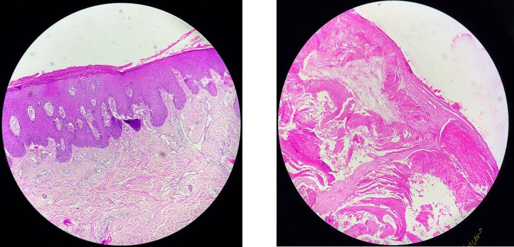
Figure 1: Psoriasis vulgaris (H&E, ×100).
Section shows psoriasiform hyperplasia with elongated rete ridges, parakeratosis, hypogranulosis,
and inflammatory infiltrate in the superficial dermis,characteristic of psoriasis vulgaris.
Figure 2: Pilomatricoma (H&E, ×100).
Section shows dermal tumor with islands of basaloid cells and areas of eosinophilic anucleate shadow (ghost) cells, characteristic of pilomatricoma
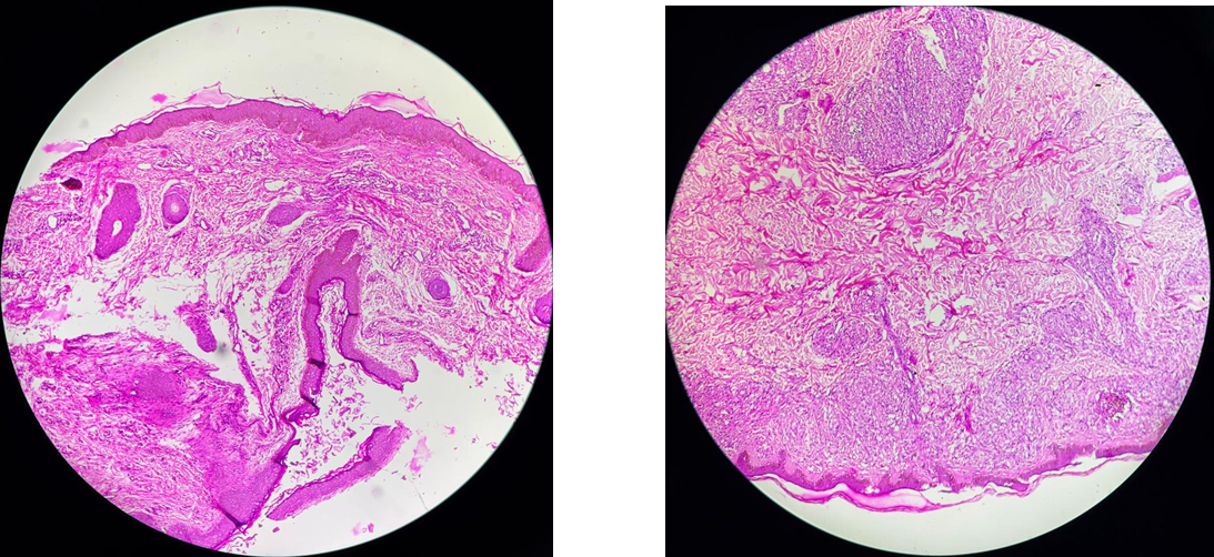
Figure 3: Epidermal inclusion cyst (H&E stain, ×100).
Photomicrograph shows a cyst lined by stratified squamous epithelium with a well-formed granular layer and lumen filled with lamellated keratin, consistent with epidermal inclusion cyst.
Figure 4: Lepromatous Hansen’s disease (H&E, ×100).
Dermis shows sheets of foamy histiocytes (Virchow cells) with sparse lymphocytes and grenz zone beneath epidermis, consistent with lepromatousHansen’s disease.
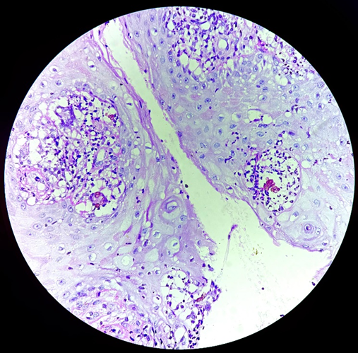
Figure 5: Chromoblastomycosis (H&E stain, ×400).
Photomicrograph shows hyperkeratosis and acanthosis of the epidermis with dermal granulomatous inflammation containing characteristic brown, thick-walled, round sclerotic (Medlar) bodies, also known as copper-
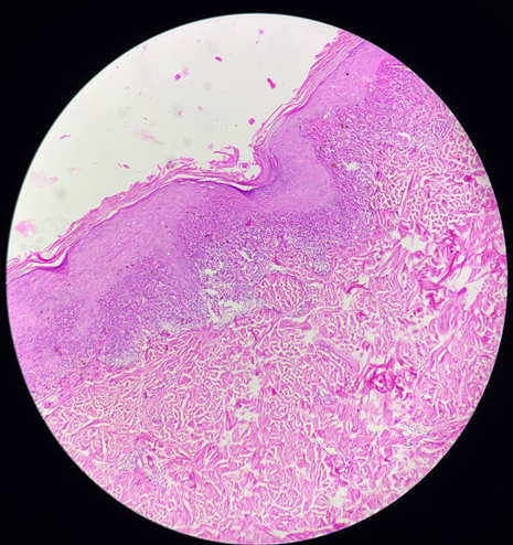
Figure 6: Lichen planus (H&E stain, ×100).
Photomicrograph shows hyperkeratosis, wedge-shaped hypergranulosis, irregular acanthosis, and dense band-like lymphocytic infiltrate at the dermoepidermal junction with basal cell degeneration, consistent with lichen planus.
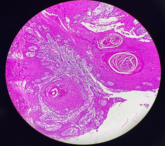
Figure 7: Squamous cell carcinoma (H&E, ×100).
Section shows invasive malignant squamous epithelial cells arranged in nests and islands with prominent keratin pearl formation, characteristic of well-differentiated squamous cell carcinoma.
DISCUSSION :
The present study evaluated the histopathological spectrum of 329 skin punch biopsies over a one-year period. Skin biopsy is widely acknowledged as the gold standard for confirmation of dermatological diagnosis because many inflammatory and infectious dermatoses share overlapping clinical features. (Goswami et al., 2022; Adhikari et al., 2019)
In the present series, non-neoplastic lesions formed the majority, similar to multiple Indian studies that report a predominance of inflammatory and infectious conditions among biopsy specimens. (Gaikwad et al., 2016; Adhikari et al., 2019; Mehar et al., 2014)
The commonest age group in our study was 46–60 years, while a mild male preponderance was observed. Prior studies show variable age peaks and sex ratios depending on referral patterns and regional epidemiology. (Goswami et al., 2022; Adhikari et al., 2019)
Infectious lesions constituted 16.4% of the total cohort, and leprosy accounted for the bulk of infectious diagnoses in our setting. This is comparable to several Indian studies where Hansen’s disease forms a major proportion of infectious skin biopsies. (Goswami et al., 2022; Adhikari et al., 2019)
Among non-infectious non-neoplastic lesions, psoriasis and lichen planus were common. Papulosquamous and eczematous dermatoses are frequent indications for biopsy in most tertiary care studies, reflecting the need for histological confirmation and exclusion of mimics. (Goswami et al., 2022; Adhikari et al., 2019 ; Agrawal et al., 2018)
Neoplastic lesions constituted a smaller fraction of the overall biopsies. In our malignant category, basal cell carcinoma was the most frequent malignancy, with squamous cell carcinoma and malignant melanoma forming a smaller subset. (Goswami et al., 2022; Adhikari et al., 2019) Similar patterns, with basal cell carcinoma and squamous cell carcinoma as common non-melanoma skin cancers, have been reported in Indian hospital-based studies. (Goswami et al., 2022; Adhikari et al., 2019; Thapa et al., 2018)
Lever’s Histopathology of the Skin emphasizes that robust clinicopathological correlation and awareness of reaction patterns are essential for accurate diagnosis, especially in interface dermatitis, psoriasiform dermatitis, vasculitis, and vesiculobullous disorders. (Elder et al., 2018)
Table 6: Comparative summary of histopathological spectrum of skin lesions in the present study and selected published studies.
|
Author (Year) |
Region |
Study period |
Sample size |
Non‑neoplastic (%) |
Benign neoplastic (%) |
Malignant neoplastic (%) |
|
Goswami et al., 2022 [1] |
India |
NR (not reported) |
— |
85–90 |
5–10 |
3–8 |
|
Bezbaruah et al., 2018 [4] |
India |
NR (not reported) |
— |
88.0 |
6.5 |
5.5 |
|
Gaikwad et al., 2016 [5] |
India |
NR (not reported) |
— |
90.2 |
5.1 |
4.7 |
|
Thapa et al., 2018 [8] |
Nepal |
NR (not reported) |
— |
82.6 |
9.4 |
8.0 |
|
Present study, 2025 |
Shivamogga |
December 2024 – December 2025 |
329 |
86.3 |
6.4 |
7.3 |
LIMITATIONS OF THE STUDY
The present study has certain limitations. First, it was a single-center hospital‑based study and therefore the findings may not represent the exact prevalence of dermatological diseases in the general population. Second, the retrospective nature of the study limited detailed clinical information and follow‑up data in some cases. Third, ancillary investigations such as special stains and immunohistochemistry were not performed in all cases, which might have further aided in characterization of certain lesions.
CONCLUSION :
Non-neoplastic lesions predominate among skin punch biopsies in a tertiary care setting. Infectious dermatoses—particularly leprosy—continue to contribute a significant burden. Psoriasis, lichen planus, and dermatitis/eczema are common non-infectious diagnoses. (Goswami et al., 2022; Adhikari et al., 2019). Although neoplastic lesions are less frequent, malignant tumors (especially basal cell carcinoma) are clinically important and highlight the value of early biopsy. Histopathological examination remains central to definitive diagnosis and management. (Singh et al., 2023)
BIBLIOGRAPHY:
1. Adhikari, R. C., Shah, M., & Jha, A. K. (2019). Histopathological spectrum of skin diseases in a tertiary skin health and referral centre. Journal of Pathology of Nepal, 9(1), 1434–1440.
2. Agrawal, S., Mishra, K. B., & Gupta, C. M. (2018). Histopathological spectrum of non‑infectious erythematous papulo‑squamous lesions. International Journal of Research in Medical Sciences, 6(6), 2072–2075.
3. Bezbaruah, R., & Baruah, M. (2018). Histopathological spectrum of skin lesions: A hospital‑based study. Indian Journal of Applied Research, 8(3), 51–52.
4. Elder, D. E., Massi, D., Scolyer, R. A., & Willemze, R. (2018). Lever’s Histopathology of the Skin (11th ed.). Wolters Kluwer.
5. Gaikwad, S. L., Kumawat, U. D., Sakhare, N. A., & D’Costa, G. F. (2016). Histopathological spectrum of skin lesions: Experience at a rural‑based hospital. International Journal of Current Research, 8(9), 36223–36227.
6. Goswami, A., Agarwal, P., Singh, K., Verma, R., Sharma, S., & Gupta, S. (2022). Histopathological spectrum of skin disorders: An observational study at a tertiary care centre. Journal of Family Medicine and Primary Care, 11(8), 4610–4616.
7. Mehar, R., Jain, R., Kulkarni, C. V., Narang, S., Mittal, M., & Patidar, H. (2014). Histopathological study of dermatological lesions: A retrospective approach. International Journal of Medical Science and Public Health, 3(9), 1082–1085.
8. Singh, B. P., Bano, S., Porte, J., Ganguly, S., & Hirani, N. (2023). Histomorphological spectrum of skin lesions in a tertiary care hospital: A 10‑year retrospective study. Indian Journal of Pathology and Oncology, 12(4), 351–357.
9. Thapa, R., Gurung, P., Hirachand, S., & Shrestha, S. B. (2018). Histomorphologic profile of skin tumors. Journal of Nepal Medical Association, 56(214), 953–957.