PULMONARY ACTINOMYCOSIS MIMICKING LUNG MALIGNANCY IN AN ELDERLY MALE: A RARE CASE REPORT
- Manasa Paramaiahgari , Dept of Respiratory Medicine, Jawaharlal Nehru Medical College and KLES Dr. Prabhakar Kore Hospital & Medical Research Center, Belagavi.
- Bhagyashri Patil , Professor & Head of the Department, Dept of Respiratory Medicine, Jawaharlal Nehru Medical College and KLES Dr. Prabhakar Kore Hospital & Medical Research Center, Belagavi.
- Jyothi Hattiholi , Associate Professor & Consultant, Dept of Respiratory Medicine, Jawaharlal Nehru Medical College and KLES Dr. Prabhakar Kore Hospital & Medical Research Center, Belagavi.
- Gautam.S , Associate Professor & Consultant, Dept of Respiratory Medicine, Jawaharlal Nehru Medical College and KLES Dr. Prabhakar Kore Hospital & Medical Research Center, Belagavi
- Kirankumar Pujar , Assistant Professor & Consultant, Dept of Respiratory Medicine, Jawaharlal Nehru Medical College and KLES Dr. Prabhakar Kore Hospital & Medical Research Center, Belagavi
- Guruprasad Antin , Assistant Professor & Consultant, Dept of Respiratory Medicine, Jawaharlal Nehru Medical College and KLES Dr. Prabhakar Kore Hospital & Medical Research Center, Belagavi.
- Ningappa Karalingannavar , Assistant Professor & Consultant, Dept of Respiratory Medicine, Jawaharlal Nehru Medical College and KLES Dr.Prabhakar Kore Hospital & Medical Research Center, Belagavi.
- Rajeev Tangella , Senior Resident, Dept of Respiratory Medicine, Jawaharlal Nehru Medical College and KLES Dr.Prabhakar Kore Hospital & Medical Research Center,Belagavi
Article Information:
Abstract:
Background: Pulmonary actinomycosis is a rare, chronic infectious disease caused by Actinomyces species, often posing a diagnostic challenge due to its nonspecific clinical and radiological features. Pulmonary infection accounts for approximately 15-20% of all actinomycosis cases with no accurate global incidence or prevalence rates reported due to its rarity. It frequently mimics pulmonary tuberculosis or lung malignancy, especially in elderly patients and in tuberculosis-endemic regions. Timely diagnosis is crucial, as the disease responds well to prolonged antibiotic therapy. Case Presentation: A 73-year-old male with a history of treated pulmonary tuberculosis and long-standing diabetes mellitus who presented with chronic cough, progressive breathlessness, hemoptysis, weight loss, and constitutional symptoms. Imaging revealed a necrotic, mass-like consolidation in the right upper lobe, raising suspicion of malignancy. Bronchoscopic evaluation with biopsy demonstrated filamentous gram-positive bacterial colonies with the Splendore–Hoeppli phenomenon, confirming pulmonary actinomycosis. Microbiological studies for tuberculosis and fungal infections were negative. Due to a documented penicillin allergy, the patient was treated with oral doxycycline for six months, resulting in marked clinical improvement and near-complete radiological resolution, with only residual fibrotic changes on follow-up imaging. This case highlights the importance of considering pulmonary actinomycosis in the differential diagnosis of chronic lung consolidations or mass lesions, particularly in patients with prior pulmonary disease. Histopathological confirmation is essential for diagnosis, and early initiation of appropriate antibiotic therapy leads to excellent outcomes while avoiding unnecessary surgical intervention.
Keywords:
Article :
INTRODUCTION:
Pulmonary actinomycosis is a rare, chronic, suppurative infectious disease caused by anaerobic or microaerophilic, gram-positive filamentous bacteria of the genus Actinomyces, which are normal commensals of the oropharynx, gastrointestinal tract, and female genital tract (1). India comes up to Asia’s 32.2% share of reported cases of pulmonary actinomycosis mimicking lung cancer in a 2024 scoping review of 48 global cases (2). Human actinomycosis most commonly involves the cervicofacial region, followed by abdominopelvic disease, while thoracic involvement accounts for approximately 10–15% of cases (3). Despite being described more than a century ago, pulmonary actinomycosis remains an underdiagnosed entity due to its indolent course, nonspecific clinical manifestations, and striking resemblance to more common pulmonary diseases such as tuberculosis, lung abscess, and bronchogenic carcinoma (4).
Pulmonary actinomycosis typically results from aspiration of oropharyngeal secretions containing Actinomyces organisms into the lower respiratory tract. Predisposing factors include poor oral hygiene, diabetes mellitus, chronic lung disease, prior pulmonary infections, alcoholism, smoking, and immunosuppression (5). Structural lung damage caused by previous infections, particularly pulmonary tuberculosis, creates a favorable environment for colonization and invasion by these organisms. Consequently, pulmonary actinomycosis is more frequently observed in elderly individuals and in males, with a male-to-female ratio of approximately 3:1 (6).
Clinically, pulmonary actinomycosis presents with chronic and progressive symptoms such as cough, expectoration, low-grade fever, chest pain, hemoptysis, weight loss, and breathlessness. These symptoms often evolve over weeks to months, leading to frequent misdiagnosis as pulmonary tuberculosis or malignancy, especially in tuberculosis-endemic regions (3). Hemoptysis and significant weight loss further reinforce suspicion of lung cancer, prompting extensive evaluation. Physical examination findings are usually nonspecific, and chest wall sinuses, though classically described, are uncommon in early disease (7).
Radiological features of pulmonary actinomycosis are highly variable and lack pathognomonic characteristics. Imaging may reveal segmental or lobar consolidation, cavitary lesions, necrotic masses, pleural thickening, or chest wall involvement (8). These findings frequently mimic neoplastic lesions or chronic infections, often leading to empirical antitubercular or antibiotic therapy without microbiological confirmation. Failure to respond to conventional treatment should raise suspicion for alternative diagnoses, including actinomycosis (9).
Definitive diagnosis of pulmonary actinomycosis is challenging and relies on histopathological demonstration of characteristic filamentous organisms forming sulfur granules or showing the Splendore–Hoeppli phenomenon. Culture isolation is difficult due to the fastidious nature of the organism and prior antibiotic exposure (10). Bronchoscopic biopsy, transthoracic biopsy, or surgical specimens are often required to establish the diagnosis. Early recognition is crucial, as pulmonary actinomycosis responds dramatically to prolonged antibiotic therapy, most commonly penicillin or suitable alternatives in cases of allergy, with excellent prognosis and avoidance of unnecessary surgical interventions (11).
Given its rarity, diagnostic complexity, and potential to mimic serious pulmonary conditions, pulmonary actinomycosis remains an important clinical entity for respiratory physicians. This case report aims to highlight the clinical, radiological, and pathological features of pulmonary actinomycosis in an elderly male with prior pulmonary tuberculosis, emphasizing the importance of maintaining a high index of suspicion in chronic lung lesions.
CASE PRESENTATION:
Patient Information: A 73-year-old male, farmer by occupation, presented to the Department of Respiratory Medicine with chronic respiratory complaints. He was an ex-smoker with a 15 pack-year smoking history and had quit smoking 20 years prior to presentation. He consumed alcohol occasionally. The patient was a known case of type 2 diabetes mellitus for the past 20 years with suboptimal glycemic control. There was no history of hypertension, chronic kidney disease, chronic liver disease, ischemic heart disease, or thyroid disorders. Family history was not significant.
Presenting Complaints: The patient presented with breathlessness on exertion for 3–4 years, which had progressively worsened over the last two months. He also complained of chronic cough with minimal whitish expectoration for one year, with an increase in severity over the preceding month. Additionally, he reported streaky hemoptysis for onemonth, intermittent low-grade fever lasting 2–3 days about one month prior to admission, loss of appetite, and significant unintentional weight loss of approximately 12 kg over one year. There were no complaints of chest pain, wheezing, hoarseness of voice, stridor, palpitations, abdominal pain, or joint pains.
Past Medical History: The patient had a significant past history of microbiologically confirmed pulmonary tuberculosis diagnosed ten years earlier, for which he completed a six-month course of antitubercular therapy with documented clinical and radiological resolution. Residual fibrotic bands were noted in the right upper lobe. He remained asymptomatic for approximately four to five years following treatment before developing insidious respiratory symptoms. There was no prior history of recurrent respiratory infections or hospitalizations.
Clinical Examination: On general physical examination, the patient was moderately built and nourished, conscious, cooperative, and oriented to time, place, and person. Vital signs were stable with a pulse rate of 92 beats per minute, blood pressure of 130/80 mmHg, respiratory rate of 18 cycles per minute, and oxygen saturation of 95% on room air. He was afebrile at presentation. There was no pallor, cyanosis, clubbing, lymphadenopathy, or peripheral edema. Examination of the oral cavity revealed good oral hygiene, and there was no paranasal sinus tenderness.
Respiratory System Examination: Inspection revealed symmetrical chest movements with no visible chest wall deformities or draining sinuses. Palpation showed equal chest expansion bilaterally. Percussion elicited resonant notes over all lung fields. Auscultation revealed vesicular breath sounds bilaterally with fine crepitations localized to the right mammary area. No bronchial breathing or wheeze was appreciated.
Systemic Examination: Cardiovascular examination revealed normal heart sounds with no murmurs. Central nervous system examination showed no focal neurological deficits. Abdominal examination revealed a soft, non-tender abdomen with no organomegaly.
Laboratory Investigations: Baseline hematological investigations showed a hemoglobin level of 12.5 g/dL and a total leukocyte count of 8,300/mm³. Renal and liver function tests were within normal limits. Viral markers were negative. D-dimer was mildly elevated at 396 ng/mL. Glycosylated hemoglobin (HbA1c) was 8.6%, indicating poor glycemic control.
Radiological Investigations: Chest radiograph revealed a right-sided pulmonary opacity. Contrast-enhanced computed tomography of the thorax demonstrated a round consolidation with internal necrosis and multiple air foci involving the anterior segment of the right upper lobe, with differential considerations of neoplastic and infective etiologies.
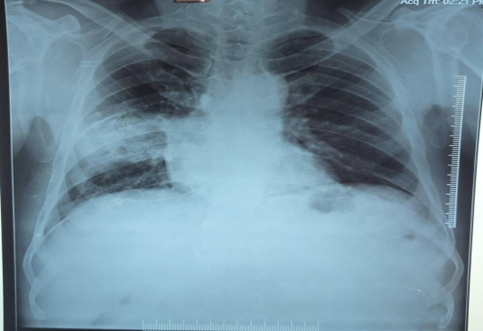
Figure 1: Chest radiograph showing right middle zone consolidation with background fibrotic changes
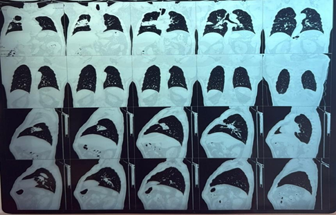
Figure 2: Contrast-Enhanced Computed Tomography (CECT) of the Thorax an year before (Lung Window)
(Necrotic consolidation with intralesional air foci involving the anterior segment of the right upper lobe, with differential considerations including infective etiology such as actinomycosis or neoplastic pathology)
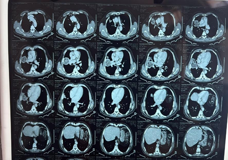
Figure 3: Axial sections of Contrast-Enhanced Computed Tomography (CECT) of the Thorax (recent)
(Reported as ‘Round consolidation with internal necrosis and multiple air foci involving anterior segment of right upper lobe with differentials being Neoplastic or Infective etiology)
Bronchoscopic Evaluation: Fiberoptic bronchoscopy was performed under local anesthesia. It revealed narrowing of the lateral segmental bronchus of the right middle lobe with the presence of thick whitish secretions. Bronchoalveolar lavage samples were sent for Gram stain, potassium hydroxide mount, bacterial and fungal cultures, GeneXpert for tuberculosis, and cytology.
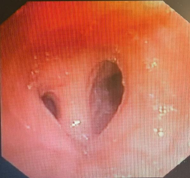
Figure 4: Fiberoptic bronchoscopy revealed narrowing of the lateral segmental bronchus of the right middle lobe with thick whitish secretions
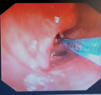
Figure 5: Bronchoscopy demonstrated narrowing of the lateral segmental bronchus of the right middle lobe, and endobronchial biopsy was performed from the inflamed mucosa
Microbiological and Histopathological Findings: Bronchoalveolar lavage was negative for bacterial, fungal, and mycobacterial pathogens, and GeneXpert testing for Mycobacterium tuberculosis was negative. Histopathological examination of bronchoscopic biopsy specimens showed dense inflammatory infiltrates composed of lymphocytes, plasma cells, and sheets of neutrophils. Filamentous bacterial colonies exhibiting the Splendore–Hoeppli phenomenon were identified. Gram staining demonstrated gram-positive filamentous organisms, while Ziehl–Neelsen staining was negative for acid-fast bacilli. No evidence of granulomatous inflammation or malignancy was observed.
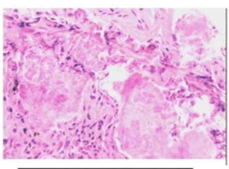
Figure 6: Ziehl–Neelsen staining of the biopsy specimen was negative for acid-fast bacilli, ruling out tuberculosis
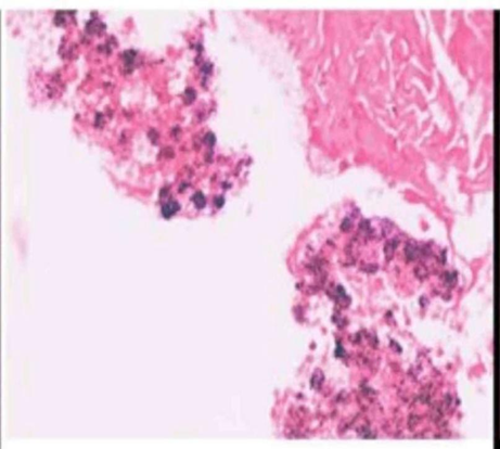
Figure 7: Gram staining of the biopsy specimen revealed gram-positive filamentous bacterial colonies consistent with Actinomyces species
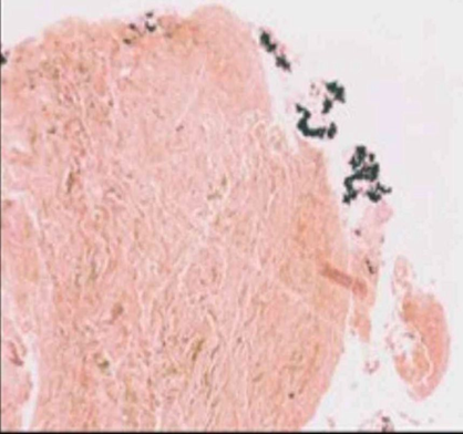
Figure 8: Gomori methenamine silver staining was negative for fungal organisms
Final Diagnosis: Based on the clinical, radiological, and histopathological findings, a definitive diagnosis of pulmonary actinomycosis was established.
Treatment and Follow-Up: In view of a documented penicillin allergy, the patient was initiated on oral doxycycline 100 mg twice daily. Antibiotic therapy was continued for six months. The patient showed significant symptomatic improvement with resolution of cough, hemoptysis, and breathlessness. Follow-up chest radiograph at the end of six months demonstrated near-complete resolution of the pulmonary consolidation with residual fibrotic bands in the right middle zone.
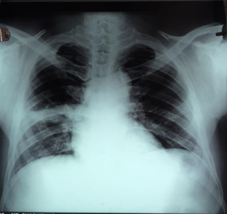
Figure 9: Follow-up chest radiograph showing resolution of right lung consolidation with residual fibrotic bands
DISCUSSION:
Pulmonary actinomycosis is an uncommon but clinically significant infectious disease that often poses a diagnostic dilemma due to its indolent course and close resemblance to more prevalent pulmonary conditions such as tuberculosis, lung abscess, and bronchogenic carcinoma (5). Actinomyces species are anaerobic, gram-positive, filamentous bacteria that are part of the normal flora of the oropharynx. Pulmonary involvement typically occurs following aspiration of contaminated oropharyngeal secretions into the lower respiratory tract, particularly in individuals with compromised local or systemic host defenses (12).
Several predisposing factors have been identified for pulmonary actinomycosis, including advanced age, male gender, diabetes mellitus, poor oral hygiene, smoking, alcoholism, and pre-existing lung disease (3). In the present case, the patient had multiple risk factors, notably long-standing diabetes mellitus and prior pulmonary tuberculosis, which likely resulted in structural lung damage and fibrosis. Such altered pulmonary architecture creates an environment conducive to colonization and invasion by Actinomyces organisms. The male predominance observed in this disease is consistent with previously reported literature, which suggests a male-to-female ratio of approximately 3:1.
The clinical presentation of pulmonary actinomycosis is often nonspecific and slowly progressive. Common symptoms include chronic cough, sputum production, hemoptysis, low-grade fever, chest pain, and significant weight loss. These manifestations frequently overlap with those of pulmonary tuberculosis and lung malignancy, particularly in elderly patients (13). In tuberculosis-endemic regions, there is a strong tendency to empirically treat such patients for tuberculosis, leading to delays in accurate diagnosis (14). In the present case, the patient’s constitutional symptoms, hemoptysis, and radiological findings raised strong suspicion of malignancy or Tuberculosis, highlighting the diagnostic challenge posed by this condition.
Radiological findings in pulmonary actinomycosis are highly variable and lack specificity. Common imaging features include segmental or lobar consolidation, cavitation, mass-like lesions, necrosis, pleural thickening, and chest wall invasion in advanced cases (15). Computed tomography often reveals heterogeneous consolidations with areas of low attenuation due to necrosis, which can be misinterpreted as neoplastic lesions (16). In this patient, the presence of a round well defined consolidation with internal necrosis and air foci in the right upper lobe closely mimicked a lung tumor, necessitating further invasive evaluation. This reinforces the importance of tissue diagnosis in patients with atypical or non-resolving pulmonary lesions.
Microbiological confirmation of pulmonary actinomycosis is challenging, as Actinomyces species are fastidious organisms that require prolonged anaerobic culture conditions. Prior antibiotic exposure further reduces the yield of cultures (12). As a result, histopathological examination remains the cornerstone of diagnosis. The demonstration of filamentous bacterial colonies surrounded by eosinophilic material, known as the Splendore–Hoeppli phenomenon, is considered characteristic (17). In the present case, bronchoscopic biopsy provided definitive evidence of actinomycosis, with gram-positive filamentous organisms and absence of granulomas or malignant cells, thereby excluding tuberculosis and malignancy.
Early and accurate diagnosis of pulmonary actinomycosis is crucial, as the disease responds favorably to prolonged antibiotic therapy. Penicillin remains the treatment of choice, typically administered for several months to ensure complete eradication and prevent relapse (3). In patients with penicillin allergy, alternative agents such as doxycycline, erythromycin, or clindamycin have shown good efficacy. The favorable clinical and radiological response observed in this patient following six months of doxycycline therapy underscores the effectiveness of medical management when instituted promptly. Surgical intervention is generally reserved for cases with complications, diagnostic uncertainty, or lack of response to medical therapy (18).
If left untreated, it leads to formation of multiple abscesses with draining sinuses that open on skin surface, known as thoracocutaneous fistlulae. Invasion to adjacent structures particularly in immunocompromised individuals leads to empyema, endocarditis, pericarditis, pericardial effusion. Very rarely, hematogenous dissemination to distant organs can result in multiple brain abscesses and meningitis, osteomyelitis, hepatic and renal actinomycosis (10).
This case highlights several important learning points for respiratory physicians. First, pulmonary actinomycosis should be considered in the differential diagnosis of chronic pulmonary consolidations or mass lesions, especially in patients with risk factors such as diabetes mellitus and prior pulmonary tuberculosis. Second, reliance solely on radiological findings may be misleading, and histopathological confirmation is essential for accurate diagnosis. Finally, timely initiation of appropriate antibiotic therapy can lead to excellent outcomes and avoid unnecessary surgical procedures.
In conclusion, pulmonary actinomycosis remains a rare but treatable condition that demands a high index of suspicion. Awareness of its varied clinical and radiological presentations is essential to facilitate early diagnosis and optimal management, particularly in tuberculosis-endemic settings.
CONCLUSION:
Pulmonary actinomycosis, though rare, should be actively considered in the evaluation of chronic pulmonary masses, particularly in elderly patients with underlying risk factors such as diabetes mellitus and prior pulmonary tuberculosis. Its ability to closely resemble lung malignancy or tuberculosis on clinical and radiological grounds often leads to diagnostic delay. This case underscores the critical role of histopathological examination in establishing a definitive diagnosis, as microbiological confirmation is frequently elusive. Prompt recognition and appropriate prolonged antibiotic therapy can result in complete clinical and radiological resolution, even in patients unable to receive penicillin. Increased awareness of this entity among clinicians is essential to prevent misdiagnosis, avoid unnecessary surgical interventions, and ensure timely, effective treatment with excellent outcomes.
REFERENCES:
1. Mabeza GF, Macfarlane J. Pulmonary actinomycosis. Eur Respir J. 2003;21(3):545-51.
2. Khoshbayan A, Amirmozafari N, Mirkalantari S. An overview of case reports and case series of pulmonary actinomycosis mimicking lung cancer: a scoping review. Front Med (Lausanne). 2024 Mar 8;11:1356390.
3. Valour F, Sénéchal A, Dupieux C, Karsenty J, Lustig S, Breton P, et al. Actinomycosis: etiology, clinical features, diagnosis, treatment, and management. Infect Drug Resist. 2014;7:183-197.
4. Yi S, Ghimire R, Sporn TA, Sutton AT, Lebron Figueroa DA, Markantonis JE. Pulmonary actinomycosis in a 65-year-old female with poor oral dentition. Diagnostics (Basel). 2024;14(13):1421.
5. Ferreira M, Ferreira L, Pereira IA, Silva AS, Ferreira IH. Pulmonary actinomycosis: a diagnostic challenge. Cureus. 2023;15(2):e35118.
6. Wu S, Qin Z. Pulmonary actinomycosis misdiagnosed as lung cancer: a case report. J Int Med Res. 2024;52(9):03000605241275375.
7. Singer ED, Faiz SA, Qdaisat A, Abdeldaem K, Dagher J, Chaftari P, et al. Hemoptysis in cancer patients. Cancers (Basel). 2023;15(19):4765.
8. Khaled D, Besma T, Kamel A, Rachid B. Pulmonary actinomycosis revealed by a solitary pulmonary nodule. Case Rep Pulmonol. 2022;2022:7877729.
9. Bonnefond S, Catroux M, Melenotte C, Karkowski L, Rolland L, Trouillier S, et al. Clinical features of actinomycosis: a retrospective, multicenter study of 28 cases of miscellaneous presentations. Medicine (Baltimore). 2016;95(24):e3923.
10. Boot M, Archer J, Ali I. The diagnosis and management of pulmonary actinomycosis. J Infect Public Health. 2023;16(4):490-500.
11. Ding X, Sun G, Fei G, Zhou X, Zhou L, Wang R. Pulmonary actinomycosis diagnosed by transbronchoscopic lung biopsy: a case report and literature review. Exp Ther Med. 2018;16(3):2554-2558.
12. Sharma S, Hashmi MF, III DJV. Actinomycosis. In: Greene’s Infectious Diseases of the Dog and Cat. 5th ed. 2023. p. 704-713.
13. Boot M, Archer J, Ali I. The diagnosis and management of pulmonary actinomycosis. J Infect Public Health. 2023;16(4):490-500.
14. Belkina TV, Khojiev DS, Tillyashaykhov MN, Tigay ZN, Kudenov MU, Tebbens JD, et al. Delay in the diagnosis and treatment of pulmonary tuberculosis in Uzbekistan: a cross-sectional study. BMC Infect Dis. 2014;14:624.
15. Gadkowski LB, Stout JE. Cavitary pulmonary disease. Clin Microbiol Rev. 2008;21(2):305-333.
16. Mathew B, Purandare N, Shah S, Puranik A, Agrawal A, Rangarajan V. Lung masses of unusual histologies mimicking malignancy: fluorodeoxyglucose positron emission tomography–computed tomography appearance. Indian J Nucl Med. 2019;34(4):295-300.
17. Gopinath D. Splendore–Hoeppli phenomenon. J Oral Maxillofac Pathol. 2018;22(2):161-165.
18. Ayaz A, Majeed S, Mendes S, Kazi A, et al. Pulmonary actinomycosis mimicking lung cancer: a case report of a rare imitator. Cureus. 2025;17(9).