Analysis of Dynamic Changes of Abdominal Fat during Rapid Weight Loss after Bariatric Surgery: A Prospective Magnetic Resonance Imaging Study
- Gohar Jehan , Senior Registrar, Department of Radiology, Jinnah Hospital, Lahore, Pakistan
- Abdul Majid Sheikh , Consultant Radiologist, Department of Radiology, Jinnah Burn and Reconstructive Surgery Center/ AIMC Lahore, Pakistan
- Dipak Chaulagain , Associate Professor, Jalal-Abad International University, Manas, Kyrgyzstan/ Uzhhorod National University, Uzhhorod, Ukraine
- Muhammad Imran Farid , Assistant Professor, Department of Electrical and Computer Engineering, Air University Islamabad, Pakistan
- Reeta Rani , Associate Professor Diagnostic Radiology, Shaheed Mohtarma Benazir Bhutto Medical College Lyari, Karachi, Pakistan
- Nadeem Bajkani , Assistant Professor, Department of Gastroenterology, Pir Abdul Qadir Shah Jeelani Institute of Medical Science (PASJIMS) /Gambat Medical College, Gambat Khairpur, Pakistan
- Syed Ehsan Ullah , Senior Registrar, Department of Radiology, Bolan Medical College, Quetta, Pakistan
Article Information:
Abstract:
Bariatric surgery induces a fast loss of weight and a remission duration of metabolic diseases, but the distribution of fat loss in the regions that causes such changes is still not adequately described. The magnetic resonance imaging (MRI) technique allows the accurate quantification of the location of abdominal fat and ectopic fat deposits. Objective: To analyze dynamic changes in abdominal fat distribution during rapid weight loss after bariatric surgery using MRI and to evaluate their association with metabolic improvement.Methodology: It was a prospective longitudinal research that involved 72 patients who have bariatric surgery between June 2024 and June 2025 at Jinnah Hospital, Lahore. Premoperative and post-operative recorded anthropometric parameters and laboratory investigations of metabolism. The baseline, 3 months and 6 months of postoperative were used to conduct MRI in order to quantify visceral adipose tissue (VAT), subcutaneous adipose tissue (SAT), hepatic fat fraction, and pancreatic fat fraction. Repeated-measures ANOVA and Pearson correlation tests were used to test the statistical significance.Results: Significant reductions were observed in body weight, BMI, and waist circumference at all follow-up intervals (p<0.001). MRI demonstrated a marked decline in visceral fat compared with subcutaneous fat, resulting in a decreased VAT/SAT ratio. Hepatic and pancreatic fat fractions also showed substantial reduction. Decrease in visceral fat strongly correlated with improvement in HbA1c and fasting glucose, while reduction in liver fat correlated with triglyceride improvement. Conclusion: Rapid metabolic recovery after bariatric surgery is primarily driven by preferential reduction of visceral and ectopic fat rather than total weight loss alone. MRI-based fat quantification serves as a valuable tool for assessing metabolic response following bariatric procedures.
Keywords:
Article :
INTRODUCTION :
relation to type 2 diabetes mellitus, cardiovascular disease, dyslipidemia, and non-alcoholic fatty liver disease. Obesity is known to have metabolic complications which are more and more becoming dependent not only on the bulk of fats but also on their distribution in the body. It has been demonstrated that the visceral fat and ectopic fat within the organs of the body like liver and pancreas play a pivotal role in insulin resistance and systemic inflammation and hence regional fat measurement is clinically significant [1-3].
Bariatric surgery is the most effective therapy in the severe obesity phenotype and it leads to a quick amelioration of glycemic control and therefore in most cases, it leads to significant weight reduction. This observation indicates that the process of metabolic recovery might be through qualitative changes in fat compartments and not by the decrease in total body mass only. Nonetheless, traditional clinical parameters like body mass index and body weight are not sufficient to identify the fat deposits that are metabolically dangerous and the ones that are comparatively harmless [4-6].
Magnetic resonance imaging has been shown to give valid and repeatable measurements of visceral and subcutaneous adipose tissue and organ fat infiltration without radiation. In comparison with anthropometric measures, MRI enables direct visualization of the redistribution of the fat and provides information on the mechanisms on the basis of which the metabolic remission occurs after bariatric surgery. The knowledge of such dynamic changes can aid in predicting the response to treatment and long-term outcomes [7-9].
Therefore, the present study aimed to analyze the dynamic changes in abdominal fat compartments during rapid postoperative weight loss using MRI and to evaluate the relationship between fat redistribution and metabolic improvement in patients undergoing bariatric surgery.
METHODOLOGY:
This was a prospective longitudinal imaging study prospectively observed at June 2024 and June 2025 at Jinnah Hospital, Lahore. to assess dynamic changes in the abdominal fat distribution in relation to abstract weight-loss surgery. The total number of morbidly obese patients who were enrolled in the study was 72. It involved both sleeve gastrectomy and the Roux-en-Y gastric bypass procedures. The institutional review committee had given ethical approval before the study was initiated, and all the participants had given written informed consent.
The inclusion criterion was 18-60 years old, body mass index (BMI) 40kg/m 2 or 35kg/m 2 with comorbidities related to obesity. Patients with a history of bariatric or abdominal surgeries, chronic liver disease other than non-alcoholic fatty liver disease, and malignancy, pregnant or unable to follow up the imaging, were not included. The preoperative baseline demographic data, anthropometric data, and metabolic data were noted.
Standard clinical assessment on all patients such as weight, height, BMI, waist circumference, hips circumference, and blood pressure was done. Laboratory tests were fasting glucose and HbA1c, lipid profile and liver test. The clinical measures were conducted at 1 month, 3 months and 6 months postoperative with the aim of tracking the fast reduction of weight and metabolic alterations.
Magnetic resonance imaging (MRI) was done on a 1.5Tesla system with a phased-array body coil. The diaphragm to the pelvic brim axial T1-weighted Dixon sequences were taken during breath-hold. The volumes of visceral adipose tissue (VAT) and subcutaneous adipose tissue (SAT) were quantified semiautomatically by using segmentation software. The fat content of the liver and pancreas was estimated using proton density fat fraction. To minimize observer bias, all the measurements were examined by a radiologist who was not aware of clinical and laboratory data. To determine reliability, 20 randomly selected scans were remeasured after two weeks and the intra-observer agreement was determined.
Data were entered and analyzed using SPSS version 26. Continuous variables were expressed as mean ± standard deviation, while categorical variables were presented as frequencies and percentages. Repeated-measures ANOVA was used to compare anthropometric and MRI parameters across follow-up intervals. Pearson correlation analysis was applied to evaluate associations between changes in fat compartments and metabolic parameters. A p-value <0.05 was considered statistically significant.
RESULTS :
This was done involving seventy two patients with severe obesity, majority of which were female with a mean age of 38.6 years. Majority of the participants had undergone sleeve gastrectomy and almost half were found to be having type 2 diabetes mellitus by the time they were enrolled. The laboratory parameters were poor in terms of metabolic control (high levels of HbA1c, fasting glucose, and triglycerides)..
Table 1. Baseline Demographic and Clinical Characteristics (n = 72)
|
Variable |
Value |
|
Age (years), mean ± SD |
38.6 ± 9.4 |
|
Female, n (%) |
49 (68.1) |
|
Male, n (%) |
23 (31.9) |
|
Sleeve gastrectomy, n (%) |
51 (70.8) |
|
Roux-en-Y gastric bypass, n (%) |
21 (29.2) |
|
Weight (kg), mean ± SD |
121.4 ± 18.2 |
|
BMI (kg/m²), mean ± SD |
44.7 ± 5.3 |
|
Waist circumference (cm) |
128.5 ± 12.6 |
|
Waist-hip ratio |
1.02 ± 0.08 |
|
Diabetes mellitus, n (%) |
34 (47.2) |
|
Hypertension, n (%) |
29 (40.3) |
|
Dyslipidemia, n (%) |
41 (56.9) |
|
HbA1c (%) |
7.8 ± 1.4 |
|
Fasting glucose (mg/dL) |
142.6 ± 34.7 |
|
Triglycerides (mg/dL) |
196.2 ± 51.3 |
|
HDL (mg/dL) |
38.4 ± 7.6 |
The improvement of weight loss in patients occurred quick and progressive over the 6-month follow-up. Considerable improvements in the BMI and waist circumference were already noted in the first postoperative month. Statistically significant changes of all anthropometric parameters over time were found (p < 0.001).
Table 2. Anthropometric Changes After Bariatric Surgery
|
Parameter |
Baseline |
1 Month |
3 Months |
6 Months |
p-value |
|
Weight (kg) |
121.4 ± 18.2 |
108.6 ± 16.5 |
94.2 ± 14.3 |
82.5 ± 13.1 |
<0.001 |
|
BMI (kg/m²) |
44.7 ± 5.3 |
39.9 ± 4.9 |
34.6 ± 4.5 |
30.4 ± 4.2 |
<0.001 |
|
% Total weight loss |
— |
10.5 ± 2.6 |
22.4 ± 4.8 |
32.1 ± 6.1 |
<0.001 |
|
Waist circumference (cm) |
128.5 ± 12.6 |
119.2 ± 11.9 |
107.8 ± 10.7 |
97.6 ± 9.4 |
<0.001 |
|
Waist-hip ratio |
1.02 ± 0.08 |
0.98 ± 0.07 |
0.94 ± 0.06 |
0.91 ± 0.05 |
<0.001 |
MRI revealed significant loss of visceral and subcutaneous fat compartment at the postoperative period. The visceral adipose tissue reduced faster than the subcutaneous fat leading to a decreasing VAT/SAT ratio. Also, there was a significant enhancement in hepatic and pancreatic fat content, which is a sign of an early metabolic recovery.
Table 3. MRI-Measured Abdominal Fat Compartments
|
MRI Parameter |
Baseline |
3 Months |
6 Months |
p-value |
|
Visceral fat volume (cm³) |
5,420 ± 1,120 |
3,480 ± 980 |
2,410 ± 760 |
<0.001 |
|
Subcutaneous fat volume (cm³) |
9,860 ± 1,540 |
7,920 ± 1,360 |
6,540 ± 1,210 |
<0.001 |
|
VAT/SAT ratio |
0.55 ± 0.11 |
0.44 ± 0.09 |
0.37 ± 0.08 |
<0.001 |
|
Liver fat fraction (%) |
21.6 ± 7.5 |
11.2 ± 5.1 |
6.8 ± 3.7 |
<0.001 |
|
Pancreatic fat (%) |
14.3 ± 4.8 |
9.1 ± 3.6 |
6.2 ± 2.9 |
<0.001 |
Reduction in visceral fat showed strong positive correlation with improvement in glycemic control. Decrease in liver fat significantly correlated with reduction in triglyceride levels. Improvements in lipid profile and blood pressure were associated with reduction in abdominal fat distribution.
Table 4. Correlation Between Fat Reduction and Metabolic Improvement
|
Variable Comparison |
r-value |
p-value |
|
Δ Visceral fat vs Δ HbA1c |
0.62 |
<0.001 |
|
Δ Visceral fat vs Δ Fasting glucose |
0.58 |
<0.001 |
|
Δ Liver fat vs Δ Triglycerides |
0.49 |
<0.001 |
|
Δ VAT/SAT ratio vs Δ Systolic BP |
0.41 |
0.002 |
|
Δ Subcutaneous fat vs Δ HDL |
−0.36 |
0.006 |
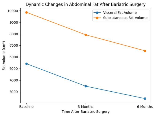
Figure 1: Line graph demonstrating progressive reduction in visceral adipose tissue (VAT) and subcutaneous adipose tissue (SAT) volumes measured by MRI at baseline, 3 months, and 6 months after bariatric surgery. Visceral fat showed a steeper decline compared to subcutaneous fat, indicating preferential metabolic fat loss
DISCUSSION :
This is a prospective MRI-based study comparing changes of the abdominal fat during early post baritric surgery and found that the visceral adipose tissue was reduced faster than subcutaneous fat. Even though, total weight loss was impressive during the initial six months of operation, the imaging showed that the enhancement of metabolism was better correlated to fat compartment redistribution other than just to decrease in total body weight. This observation corroborates the idea that the obesity-related metabolic risk is heavily dependent on the location of fat and not the mass of fat [10-12].
The most significant finding was the significant reduction in the visceral adipose tissue volume and VAT/SAT ratio. Visceral fat is also metabolically active and closely linked to insulin resistance, production of inflammatory cytokines, and cardiometabolic disease thus its early loss explains the rapid glycemic parameter changes that occurs following bariatric surgery. The positive relationship between the reduction in visceral fat and the fall in HbA1c and fasting glucose in our cohort emphasizes the fact that bariatric surgery triggers metabolic remission using hormonal and adipokine-mediated effects: even prior to the achievement of the maximal weight reduction [13-15].
The other significant observation was the large decrease in hepatic fat fraction. Non-alcoholic fatty liver disease is also associated with morbid obesity and is directly related to the viscerality fat deposition and insulin insensitivity. The liver fat in the current study decreased significantly after three months, which is matched with the changes in triglycerides, indicating the recovery of the insulin sensitivity of the liver and lipid metabolism. This is in line with the theory that bariatric surgery can quickly reverse the ectopic fat deposition and not just reducing the peripheral adiposity [16, 17].
The subcutaneous adipose tissue also reduced but at a lower rate than the visceral fat. This selective fat mobilization could be a manner of preferential lipolysis of metabolically detrimental adipose reserves during the initial postoperative catabolic period. This decrease in VAT/SAT ratio noted in this research also underlines the point that better metabolic profile is determined by qualitative redistribution of fat. MRI was useful in detecting these compartment-specific alterations that cannot be detected using anthropometric measures only [18-20].
Lastly, the research paper identifies the clinical significance of MRI as an example of non-invasive biomarker to measure treatment response following bariatric surgery. Traditional measures like BMI are inadequate to determine the degree of metabolic recovery and imaging fat measures can give objective results which indicate reduction of cardiometabolic risk. Thus, radiological evaluation can be involved in the determination of diabetes remission and postoperative follow-up method.
CONCLUSION:
Bariatric surgery leads to rapid metabolic improvement primarily through preferential reduction of visceral and ectopic fat rather than overall weight loss alone. MRI demonstrates significant early decreases in visceral, hepatic, and pancreatic fat that strongly correlate with glycemic and lipid improvement. Imaging-based fat quantification may serve as a reliable biomarker for evaluating metabolic recovery after bariatric surgery.
BIBLIOGRAPHY:
1. participants with obesity following bariatric surgery–A prospective longitudinal fMRI study. 2021. 163: p. 105237.
2. Blaho, M., et al., Use of magnetic resonance imaging to quantify fat and steatosis in the pancreas in patients after bariatric surgery: a retrospective study. 2022. 32(11): p. 3666-3674.
3. Fiorillo, C., et al., Effects of laparoscopic sleeve gastrectomy on gastric structure and function documented by magnetic resonance imaging are strongly associated with post-operative weight loss and quality of life: a prospective study. 2020. 30(12): p. 4741-4750.
4. Li, G., et al., Brain functional and structural magnetic resonance imaging of obesity and weight loss interventions. 2023. 28(4): p. 1466-1479.
5. Quero, G., et al., The causes of gastroesophageal reflux after laparoscopic sleeve gastrectomy: quantitative assessment of the structure and function of the esophagogastric junction by magnetic resonance imaging and high-resolution manometry. 2020. 30(6): p. 2108-2117.
6. Bai, L., et al., Abdominal magnetic resonance imaging assessment and quantification of pancreatic and liver adipose tissues in obesity before and after laparoscopic sleeve gastrectomy. 2023. 16(3): p. 100609.
7. Sivakumar, J., et al., Body composition changes following a very-low-calorie pre-operative diet in patients undergoing bariatric surgery. 2020. 30(1): p. 119-126.
8. Felsenreich, D.M., et al., Swallow magnetic resonance imaging compared to 3D-computed tomography for pouch assessment and hiatal hernias after Roux-en-Y gastric bypass. 2020. 30(11): p. 4192-4197.
9. Bredella, M.A., et al., Effects of sleeve gastrectomy on bone marrow adipose tissue in adolescents and young adults with obesity. 2020. 105(11): p. e3961-e3970.
10. Puliani, G., et al., Pituitary T1 signal intensity at magnetic resonance imaging is reduced in patients with obesity: results from the CHIASM study. 2023. 47(10): p. 948-955.
11. Toemen, L., et al., Body fat distribution, overweight, and cardiac structures in school‐age children: a population‐based cardiac magnetic resonance imaging study. 2020. 9(13): p. e014933.
12. Dadson, P., et al., Change in abdominal, but not femoral subcutaneous fat CT-radiodensity is associated with improved metabolic profile after bariatric surgery. 2020. 30(12): p. 2363-2371.
13. Wang, G.-J. and Y. Zhang, Brain functional and structural magnetic resonance imaging of obesity and weight loss interventions.
14. Esparham, A., et al., The impact of bariatric surgery on cardiac structure, and systolic and diastolic function in patients with obesity: a systematic review and meta-analysis. 2023. 33(1): p. 345-361.
15. Martínez‐Montoro, J.I., et al., Obesity‐related glomerulopathy: current approaches and future perspectives. 2022. 23(7): p. e13450.
16. Yurista, S.R., et al., Magnetic resonance imaging of cardiac metabolism in heart failure: how far have we come? 2022. 23(10): p. 1277-1289.
17. Grymyr, L.M., et al., One-year impact of bariatric surgery on left ventricular mechanics: results from the prospective FatWest study. 2021. 1(2): p. oeab024.
18. Heinrichs, H.S., et al., Effects of bariatric surgery on functional connectivity of the reward and default mode network: A pre‐registered analysis. 2021. 42(16): p. 5357-5373.
19. Saari, T., et al., Obesity-associated blunted subcutaneous adipose tissue blood flow after meal improves after bariatric surgery. 2022. 107(7): p. 1930-1938.
20. Moura Cunha, G., et al., Quantitative magnetic resonance imaging for chronic liver disease. 2021. 94(1121): p. 20201377.