Molecular Mechanisms of Myocardial Fibrosis in Heart Failure with Preserved Ejection Fraction (HFpEF)
- Muhammad Hussain , Assistant Professor Cardiology, Abwa Medical College Faisalabad, Pakistan
- Imtiaz Ali , Professor of Medical Imaging, ACE Institute of Technology, New York
- Anum Arshad , Senior Registrar Cardiology, Sheikh Zayed Hospital, Rahim Yar Khan, Pakistan
- Fawwad Fazal ,
- Sanjay Kumar , Assistant Professor, Department of Cardiology, Suleman Roshan Medical College and Hospital Tando Adam, Pakistan.
Article Information:
Abstract:
HFpEF is a fibrosis-based syndrome that is being increasingly recognised as one with diastolic dysfunction, occurring in the presence of preserved systolic function. It is believed that diffuse myocardial fibrosis and extracellular matrix remodeling play a major part in the stiffening of the ventricles and increased filling pressures. Purpose: The purpose of the study was to examine the molecular pathways of myocardial fibrosis in HFpEF, combining imaging, histological, transcriptomic, and hemodynamic data. Methodology: A prospective observational study was carried out with HFpEF patients and age-matched controls. The study involved the subject of echocardiography, cardiac magnetic resonance imaging with extracellular volume determination, and invasive hemodynamic assessment. The analysis of endomyocardial biopsies was done using histological staining, collagen cross-linking, quantitative PCR, western blotting, and single-cell RNA sequencing. Multivariate regression equations determined independent predictors of the degree of fibrosis. Findings: HFpEF patients exhibited higher extracellular volume, collagen volume fraction and collagen cross-linking than controls. TGF-bi signaling, LOXL2, and inflammatory mediator upregulation at the macro level was seen at both the mRNA and protein levels. Left ventricular filling pressure had a significant correlation with extracellular volume. Predictors of fibrosis burden independently were LOXL2, TGFB1, IL6 expression, and hemodynamic stress. Conclusion: Accurate myocardial fibrosis in HFpEF is defined as the coordinated signaling of profibrotic and inflammatory mechanisms that contributes to diastolic dysfunction. Activation of collagen cross-linking and TGF-B could also be a useful therapeutic target.
Keywords:
Article :
INTRODUCTION :
Heart failure with preserved ejection fraction (HFpEF) has emerged as a predominant ischemic form of heart failure across the globe and is clinically characterized by signs and symptoms of heart failure with a left ventricular ejection fraction usually ≥50 and evidence of cardiac structural or functional abnormalities and increased filling pressures.[1]. Although systolic pump fraction has been maintained, morbidity, frequent hospitalization, and mortality in patients have not been reduced as much as in heart failure with reduced ejection fraction[2]. HFpEF is not a disease, but a syndrome which is the result of concurrent pathobiologic processes and varies depending on age, sex, burden of comorbidity, and hemodynamic profile[3].
Clinically, this heterogeneity is apparent in studies of data-driven phenotyping data in which unsupervised clustering of dense clinical and echocardiographic variables recognizes reproducible HFpEF subgroups with unique comorbidities, congestion patterns, and outcomes.[4]. This type of work is consistent with the idea of mechanistic endotypes, some having a subgroup with increased concentric remodeling and biomarker signatures of increased extracellular matrix (ECM) remodeling, to whom myocardial fibrosis is a potentially valuable therapeutic target.
Myocardial fibrosis has become identified as a key factor in ventricular stiffening, loss of diastolic reserve and unsynchronized ventricular-vascular coupling among the structural abnormalities linked with HFpEF, typically diffuse and interstitial, accompanied by perivascular collagen deposition, instead of massive replacement scars. Notably, fibrotic remodeling modifies passive stiffness of myocardial cells and decreases compliance, which perpetuates undesirable remodelling and functional impairment, establishes a feed-forward mechanism[5,7] that maintains adverse remodeling and functional restriction.
Myocardial studies in human beings have revealed that HFpEF is also a condition where cardiomyocytes and ECM are abnormal to systolic heart failure conditions[6]. Passive stiffness culminating in high-passive stiffness in HFpEF may occur through titin-based cardiomyocyte stiffness and collagen-based ECM remodeling, but these processes are more equally contributed to by individual patients and at different stages within the disease process [9]. Such observations can be used to support a mechanistic model in which cardiomyocyte and interstitial pathways together set the stage of diastolic dysfunction, and help explain the apparent failure of therapies which ameliorate hemodynamics to restore symptoms in the presence of advanced structural stiffening.
There is a large literature that connects the high comorbidity burden characteristic of HFpEF, such as obesity, diabetes, hypertension, chronic kidney disease, and aging, to systemic inflammation contributing to coronary microvascular endothelial activation.[10]. This microvascular hypothesis is supported by endothelial inflammation and oxidative stress which depletes the bioavailability of nitric oxide, impairs cyclic GMP and protein kinase G signaling, and promotes cardiomyocyte hypertrophy and stiffness and activates interstitial remodelling and reserve limitation [10,7]. In addition to decreased perfusion reserve, there exists direct interaction between inflammatory endothelial signaling and the behaviour of fibroblasts via paracrine mediators, extracellular vesicles, and modification of capillary permeability which link systemic comorbidities with myocardial ECM remodelling [8,10].
Molecular mechanisms Cardiac fibrosis is thought to be an imbalance of ECM synthesis and degradation that occurs as a consequence of resident cardiac fibroblast activation, recruiting or transdifferentiating other mesenchymal-like populations via transcriptional upregulation of collagen and other ECM genes using Smad and noncanonical signaling pathways[8,11]. Mechanical stress due to hypertension, neurohormonal signals, metabolic inflammation, and reactive oxygen species, among others, in upstream stages increase profibrotic gene programs and inhibit ECM degradation, are part of the upstream stimuli of HFpEF, and have been explored in phenotyping and risk assessment of HFpEF[8,11]. Crosstalk between immune and fibroblastic signaling/mediators in the downstream drives the fibrotic response, and biomarkers that reflect the inflammation
Fibrosis HFpEF is not simply identified by the amount of collagen, but also by collagen quality, especially cross-linking that elevates ECM stiffness and elevates resistance to proteolytic degradation. Cross-linking through lysyl oxidase family enzymes has been shown to form more resistant and stiffer collagen networks and overproduction of myocardial collagen cross-linking has also been associated with the risk of hospitalization due to heart failure in hypertensive heart failure, a precondition and overlap of HFpEF[13]. Previous studies in patients with diastolic heart failure caused by hypertension reported that collagen metabolism is robust and that functional impairment, supporting the idea that fibrosis is a dynamic and not a static process of scarring. Nonenzymatic cross-linking can be further enhanced by metabolic comorbidities via use of advanced glycation end products, which are another source of stiffness mechanism especially in the context of diabetes-associated HFpEF phenotypes.[3].
Myocardial fibrosis may be a target of intervention, so it has now taken center stage in the phenotyping of HFpEF and clinical studies aiming to treat it as well as to measure it.[15]. Noninvasive estimates of diffuse interstitial fibrosis using cardiovascular magnetic resonance T1 mapping and extracellular volume quantification have been consistent across the spectrum of ejection fraction and linked to later heart failure hospitalization and death, indicating that myocardial fibrosis is a clinically relevant vulnerability phenomenon [15]. Nonetheless, there are significant gaps in the way fibrosis biology is translated into effective HFpEF therapies. Such gaps are filling could be answering the questions of what molecular pathways predominate in particular endotypes of HFpEF, temporal relationships between microvascular dysfunction, cardiomyocyte stiffness and ECM remodeling, and the identification of tractable targets that could reduce fibrosis without interfering with required wound-healing and immune responses[3,8]. This accuracy is critical towards enhancing HFpEF outcomes.
METHODOLOGY:
Study Design and Population
This research is planned as a prospective, observational, translational research project that aims at analyzing the molecular factors that determine myocardial fibrosis in heart failure patients with preserved ejection fraction (HFpEF). The study was conducted in Abwa Medical College Faisalabad from May 2024 to May 2025. A tertiary cardiovascular center will recruit grown-up patients aged between 45 to 85 years with a definite diagnosis of HFpEF, which is characterized by left ventricular ejection fraction 50 years, heart failure symptoms, and objective data of increased filling pressures. The control group will consist of age- and sex-matched patients who do not have clinical heart failure but who are undergoing the elective cardiac procedures due to noninflammatory conditions. The following criteria will be held as exclusion criteria: active myocarditis, severe valvular disease that needs urgent surgery, infiltrative cardiomyopathies and recent acute coronary syndrome. Informed consent will be done in written form and the research will be ethically sanctioned by the institutional ethics committee.
Clinical and Imaging Assessment
Extensive clinical assessment will entail medical history, comorbidity profiling, anthropometric measures, and generic laboratory assessments. To determine the diastolic parameters, left atrial volume, and the ventricle wall thickness, the Echocardiography will be conducted. T1 mapping of cardiac magnetic resonance imaging will measure surrogate diffusion measurement of extracellular volume fraction in the presence of diffuse interstitial fibrosis. Measures of circulating fibrosis-related biomarkers will be done by taking blood samples which include procollagen peptides, galactin-3, and matrix metalloproteinases. Right heart catheterization will be performed on a subgroup of participants to ensure that hemodynamic data is obtained to verify increased filling pressures and align invasive parameters with molecular data.
Myocardial Tissue Collection and Histological Analysis
Sample Endomyocardial biopsy will be collected with the consent of HFpEF patients undergoing procedures that are clinically necessary and in ethically possible cases of the control group. Tissue specimens will be separated instantly into histology, molecular and transcriptomic. Masson trichrome and picrosirius red histological staining will be used to quantify volume fraction of collagen and organization of collagen fibres. The evaluation of collagen cross-linking will be done using biochemical tests that determine the content of hydroxylysyl pyridinoline. To characterize fibroblast stimulation, and inflammatory infiltration, immunohistochemistry will be used to measure α-smooth muscle actin, TGF- 0 signaling intermediates and inflammatory markers.
Molecular and Transcriptomic Profiling
Myocardial samples will be able to extract total RNA and protein to determine gene and protein expression with regard to fibrosis pathways. Quantitative polymerase chain reaction is the procedure that will be used to measure transcripts related to TGF-B signaling, lysyl oxidase family enzymes, extracellular matrix proteins, and inflammatory mediators. The activation of pathways will be quantified by western blotting on a protein level. A smaller group of samples will undergo single-cell RNA sequencing in order to establish fibroblast subpopulations and immune cell interactions that play a role in ECM remodeling. Transcriptomic signatures will be incorporated into factors of bioinformatical analysis of imaging-derived fibrosis.
Statistical Analysis
Continuous variables will be described in terms of mean standard deviation or interquartile range. Independent t-tests or Mann Whitney U tests will be used to carry out comparison between the HFpEF and the control groups. Correlation tests will be conducted to evaluate association of molecular markers, extracellular volume as viewed by imaging and hemodynamic parameters. Independent predictors of the severity of myocardial fibrosis will be determined by multivariate regression analysis on independent molecular predictors. Two-sided p= <0.05 will be taken as statistically significant.
RESULTS :
Participant Characteristics
Data on 100 individuals (60 having HFpEF and 40 as controls) were analyzed. Baseline features (Table 1): HFpEF has a greater body mass index and systolic blood pressure whereas ejection fraction of the left ventricle was conserved in both groups. Compared to HFpEF, NT-proBNP was significantly more in the HFpEF group, which is in line with increased hemodynamic stress.
Table 1. Baseline Demographic and Clinical Characteristics
|
Variable |
HFpEF (n=60) |
Controls (n=40) |
p-value |
|
Age (years) |
71.4 ± 8.2 |
69.1 ± 7.9 |
0.18 |
|
Female (%) |
38 (63%) |
22 (55%) |
0.42 |
|
BMI (kg/m²) |
31.8 ± 4.6 |
26.3 ± 3.9 |
<0.001 |
|
Systolic BP (mmHg) |
146 ± 18 |
128 ± 14 |
<0.001 |
|
Heart Rate (bpm) |
78 ± 11 |
72 ± 9 |
0.01 |
|
LVEF (%) |
58.6 ± 4.2 |
60.2 ± 3.8 |
0.07 |
|
NT-proBNP (pg/mL) |
984 (650–1480) |
132 (85–210) |
<0.001 |
The comorbidity and medication (Table 2) figures imply more hypertension, diabetes, atrial fibrillation, and chronic kidney disease in HFpEF, more ACEi/ARB, beta-blockers, and diuretics. These disparities conceptualize fibrosis as a myocardial reaction that is produced by metacardiac and vascular comorbidity burdens and not an independent cardiac pathology.
Table 2. Comorbidity Distribution and Medication Profile
|
Variable |
HFpEF (n=60) |
Controls (n=40) |
p-value |
|
Hypertension (%) |
52 (87%) |
16 (40%) |
<0.001 |
|
Diabetes Mellitus (%) |
34 (57%) |
8 (20%) |
<0.001 |
|
Chronic Kidney Disease (%) |
18 (30%) |
4 (10%) |
0.02 |
|
Atrial Fibrillation (%) |
22 (37%) |
3 (7%) |
<0.001 |
|
ACEi/ARB use (%) |
45 (75%) |
14 (35%) |
<0.001 |
|
Beta-blocker use (%) |
41 (68%) |
12 (30%) |
<0.001 |
|
Diuretics (%) |
49 (82%) |
2 (5%) |
<0.001 |
Echocardiography, Hemodynamics, and Tissue-Level Fibrosis Burden
The diastolic measurements of the functions were always not normal in HFpEF. Table 3 indicated significantly increased E/e L and left atrial volume index in favor of chronically high filling pressures and failure of relaxation. In the catheterization group LV end-diastolic pressure, and capillary wedge pressure were increased thus proving invasive congestion physiology.
Table 3. Echocardiographic and Hemodynamic Parameters
|
Parameter |
HFpEF |
Controls |
p-value |
|
E/e′ ratio |
17.8 ± 4.1 |
9.6 ± 2.2 |
<0.001 |
|
Left Atrial Volume Index (mL/m²) |
44.2 ± 9.8 |
28.5 ± 6.4 |
<0.001 |
|
LV Mass Index (g/m²) |
122 ± 24 |
94 ± 18 |
<0.001 |
|
LV End-Diastolic Pressure (mmHg)* |
22.4 ± 5.2 |
11.3 ± 3.1 |
<0.001 |
|
Pulmonary Capillary Wedge Pressure (mmHg)* |
24.8 ± 6.3 |
12.6 ± 3.5 |
<0.001 |
*Subset n=35 HFpEF, n=20 controls
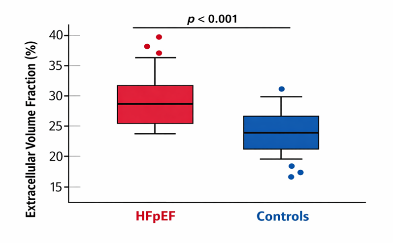
Table 4 results of cardiac magnetic resonance reveal an augmentation of native T1 and extra-cellular volume (ECV) in HFpEF, which is related to an expansion of the interstitial matrix diffusely. The separation of the groups in the ECV is reviewed in figure 1 where HFpEF has a high median and wider distribution, indicating that there is a non-uniformity in the burden of fibrosis. Minority (Table 4) had late gadolinium enhanced, which suggests that focal scar may co-relate with diffuse fibrosis.
Table 4. Cardiac Magnetic Resonance–Derived Fibrosis Indices
|
Parameter |
HFpEF |
Controls |
p-value |
|
Native T1 (ms) |
1086 ± 42 |
1018 ± 35 |
<0.001 |
|
Extracellular Volume (%) |
32.4 ± 4.3 |
24.7 ± 3.1 |
<0.001 |
|
Late Gadolinium Enhancement (%) |
18 (30%) |
4 (10%) |
0.02 |
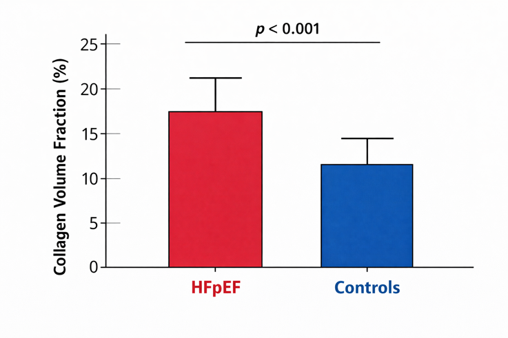
Imaging was supported by direct myocardial assessment. Biochemical analyses and histology (Table 5) indicate that HFpEF biopsies were characterized by increased volume fraction of collagen and increased collagen fibers in terms of size and cross-linking measured by hydroxylysyl pyridinoline. The example Masson trichrome and picrosirius red Masson staining patterns as per Figure 3 indicate that the trichrome and picrosirius red were used as representative of greater collagen deposition, and fibrous architecture changes, when compared to between-group difference in collagen volume fraction quantified in Figure 4. The trend direction of CMR-fertilized ECV (Table 4, Figure 1) as compared to tissue fibrosis markers (Table 5, Figures 3 and 4) can be regarded as the evidence in favor of ECV being a noninvasive surrogate of interstitial remodeling.
Table 5. Histological and Biochemical Fibrosis Measurements
|
Parameter |
HFpEF (n=25) |
Controls (n=15) |
p-value |
|
Collagen Volume Fraction (%) |
18.6 ± 5.2 |
9.4 ± 3.1 |
<0.001 |
|
Collagen Fiber Thickness (µm) |
4.8 ± 1.2 |
2.6 ± 0.9 |
<0.001 |
|
Hydroxylysyl Pyridinoline (pmol/mg) |
0.84 ± 0.21 |
0.41 ± 0.12 |
<0.001 |
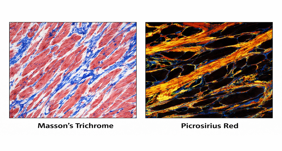
Figure 3. Representative Masson’s Trichrome and Picrosirius Red Staining Images
Relationship between Fibrosis and Filling Pressure
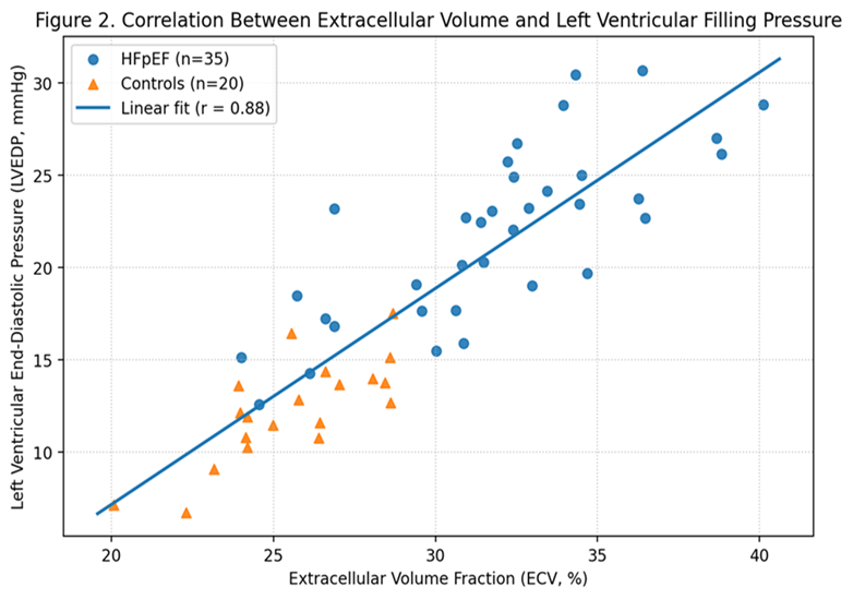
Hemodynamic stress followed with fibrosis. Figure 2 demonstrates that there is significant evidence of positive relations between ECV and LV end-diastolic pressure in individuals, which suggests that maximum expansion of the matrix is associated with high filling pressures. The relationship gives evidence of a mechanistic explanation where ECM remodeling is involved in making ventricular compliance lower, and hence, a small variation in filling volume induces a greater increase in pressure. The correlation also implies the presence of a feed-forward relationship where loading conditions can speed up the activation of fibroblasts and collagen remodelling.
Profibrotic Gene and Protein Activation
Profibrotic programs were shown to have been activated by molecular profiling. Results of differential gene expression (Table 6) indicate that TGFB1, COL1A1, COL3A1, LOX, LOXL2, TIMP1, and IL6 are upregulated in HFpEF compared to controls with moderate increase in MMP2. These relative changes in mRNA levels are depicted in Figure 5 and revealed parallel amplification in collagen synthesis movements, cross-linking apparatus and inflammatory mediators. The increase in both MMP2 and TIMP1 indicates that there is active matrix turnover and net ECM deposition, which is in line with ongoing remodeling and not a fixed scar.
Table 6. Differential Gene Expression Profiles (Fold Change vs Control)
|
Gene |
Fold Change |
p-value |
|
TGFB1 |
2.8 ± 0.6 |
<0.001 |
|
COL1A1 |
3.4 ± 0.9 |
<0.001 |
|
COL3A1 |
2.7 ± 0.7 |
<0.001 |
|
LOX |
2.5 ± 0.5 |
<0.001 |
|
LOXL2 |
3.1 ± 0.8 |
<0.001 |
|
MMP2 |
1.8 ± 0.4 |
0.002 |
|
TIMP1 |
2.2 ± 0.6 |
<0.001 |
|
IL6 |
2.6 ± 0.7 |
<0.001 |
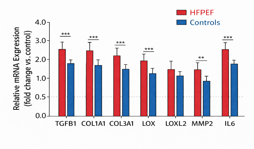
Figure 5. Relative mRNA Expression of Profibrotic Genes
Findings on the protein level were concordant. Table 7 indicates greater TGF-B1, phosphorylated Smad2/3, α-SMA, and LOXL2 protein concentration in HFpEF. Figure 6 shows the typical western blots and densitometry, which shows stronger signals of p-Smad2/3 and TGF-B1 in HFpEF with a uniform loading factor. Collectively, these data suggest active TGF-B signaling and differentiation of myofibroblasts as proximal mediators of matrix accumulation, and LOXL2 suggests an additional role of stiffness mediated by cross-linking collagen as the biochemical complement of Table 5.
Table 7. Protein Expression Levels (Relative Densitometry Units)
|
Protein |
HFpEF |
Controls |
p-value |
|
TGF-β1 |
1.95 ± 0.42 |
1.00 ± 0.28 |
<0.001 |
|
p-Smad2/3 |
2.21 ± 0.51 |
1.05 ± 0.33 |
<0.001 |
|
α-SMA |
1.88 ± 0.47 |
0.92 ± 0.25 |
<0.001 |
|
LOXL2 |
2.03 ± 0.44 |
1.07 ± 0.29 |
<0.001 |
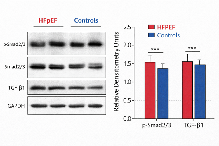
Figure 6. Western Blot Analysis of TGF-β Signaling Activation
Single-Cell Landscape and Pathway Enrichment
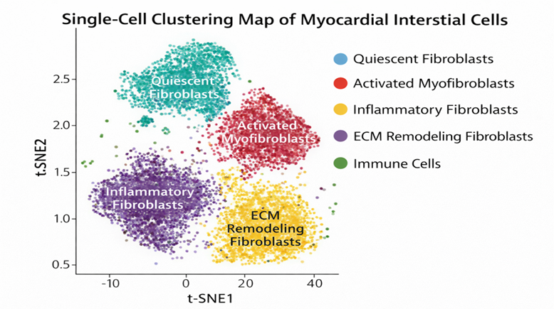
The changes in the states of interstitial cells to single-cell RNA sequencing showed changes in the interstitial cell state. Table 8 presents an increase of activated myofibroblasts and ECM remodeling fibroblasts in HFpEF when compared to controls, as well as an increased compartment of inflammatory fibroblasts. These subpopulations are represented as discrete clusters in Fig. 7, and they give credence to the possibility of more than one uniform state through the existence of a number of fundamentally different fibroblast activation patterns. Interestingly the cluster of inflammatory fibroblasts coincides with the high IL6 production in Table 6, which correlates the state of the immune tone with the remodeling of the matrices. Higher LOXL2 signals are also accompanied by the expansion of ECM remodeling fibroblasts, which hints at the idea that cross-linking is cell-state but not systemic.
Table 8. Identified Fibroblast Subpopulations (Single-Cell RNA-seq)
|
Cluster |
Cell Count (HFpEF) |
Cell Count (Control) |
Key Marker Genes |
|
Quiescent Fibroblasts |
1,245 |
1,102 |
PDGFRA, DCN |
|
Activated Myofibroblasts |
2,384 |
612 |
ACTA2, COL1A1 |
|
Inflammatory Fibroblasts |
1,102 |
310 |
IL6, CCL2 |
|
ECM Remodeling Fibroblasts |
1,876 |
508 |
LOXL2, MMP2 |
 ,
,
Figure 7. Single-Cell Clustering Map of Myocardial Interstitial Cells
Figure 8 summarizes an analysis of enrichment of activated fibroblast clusters with pathways like ECM-receptor interaction, focal adhesion, PI3K-Akt signaling, TGF-b signaling, cytokine-cytokine receptor and inflammatory pathways. This tendency helps to bring mechanical clues, cytokines, and profibrotic gene expression in activated fibroblast states that are supportive of matrix deposition and stabilization.
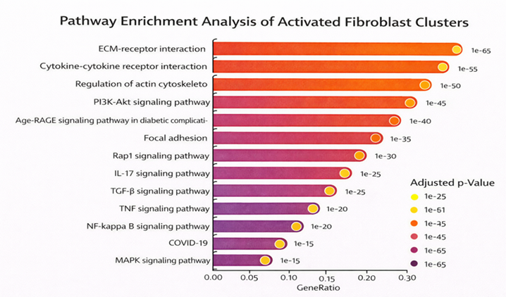
Figure 8. Pathway Enrichment Analysis of Activated Fibroblast Clusters
Integrated Associations and Predictors of Fibrosis Severity
The data from correlation analyses (Table 9) provide strong indications that ECV, collagen volume fraction, expression of LOXL2, and expression of TGFB1 are interrelated, and all of them create a coherent fibrosis axis. Multivariate analysis (Table 10) is able to find that LOXL2, TGFB1, IL6 expression, and LV end-diastolic pressure are independent predictors of ECV, and age contributes less.
Table 9. Correlation Matrix (r values)
|
Variable |
ECV |
Collagen Volume Fraction |
LOXL2 Expression |
TGFB1 Expression |
|
Extracellular Volume |
1.00 |
0.74 |
0.69 |
0.65 |
|
Collagen Volume Fraction |
0.74 |
1.00 |
0.71 |
0.68 |
|
LOXL2 Expression |
0.69 |
0.71 |
1.00 |
0.62 |
|
TGFB1 Expression |
0.65 |
0.68 |
0.62 |
1.00 |
All correlations p < 0.01
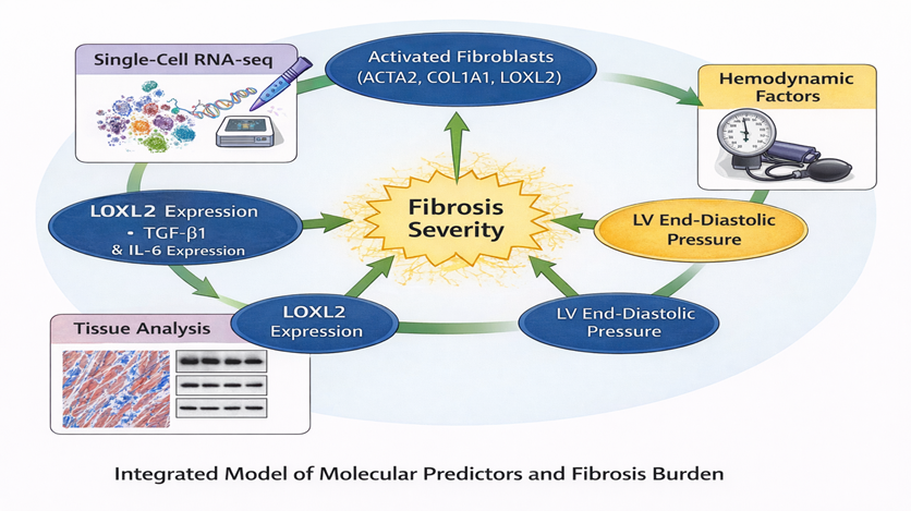
Figure 9. Integrated Model of Molecular Predictors and Fibrosis Burden
Figure 9 combines transcriptomic activation, tissue-level metrics of fibrosis, and hemodynamic stress as convergent predictors of the fibrosis severity in favor of a model of HFpEF that is characterized by diffuse ECM expansion, enhanced cross-linking and activated fibroblast programs that correlate with higher filling pressures.
Table 10. Multivariable Regression Analysis for Predictors of Fibrosis Severity
Dependent Variable: Extracellular Volume (%)
|
Predictor |
β Coefficient |
Standard Error |
p-value |
|
LOXL2 Expression |
0.42 |
0.08 |
<0.001 |
|
TGFB1 Expression |
0.31 |
0.07 |
0.002 |
|
IL6 Expression |
0.26 |
0.09 |
0.01 |
|
LV End-Diastolic Pressure |
0.38 |
0.11 |
0.004 |
|
Age |
0.12 |
0.05 |
0.08 |
Model R² = 0.62
malformations. Clinical and radiological correlation was advised. The patient was planned for multidisciplinary evaluation involving paediatric surgery, dermatology, and radiology for further management and follow-up.
DISCUSSION :
The current research paper indicates that myocardial fibrosis in HFpEF is associated with the coordinated structural, molecular, and hemodynamic changes. Increased extracellular volume and collagen volume fraction was accompanied with increased TGF-B signaling, LOXL2-induced cross-linking pathways and inflammatory gene expression. These conclusions are consistent with the increasing opinion that HFpEF is essentially a disease of myocardial stiffening that is partially mediated by maladaptive extracellular matrix remodeling but not necessarily cardiomyocyte-
specific dysfunction. The findings presented in our study build on the initial experimental findings as a combination of imaging, histological, transcriptomic, and invasive hemodynamic information in one cohort.
Multiple clinical studies have identified an increase in diffuse fibrosis in HFpEF with cardiac magnetic resonance, and extracellular volume fraction has been associated with poor clinical outcomes.[17]. These findings are in line with our observation that there is a strong correlation between extracellular volume and left ventricular end-diastolic pressure, which reinforces the idea that the left ventricular end-diastolic pressure is directly affected by matrix expansion. Furthermore, the augmented interstitial deposition of collagen and an abnormal matrix organization in HFpEF have been characterized through autopsy and likewise biopsy studies, which tend to be extremely higher than collagen deposition and matrix organization seen in hypertensive controls[18]. The current evidence supports these changes of structures and indicate that they are complemented by molecular activation of profibrotic pathways, indicating continuous dynamic remodeling and not scar-quiet development.
The TGF-B signaling upregulation detected in the composition of mRNAs and protein agrees with experimental paradigms that TGF-B is a key node of fibroblast stimulation and myofibroblast differentiation.[19]. Notably, greater LOXL2 expression and greater collagen cross-linking are also noted in our findings. Earlier translational studies have also stressed that collagen cross-linking might be the most vital factor in the total collagen amount in the determination of myocardial stiffness[20]. Heightened cross-linking makes collagen fibers difficult to disappear, and tissue stiffer thus possible reasons why certain patients have over-representative high filling pressures compared to collagen volume fraction alone. This mechanistic relationship promotes therapeutic interventions on cross linking enzymes besides inhibiting the formation of collagen production.
Transcriptomic profiling of single cells showed the proliferation of activated and inflammatory fibroblast subsets, in line with the reports that interstitial cell heterogeneity is a major property of cardiac remodelling.[21]. Pathway enrichment of ECM-receptor interaction, focal adhesion, and PI3K-Akt indicates the importance of mechano-transduction in the maintenance of fibroblast activation. Similar results have previously been obtained in the literature on pressure-overload models of metabolic heart disease, which have found mechanical stress and inflammatory cytokines interacting synergistically to cause phenotypic shifts in fibroblasts [22]. The inflammatory fibroblast cluster phenotype, as well as the high IL-6 expression level in our cohort, supports the theory of systemic comorbidities with HFpEF as leading to the localization of inflammatory-fibrotic coupling in the myocardium.[23].
In comparison with the previous studies where the main interest was on imaging forms or circulating biomarkers, the combination of tissue level molecular information with invasive hemodynamics offered in this study offers a more holistic mechanistic approach to the problem. Although the previous literature proposed that cardiomyocyte stiffness associated with titin alterations may dominate in the early HFpEF,[24] our results demonstrate that cross-linking and extracellular matrix remodeling are key factors in a disease that has been diagnosed. This finding leads to the use of a stage-specific model, where cardiomyocytes abnormalities could eventually progress to a model dominated by stiffness in the matrix, where fibroblast activation continues.
Clinical Implications
The implication of the finding of LOXL2 and TGF-b signaling as independent predictors of fibrosis burden is the possibility of using antifibrotic treatment in HFpEF. TGF-B signaling has been pharmacologically modulated in other fibrotic diseases, and selective inhibition of cross-linking enzymes of collagen is being studied in cardiovascular disease.[25]. Additionally, extracellular volume was highly associated with hemodynamic parameters which means that the noninvasive imaging may be used as a surrogate endpoint to carry out therapeutic trials based on the looting about the matrix remodeling. Molecular and imaging biomarkers should be used to determine the precise phenotype of HFpEF, which could be used to select HFpEF subgroups that are most likely to respond to antifibrotic interventions.
Study Limitations
There are a number of restrictions that should be considered. The biopsy group was relatively small and can not possibly reflect the entire range of HFpEF phenotypes. Though the correlations between molecular markers and the severity of fibrosis were strong, the causality cannot be proven in this observational design. Having cross-sectional tissue sampling does not allow one to assess the temporal course or reversibility of fibrosis. Furthermore, although the single-cell RNA sequencing study offered the evidence of heterogeneity of fibroblasts, the spatial and transcriptomic data was lacking to establish the microanatomic context. Lastly, comorbid conditions like diabetes and obesity can play a role of their own in the fibrotic signaling and makes it more difficult to pin down HFpEF pathophysiology only.
CONCLUSION :
Overall, this analysis shows that HFpEF is accompanied by diffuse myocardial fibrosis with an elevated extracellular volume, increased cross-linking of collagen, and TGF-p2, and inflammatory signal activation. Loxl2 and Il-6 are molecular predictors, which are associated with the fibrosis load and increased filling pressures, which are strong evidence that fibroblast activity and diastolic dysfunction are connected mechanistically. These results support the idea that extracellular matrix re-modeling is at the heart of the pathogenesis of HFpEF and emphasize the possibilities of the specific antifibrotic approach that should be informed by integrated imaging and molecular profiling.
BIBLIOGRAPHY:
1. Heidenreich PA, Bozkurt B, Aguilar D, et al. 2022 AHA/ACC/HFSA guideline for the management of heart failure. Circulation. 2022;145(18):e895-e1032.
2. Owan TE, Hodge DO, Herges RM, Jacobsen SJ, Roger VL, Redfield MM. Trends in prevalence and outcome of heart failure with preserved ejection fraction. N Engl J Med. 2006;355(3):251-259.
3. Borlaug BA, et al. Heart failure with preserved ejection fraction: JACC scientific statement. J Am Coll Cardiol. 2023;81(18):1810-1834.
4. Shah SJ, Katz DH, Selvaraj S, et al. Phenomapping for novel classification of heart failure with preserved ejection fraction. Circulation. 2015;131(3):269-279.
5. Zile MR, Baicu CF, Ikonomidis JS, et al. Myocardial stiffness in patients with heart failure and a preserved ejection fraction: contributions of collagen and titin. Circulation. 2015;131(14):1247-1259.
6. van Heerebeek L, Borbely A, Niessen HW, et al. Myocardial structure and function differ in systolic and diastolic heart failure. Circulation. 2006;113(16):1966-1973.
7. Mohammed SF, Hussain S, Mirzoyev SA, Edwards WD, Maleszewski JJ, Redfield MM. Coronary microvascular rarefaction and myocardial fibrosis in heart failure with preserved ejection fraction. Circulation. 2015;131(6):550-559.
8. Frangogiannis NG. Cardiac fibrosis: cell biological mechanisms, molecular pathways and therapeutic opportunities. Mol Aspects Med. 2019;65:70-99.
9. Franssen C, González Miqueo A. The role of titin and extracellular matrix remodelling in heart failure with preserved ejection fraction. Neth Heart J. 2016;24(4):259-267.
10. Paulus WJ, Tschöpe C. A novel paradigm for heart failure with preserved ejection fraction: comorbidities drive myocardial dysfunction and remodeling through coronary microvascular endothelial inflammation. J Am Coll Cardiol. 2013;62(4):263-271.
11. Dobaczewski M, Chen W, Frangogiannis NG. Transforming growth factor (TGF)-β signaling in cardiac remodeling. J Mol Cell Cardiol. 2011;51:600-606.
12. de Boer RA, Lok DJA, Jaarsma T, van der Meer P, Voors AA, Hillege HL, van Veldhuisen DJ. Galectin-3 in heart failure with preserved ejection fraction. Eur J Heart Fail. 2013;15(10):1095-1101.
13. López B, Ravassa S, González A, Zubillaga E, Bonavila C, Bergés M, et al. Myocardial collagen cross-linking is associated with heart failure hospitalization in patients with hypertensive heart failure. J Am Coll Cardiol. 2016;67(3):251-260.
14. Martos R, Baugh J, Ledwidge M, O'Loughlin C, Conlon C, Patle A, Donnelly SC, McDonald K. Diastolic heart failure: evidence of increased myocardial collagen turnover linked to diastolic dysfunction. Circulation. 2007;115(7):888-895.
15. Schelbert EB, Piehler KM, Zareba KM, et al. Myocardial fibrosis quantified by extracellular volume is associated with subsequent hospitalization for heart failure, death, or both across the spectrum of ejection fraction and heart failure stage. J Am Heart Assoc. 2015;4(12):e002613.
16. Lam CSP, Voors AA, de Boer RA, Solomon SD, van Veldhuisen DJ. Heart failure with preserved ejection fraction. Nat Rev Dis Primers. 2018;4:16:02.
17. Puntmann VO, Carr-White G, Jabbour A, et al. T1 mapping and outcome in heart failure with preserved ejection fraction. JACC Cardiovasc Imaging. 2016;9(1):40-50.
18. Westermann D, Lindner D, Kasner M, et al. Cardiac inflammation contributes to diastolic dysfunction in HFpEF. Circulation. 2011;124(6):677-688.
19. Bujak M, Kweon HJ, Chatila K, et al. TGF-β signaling in cardiac remodeling. Circ Res. 2007;101(9): 941-953.
20. Díez J, González A, López B, Querejeta R. Mechanisms of disease: collagen cross-linking in cardiac fibrosis. Nat Clin Pract Cardiovasc Med. 2005;2(4):209-215.
21. Farbehi N, Patrick R, Dorison A, et al. Single-cell expression profiling reveals dynamic fibroblast states. Nat Commun. 2019;10:4018.
22. Travers JG, Kamal FA, Robbins J, Yutzey KE, Blaxall BC. Cardiac fibrosis pathogenesis. Circ Res. 2016;118(6):1021-1040.
23. Van Linthout S, Tschope C. Inflammation in HFpEF. Cardiovasc Res. 2017;113(12): 1399-1412.
24. Borbély A, Falcao-Pires I, van Heerebeek L, et al. Cardiomyocyte stiffness in diastolic heart failure. Circulation. 2009;119(1): 89-96.
25. López B, González A, Ravassa S, et al. Therapeutic targeting of myocardial fibrosis. J Am Coll Cardiol. 2021;78(3): 300-312.