The Role of Oral and Maxillofacial Surgeons in the Early Detection of Multiple Myeloma: A Systematic Review
- Dr Maryam Shamin , Crusader Community Health (DA), I l l i n o i s, United States of America
- Dr Mahnoor Mansoor , Ex-House Officer, Department of Oral Surgery, Riphah International University, Islamabad, Pakistan
- Dr Bela Badar Ali Shah M. Hamza , Postgraduate, Ex-Lecturer, Department of Dental Materials, Liaquat College of Medicine and Dentistry, Karachi, Pakistan
- Dr Alona Joppan , Grace Dental Hillcroft, (RDA), Texas, United States of America
- Dr Ushba Jamal , Ex-Lecturer, Postgraduate Student, Concordia University, Chicago, I l l i n o i s, United States of America
- Dr Irfan Qureshi , Prosthodontist, Diplomate, American Board of Oral Implantology/Implant Dentistry, Dr Irfan Qureshi’s Team of Professionals, Karachi, Pakistan.
Article Information:
Abstract:
Background: Multiple myeloma (MM) is a malignant plasma cell neoplasm with both systemic and oral manifestations. Oral lesions may precede systemic symptoms, providing an opportunity for early detection. Objective To systematically summarize the role of OMFS in the early recognition of MM through clinical and radiographic oral findings. Methods A systematic literature search of PubMed (2005–2025) was conducted using the keywords “multiple myeloma” AND “oral manifestation.” Inclusion criteria were case reports, cohort studies, and cross-sectional studies reporting oral and radiographic manifestations of MM. Reviews, narrative articles, non-English publications, and studies lacking validated clinical or radiographic descriptions were excluded. Data on oral features, radiographic findings, systemic CRAB criteria, and OMFS interventions were extracted. Methodological quality was assessed using a standardized risk of bias tool, following PRISMA 2020 guidelines. Results Twenty studies were included. Oral manifestations were diverse, with swelling (55%), jaw pain (30%), soft tissue masses (25%), gingival enlargement (25%), dental mobility (20%), and paresthesia (15%) most frequently reported. Radiographically, punched-out osteolytic lesions were observed in 12 studies, predominantly affecting the posterior mandible. Systemic features frequently aligned with CRAB criteria, including bone pain, anemia, fatigue, and recurrent infections. OMFS interventions facilitated timely hematology referral, imaging, biopsy, and prevention of bisphosphonate-related osteonecrosis. Conclusions Early recognition of oral and radiographic manifestations by OMFS enables prompt referral and multidisciplinary management, improving patient outcomes. Incorporating vigilant oral assessments into routine practice enhances early diagnosis and prognosis in MM patients.
Keywords:
Article :
INTRODUCTION :
The term "multiple myeloma" (MM) originated by J. Von Rustizky in 1873 after he identified eight distinct bone marrow malignancies, while Samuel Solley documented the earliest well-characterized case in 1844 (1). Multiple myeloma is a malignant B-cell neoplasm characterized by clonal proliferation of plasma cells within the bone marrow, leading to abnormal immunoglobulin production and secretion (2). This disorder is associated with diverse complications, including immunodeficiency, renal failure, anemia, and bone destruction (3). The disease predominantly affects individuals over the age of 40, with a higher incidence in males (4,5).
Multiple myeloma is defined by multifocal proliferation of atypical plasma cells and the presence of monoclonal immunoglobulins, commonly referred to as M-proteins or myeloma proteins (6). These M-proteins consist of heavy and light chains, with IgG being the most frequent (52%), followed by IgA (21%), IgD (2%), IgM (0.5%), and light chains alone (16%) (7). Osteolytic lesions occur in over 30% of patients, and oral manifestations may serve as the initial presentation in approximately 14% of cases (8). The prevalence of primary jaw involvement ranges from 8% to 15%, predominantly affecting the mandibular molar region, ramus, and angle due to their high hematopoietic activity, while maxillary lesions are more commonly observed in the posterior regions (9,10).
Clinically, patients with MM frequently present with persistent bone pain, recurrent infections, fatigue, and multisystem involvement (11) The disease is further characterized by end-organ damage, summarized by the CRAB criteria, which includes hypercalcemia, renal insufficiency, anemia, and bone lesions (12). Despite being the second most frequent bone marrow malignancy, MM constitutes only 2% of all cancers and 10% to 15% of hematologic malignancies (5). The disease may present as a solitary plasmacytoma or as multiple lesions affecting various skeletal sites, including the spine, skull, pelvis, sternum, vertebrae, and ribs (13). Unlike other metastatic bone tumors, osteolytic lesions in MM do not trigger reactive bone formation (14). Common clinical manifestations include bone pain, pathologic fractures, fatigue, recurrent infections, and secondary amyloidosis, with radiographs often revealing diffuse or localized osteolytic lesions or ‘punched out’ lesions (15). Early diagnosis facilitates timely initiation of therapy, improving patient prognosis. Current treatment strategies include corticosteroids, bisphosphonates, chemotherapy, and hematopoietic stem-cell transplantation, with disease staging and prognosis guided by the International Staging System. Oral health is a critical yet often overlooked aspect of MM management, as systemic symptoms may adversely impact the oral cavity, particularly the gingival tissues. Dentists should be vigilant for oral manifestations, as it will help to identify early indication of disease and its progression.
MATERIAL AND METHODS:
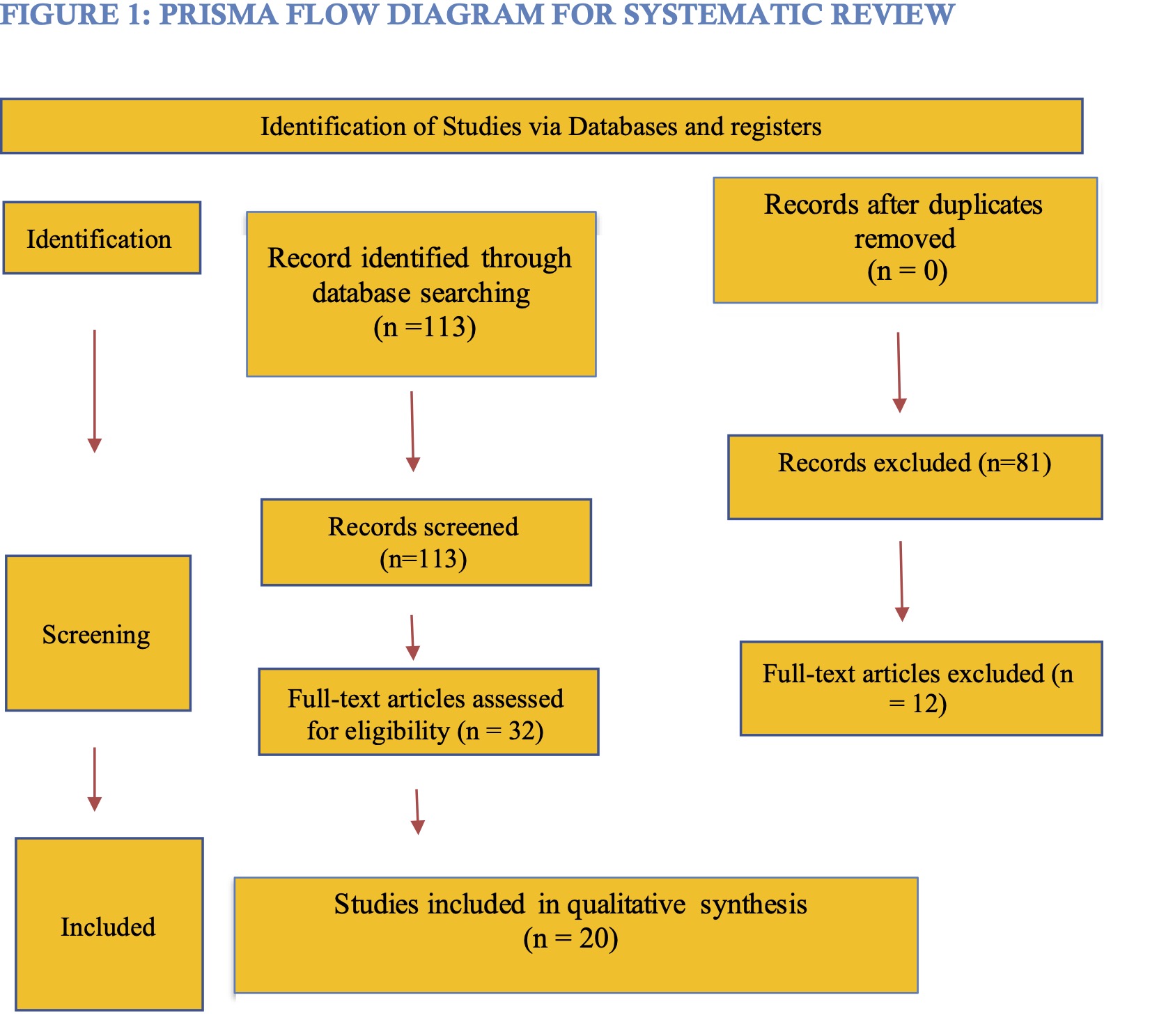
This systematic review followed the PRISMA 2020 guidelines (16). A comprehensive literature search was conducted in PubMed. Hence, studies published between 2005 and 2025 were included to ensure contemporary diagnostic, radiographic, and therapeutic relevance, using the keywords “multiple myeloma” AND “oral manifestation.” Boolean operators and MeSH terms were applied to enhance sensitivity. After retrieval, duplicates were removed, and titles and abstracts were screened. Exclusion criteria included reviews, systematic reviews, narrative articles, non-English language publications, and studies lacking validated clinical or radiographic descriptions. Full texts of potentially eligible studies were assessed for inclusion. Data extraction included author, year, country, study type, oral manifestations, radiographic findings, systemic symptoms, and diagnostic outcomes. Methodological quality was assessed using a standardized risk of bias tool, with studies categorized as high, moderate, or low quality as shown in figure 1.
RESULTS:
Following screening and eligibility assessment, 20 studies (2005–2025) met the inclusion criteria and were included in the qualitative synthesis. Most were case reports (70%), followed by cohort and cross-sectional studies (10% each), with one retrospective cohort study and one case series (5% each). No randomized controlled trials were identified, highlighting the rarity of oral-first presentations of Multiple Myeloma. Oral manifestations were diverse and frequently represented the initial sign of systemic disease. The most common presentation was jaw swelling, predominantly in the posterior mandible. Other findings included gingival enlargement, soft tissue masses, paresthesia, dental mobility, ulceration, gingival bleeding, and epistaxis often mimicking common odontogenic conditions. Radiographically, multiple osteolytic “punched out” lesions were the characteristic feature, mainly affecting the mandible. Additional findings included localized radiolucency’s, diffuse bone destruction, and occasional temporomandibular joint involvement as shown in table 1.
Table 1. Characteristics of Included Studies Reporting Oral Manifestations of Multiple Myeloma (n = 20)
|
S. No |
First Author (Year) |
Study Design |
Key Oral Manifestations |
|
1 |
Feitosa et al., (2020) (17) |
Cross-sectional |
• Pale mucosa |
|
2 |
Almeida et al., (2018) (18) |
Case report/series |
• Pain |
|
3 |
Pereira Hansen et al., (2018) (19) |
Case report |
• Gingival mass |
|
4 |
Thomas et al., (2015) (20) |
Case report |
• Facial swelling |
|
5 |
Bezerra et al., (2021) (21) |
Cross-sectional |
• Oral health alterations in MM patients |
|
6 |
Romano et al., (2014) (22) |
Case report |
• Oral lesion as first manifestation |
|
7 |
Ali et al., (2018) (23) |
Case report |
• Swelling |
|
8 |
Vieira-Leite et al., (2008) (24) |
Case report |
• Mandibular lesion as initial presentation |
|
9 |
Goetze et al., (2015) (25) |
Case report |
• Osseous mandibular plasmacytoma |
|
10 |
Beaumont et al., (2021) (26) |
Case series |
• Oral masses mimicking benign lesions |
|
11 |
Vinayachandran et al., (2013) (27) |
Case report |
• Multiple osteolytic lesions |
|
12 |
Pushpanshu et al., (2014) (28) |
Case report |
• Mandibular mass |
|
13 |
Souza et al., (2021) (29) |
Cohort study |
• Pain |
|
14 |
Kasamatsu et al., (2015) (30) |
Case report |
• Diffuse swelling |
|
15 |
Owotade et al., (2005) (31) |
Cohort study |
• Swelling |
|
16 |
Abboud et al., (2016) (32) |
Retrospective cohort |
• TMJ lesions |
|
17 |
Zhao et al., (2014) (33) |
Case report |
• Painful ulcer-like maxillary mass |
|
18 |
Crowley et al., (2016) (34) |
Case report |
• Mandibular pain |
|
19 |
Jain et al., (2013) (35) |
Case report |
• Generalized gingival enlargement |
|
20 |
Ramaiah et al., (2015) (36) |
Case report |
• Maxillary alveolar mass |
Swelling was the most reported oral feature, observed in over half of studies. Other frequent findings included jaw pain, soft tissue masses, gingival enlargement, dental mobility, and paresthesia. Rare manifestations, such as osteonecrosis, TMJ involvement, and severe intraoral hemorrhage, were reported in only one study each, highlighting the spectrum of oral presentations as shown in table 2.
Table 2. Frequency Distribution of Reported Oral Manifestations (n = 20 studies)
|
Oral Manifestation |
Number of Studies Reporting |
Percentage (%) |
|
Swelling (mandible/maxilla) |
11 |
55 |
|
Gingival enlargement / hyperplasia |
5 |
25 |
|
Gingival bleeding |
4 |
20 |
|
Soft tissue mass |
5 |
25 |
|
Dental mobility |
4 |
20 |
|
Paresthesia |
3 |
15 |
|
Jaw pain |
6 |
30 |
|
Ulceration |
2 |
10 |
|
Osteonecrosis of jaw |
1 |
5 |
|
TMJ involvement |
1 |
5 |
|
Intraoral hemorrhage |
1 |
5 |
Punched-out osteolytic lesions were the main radiographic finding, mainly in the posterior mandible. Other features, including cortical bone destruction, localized radiolucency, diffuse radiolucent lesions, TMJ involvement, and larger lytic lesions, were less frequent. Panoramic radiographs and CBCT were the primary imaging modalities as shown in table 3.
Revised Table 3: Radiographic Findings in Multiple Myeloma Affecting the Jaw (n = 20 studies)
|
Radiographic Feature |
Number of Studies |
Common Location |
Frequency |
|
Punched-out osteolytic lesions |
12 |
Posterior mandible |
High |
|
Localized radiolucency |
5 |
Mandible |
Moderate |
|
Cortical bone destruction |
4 |
Mandibular angle/ramus |
Moderate |
|
TMJ lesions |
1 |
TMJ |
Rare |
|
Diffuse radiolucent lesion |
2 |
Maxilla |
Rare |
|
Lytic Lesion (≥2 cm) |
3 |
Mandible |
Low |
Systemic manifestations accompanying oral lesions frequently aligned with CRAB criteria. Bone pain and anemia were the most reported, followed by fatigue, recurrent infections, hypercalcemia, renal insufficiency, and weight loss, underscoring the systemic impact of MM as shown in table 4.
Table 4. Systemic Manifestations Associated with Oral Presentation (CRAB Criteria, n = 20 studies)
|
Systemic Feature |
Frequency (Studies) |
|
Bone pain |
7 |
|
Anemia |
5 |
|
Fatigue |
4 |
|
Hypercalcemia |
2 |
|
Recurrent infections |
3 |
|
Renal insufficiency |
2 |
|
Weight loss |
1 |
The posterior mandible was the most frequently affected site, reflecting high hematopoietic marrow activity. Other less frequent sites included the mandibular angle/ramus, posterior maxilla, gingiva, TMJ, and diffuse jaw involvement, demonstrating variable lesion distribution as shown in table 5.
Table 5. Anatomical Distribution of Oral Lesions (n = 20 studies)
|
Site of Involvement |
Number of Studies |
Percentage (%) |
|
Posterior mandible |
11 |
55 |
|
Mandibular angle/ramus |
5 |
25 |
|
Maxilla (posterior) |
6 |
30 |
|
Gingiva |
5 |
25 |
|
TMJ |
1 |
5 |
|
Diffuse jaw involvement |
3 |
15 |
Most studies were case reports, reflecting the limited high-level evidence available on oral-first presentations of MM as shown in table 6.
Table 6. Distribution of Study Designs (n = 20)
|
Study Type |
Number of Studies |
Percentage (%) |
|
Case reports |
14 |
70 |
|
Case series |
1 |
5 |
|
Cohort studies |
2 |
10 |
|
Cross-sectional studies |
2 |
10 |
|
Retrospective cohort |
1 |
5 |
Overall, the included studies demonstrated moderate risk of bias due to small sample size, lack of controls, retrospective designs, and limited follow-up, which may affect generalizability as shown in table 7.
Table 7. Risk of Bias Assessment
|
Study Type |
Risk of Bias Level |
Common Limitation |
|
Case reports |
Moderate–High |
Small sample size |
|
Case series |
Moderate |
Lack of control group |
|
Cohort studies |
Moderate |
Retrospective design |
|
Cross-sectional studies |
Moderate |
Limited longitudinal data |
|
Retrospective cohort |
Moderate |
Limited sample size, retrospective |
Early recognition of unexplained swelling, punched-out lesions, gingival bleeding, and paresthesia by OMFS facilitated timely referral, imaging, biopsy, and lab evaluation. Pre-bisphosphonate dental assessments helped prevent osteonecrosis, highlighting the critical contribution of dental specialists in MM diagnosis and management as shown in table 8.
Table 8. Role of Oral & Maxillofacial Surgeons (OMFS) in Diagnostic Pathway
|
OMFS Intervention |
Clinical Impact |
|
Recognition of unexplained swelling |
Triggered hematology referral |
|
Identification of punched-out lesions |
Prompted imaging & biopsy |
|
Detection of gingival bleeding |
Laboratory evaluation |
|
Recognition of paresthesia |
Consideration of MM in differential |
|
Pre-bisphosphonate dental assessment |
Prevention of osteonecrosis |
DISCUSSION:
Multiple myeloma (MM) often presents with oral and maxillofacial manifestations that may precede systemic diagnosis (37). In this review of 20 studies, jaw swelling was the most frequently reported feature (55%), followed by jaw pain (30%), soft tissue masses (25%), and gingival enlargement (25%) (38, 39). Dental mobility (20%) and paresthesia (15%) were also observed, highlighting the diverse oral presentations of MM. Rare manifestations, including osteonecrosis, TMJ involvement, and severe intraoral hemorrhage, were reported in individual studies, reflecting the spectrum of clinical findings.
Radiographically, punched out osteolytic lesions were the main finding, identified in 12 studies, primarily affecting the posterior mandible (39, 40). Additional imaging findings included localized radiolucency’s, diffuse bone destruction, TMJ lesions, and larger lytic lesions, demonstrating the importance of panoramic radiography and CBCT for early detection. Anatomically, the posterior mandible was the most frequently affected site (55%), followed by posterior maxilla (30%) and mandibular angle/ramus (25%) (41).
Systemic features accompanying oral manifestations were consistent with CRAB criteria. Bone pain (7 studies), anemia (5), fatigue (4), and recurrent infections (3) were the most frequently reported systemic signs (38, 40). These findings emphasize the need for oral health professionals to consider MM in the differential diagnosis when patients present with unexplained oral lesions, particularly in the posterior jaw.3
This systematic review provides a comprehensive synthesis of oral and radiographic manifestations of multiple myeloma, emphasizing the critical role of OMFS in early detection. Strengths of this review include the inclusion of contemporary studies spanning 2005–2025, strict adherence to PRISMA 2020 guidelines, detailed extraction of clinical, radiographic, and systemic features, and structured presentation of data through tables summarizing prevalence, anatomical distribution, and OMFS interventions. The review also highlights practical implications for timely diagnosis, referral, and prevention of complications such as bisphosphonate-related osteonecrosis. However, several limitations must be acknowledged. Most included studies were case reports or cohorts, limiting generalizability and introducing potential publication bias. Heterogeneity in study designs, reporting standards, and diagnostic criteria restricted the ability to perform quantitative synthesis. Moreover, the absence of randomized controlled trials and longitudinal studies limits conclusions regarding the predictive value of oral manifestations for systemic disease progression. Despite these limitations, this review provides valuable insights into early oral presentations of MM and underscores the importance of vigilant oral assessments in clinical practice.
Conclusion:
Oral manifestations often precede systemic signs of multiple myeloma. Early recognition by oral and maxillofacial surgeons facilitates timely diagnosis, referral, and prevention of treatment complications, underscoring the critical role of dental professionals in multidisciplinary management.
References:
1. Barwick BG, Gupta VA, Vertino PM, Boise LH. Cell of origin and genetic alterations in the pathogenesis of multiple myeloma. Frontiers in immunology. 2019 May 21;10:1121. http://doi:10.3389/fimmu.2019.01121. PMID: 31231360; PMCID: PMC6558388
2. Giannakoulas N, Ntanasis-Stathopoulos I, Terpos E. The role of marrow microenvironment in the growth and development of malignant plasma cells in multiple myeloma. International journal of molecular sciences. 2021 Apr 24;22(9):4462. http://doi:10.3390/ijms22094462. PMID: 33923357; PMCID: PMC8123209.
3. Dimopoulos MA, Merlini G, Bridoux F, Leung N, Mikhael J, Harrison SJ, Kastritis E, Garderet L, Gozzetti A, Van De Donk NW, Weisel KC. Management of multiple myeloma-related renal impairment: recommendations from the International Myeloma Working Group. The Lancet Oncology. 2023 Jul 1;24(7):e293-311. http://doi:10.1016/S1470-2045(23)00223-1. PMID: 37414019.
4. Zhang N, Wu J, Wang Q, Liang Y, Li X, Chen G, Ma L, Liu X, Zhou F. Global burden of hematologic malignancies and evolution patterns over the past 30 years. Blood Cancer Journal. 2023 May 17;13(1):82. http://doi:10.1038/s41408-023-00853-3. PMID: 37193689; PMCID: PMC10188596.
5. Padala SA, Barsouk A, Barsouk A, Rawla P, Vakiti A, Kolhe R, Kota V, Ajebo GH. Epidemiology, staging, and management of multiple myeloma. Medical Sciences. 2021 Jan 20;9(1):3. http://doi:10.3390/medsci9010003. PMID: 33498356; PMCID: PMC7838784.
6. Moore AR. Diagnosing multiple myeloma and related disorders. Veterinary Clinics: Small Animal Practice. 2023 Jan 1;53(1):101-20. http://doi:10.1016/j.cvsm.2022.07.009. Epub 2022 Oct 19. PMID: 36270842.
7. Girard LP, Soekojo CY, Ooi M, Poon LM, Chng WJ, de Mel S. Immunoglobulin M paraproteinaemias. Cancers. 2020 Jun 25;12(6):1688.http://doi:10.3390/cancers12061688. PMID: 32630470; PMCID: PMC7352433.
8. Dao A, McDonald MM, Savage PB, Little DG, Schindeler A. Preventing osteolytic lesions and osteomyelitis in multiple myeloma. Journal of bone oncology. 2022 Dec 1;37:100460. http://doi:10.1016/j.jbo.2022.100460. PMID: 36388641; PMCID: PMC9640986.
9. Krishnan P, Narayan M, Divya B, Kumar TD, Kumar AR, Krishnan R. Oral manifestations of multiple myeloma–A systematic review. Oral Oncology Reports. 2024 Jun 1;10:100485.
10. Lu SY, Ma MC, Wang MC, Hsue SS. The status of jaw lesions and medication-related osteonecrosis of jaw in patients with multiple myeloma. Journal of the Formosan Medical Association. 2021 Nov 1;120(11):1967-76. http://doi:10.1016/j.jfma.2021.01.015. Epub 2021 Feb 4. PMID: 33551311.
11. Wang Y, Xu Q, Li Y, Su Y, Wang L, Wang X, Ge J, Jing H, Guo Y, Chen Y, Li X. Expert consensus on a multidisciplinary approach for the management of multiple myeloma-related bone disease☆. Cancer Pathogenesis and Therapy. 2025 Jul 30;3(04):282-94. http://doi:10.1016/j.cpt.2024.12.002. PMID: 40697511; PMCID: PMC12277822.
12. Moritz DY. Multiple myeloma and other plasma cell diseases (Doctoral dissertation, Sveučilište u Zagrebu, Sveučilište u Zagrebu, Medicinski fakultet, Katedra za internu medicinu).
13. Bahk WJ. Plasma Cell Myeloma/Multiple. Diagnosis and Management of Primary Bone Tumors: Volume 2. 2023 Nov 28;2:87.
14. Mutlu U, Balci A, Özsan GH, Özkal S, Şeyhanli A, Özgül HA. Computed tomography characteristics of multiple myeloma and other osteolytic metastatic bone lesions. Acta Radiologica. 2021 Dec;62(12):1639-47. http://doi:10.1177/0284185120977035. Epub 2020 Dec 3. PMID: 33269940.
15. Klein MJ. Myeloma. InTumors and Tumor-Like Lesions of Bone 2020 May 6 (pp. 439-448). Cham: Springer International Publishing.
16. Page MJ, McKenzie JE, Bossuyt PM, Boutron I, Hoffmann TC, Mulrow CD, Shamseer L, Tetzlaff JM, Moher D. Updating guidance for reporting systematic reviews: development of the PRISMA 2020 statement. Journal of clinical epidemiology. 2021 Jun 1;134:103-12. http://doi:10.1016/j.jclinepi.2021.02.003. Epub 2021 Feb 9. PMID: 33577987.
17. Feitosa ÉF, Magalhães RJ, de Melo Barbosa CA, Guedes FR, Maiolino A, Torres SR. Oral health status of patients with multiple myeloma. Hematology, Transfusion and Cell Therapy. 2020 Apr 1;42(2):166-72. http://doi:10.1016/j.htct.2019.01.012. Epub 2019 May 15. PMID: 31582338; PMCID: PMC7248491.
18. de Almeida TM, Cavalcanti ÉF, da Silva Freitas A, de Magalhães RJ, Maiolino A, Torres SR. Can dentists detect multiple myeloma through oral manifestations. Hematology, Transfusion and Cell Therapy. 2018 Jan 1;40(1):43-9. http://doi:10.1016/j.bjhh.2017.08.006. Epub 2017 Nov 24. PMID: 29519372; PMCID: PMC6003103.
19. Pereira Hansen CD, de Mattos Nascimento M, Alves FA, Neotti T, Costa FD. Extramedullary Manifestation of Multiple Myeloma in the Oral Cavity. Archives of Iranian Medicine (AIM). 2018 Dec 1;21(12). PMID: 30634861.
20. Thomas AE, Kurup S, Jose R, Soman C. Facial swelling as a primary manifestation of multiple myeloma. Case Reports in Dentistry. 2015;2015(1):319231. http://doi:10.1155/2015/319231. Epub 2015 Jul 1. PMID: 26229694; PMCID: PMC4502313.
21. Bezerra ML, Alves LC, Tabosa RA, Dantas SL, da Rocha TJ, Nigri FM, Neri JR, Lima DL. Oral Conditions of Elders and Middle-aged Individuals with Multiple Myeloma. The Journal of Contemporary Dental Practice. 2021 Aug 9;22:610-4. PMID: 34393115.
22. Romano A, Marescalco MS, Liardo C, Villari L, Vetro C, Conticello C, Di Raimondo F, Ferlito S. Oral lesion as unusual first manifestation of multiple myeloma: case reports and review of the literature. Case Reports in Hematology. 2014;2014(1):529452. http://doi:10.1155/2014/529452. Epub 2014 Nov 25. PMID: 25506443; PMCID: PMC4260434.
23. Ali IK, Parate AR, Kasat VO, Dora A. Multiple myeloma with primary manifestation in the mandible. Cureus. 2018 Mar 3;10(3). http://doi:10.7759/cureus.2265. PMID: 29732272; PMCID: PMC5933576.
24. Segundo AV, Falcão MF, Correia-Lins Filho R, Marques MS. Multiple Myeloma with primary manifestation in the mandible: A case report. Medicina oral, patología oral y cirugía bucal. Ed. inglesa. 2008 Apr 1;13(4):5. PMID: 18379446.
25. Goetze E, Walter C, Kämmerer PW. Osseous plasma cell neoplasm of the mandible for initial diagnosis of multiple myeloma: case report and literature review. Journal of Maxillofacial and Oral Surgery. 2015 Mar;14(Suppl 1):469-74. http://doi:10.1007/s12663-014-0688-7. Epub 2014 Sep 9. PMID: 25861199; PMCID: PMC4379288.
26. Beaumont S, Koo RM, Harrison SJ. A Wolf in Sheep’s clothing: A case report series of oral manifestations of multiple myeloma. Australian Dental Journal. 2021 Sep;66(3):324-31. http://doi:10.1111/adj.12808. Epub 2020 Dec 7. PMID: 33217006.
27. Vinayachandran D, Sankarapandian S. Multiple osteolytic lesions. Journal of Clinical Imaging Science. 2013 Aug 31;3(Suppl 1):6. http://doi:10.4103/2156-7514.117460. PMID: 24516769; PMCID: PMC3906660
28. Pushpanshu K, Punyani S, Kaushik R. Mandibular mass as the primary manifestation of multiple myeloma. Brazilian Journal of Otorhinolaryngology. 2014;80(03):266-7. http://doi:10.1016/j.bjorl.2013.01.001. PMID: 25153114; PMCID: PMC9535485.
29. Souza LL, Caceres CV, Vargas PA, Lopes MA, Santos‐Silva AR, Silva WG, Prado‐Ribeiro AC, Brandao TB, Pereira J, Mesquita RA, Pontes FS. Plasmacytoma and plasma cell myeloma affecting the jaws: A multi‐institutional collaborative study. Journal of Oral Pathology & Medicine. 2021 Jul;50(6):613-21.http://doi:10.1111/jop.13213. Epub 2021 Jun 20. PMID: 34089204.
30. Kasamatsu A, Kimura Y, Tsujimura H, Kanazawa H, Koide N, Miyamoto I, Endo-Sakamoto Y, Shiiba M, Tanzawa H, Uzawa K. Maxillary swelling as the first evidence of multiple myeloma. Case Reports in Dentistry. 2015;2015(1):439536. http://doi:10.1155/2015/439536. Epub 2015 Nov 12. PMID: 26640721; PMCID: PMC4659956.
31. Owotade F, Ugboko V, Ajike S, Salawu L, Amusa Y, Omole M. Head and neck manifestations of myeloma in Nigerians. International journal of oral and maxillofacial surgery. 2005 Oct 1;34(7):761-5. http://doi:10.1016/j.ijom.2005.02.007. PMID: 15979285.
32. Abboud W, Yahalom R, Leiba M, Greenberg G, Yarom N. Temporomandibular joint involvement in patients with multiple myeloma retrospective study. International Journal of Oral and Maxillofacial Surgery. 2016 Dec 1;45(12):1545-50.http://doi:10.1016/j.ijom.2016.06.014. Epub 2016 Jul 1. PMID: 27377681.
33. Zhao XJ, Sun J, Wang YD, Wang L. Maxillary pain is the first indication of the presence of multiple myeloma: a case report. Molecular and Clinical Oncology. 2014 Jan;2(1):59-64. http://doi:10.1016/j.ijom.2016.06.014. Epub 2016 Jul 1. PMID: 27377681.
34. Crowley M. Multiple myeloma presenting as mandibular pain. PMID: 29797838.
35. Jain S, Kaur H, Kansal G, Gupta P. Multiple myeloma presenting as gingival hyperplasia. Journal of Indian Society of Periodontology. 2013 May 1;17(3):391-3. http://doi:10.4103/0972-124X.115652. PMID: 24049344; PMCID: PMC3768194.
36. Ramaiah KK, Joshi V, Thayi SR, Sathyanarayana P, Patil P, Ahmed Z. Multiple myeloma presenting with a maxillary lesion as the first sign. Imaging science in dentistry. 2015 Mar 13;45(1):55. http://doi:10.5624/isd.2015.45.1.55. Epub 2015 Mar 13. PMID: 25793184; PMCID: PMC4362992.
37. Avet-Loiseau H, Davies FE, Samur MK, Corre J, D'Agostino M, Kaiser MF, Raab MS, Weinhold N, Gutierrez NC, Paiva B, Neri P. International Myeloma Society/International Myeloma Working Group consensus recommendations on the definition of high-risk multiple myeloma. Journal of Clinical Oncology. 2025 Aug 20;43(24):2739-51. http://doi:10.1200/JCO-24-01893. Epub 2025 Jun 9. Erratum in: J Clin Oncol. 2025 Aug;43(22):2553. doi: 10.1200/JCO-25-01367. PMID: 40489728.
38. Terpos E, Zamagni E, Lentzsch S, Drake MT, García-Sanz R, Abildgaard N, Ntanasis-Stathopoulos I, Schjesvold F, de la Rubia J, Kyriakou C, Hillengass J. Treatment of multiple myeloma-related bone disease: recommendations from the Bone Working Group of the International Myeloma Working Group. The Lancet Oncology. 2021 Mar 1;22(3):e119-30. http://doi:10.1016/S1470-2045(20)30559-3. Epub 2021 Feb 2. PMID: 33545067.
39. Cao W, Qin K, Li F, Chen W. Comparative study of cancer profiles between 2020 and 2022 using global cancer statistics (GLOBOCAN). Journal of the National Cancer Center. 2024 Jun 1;4(2):128-34. http://doi:10.1016/j.jncc.2024.05.001. PMID: 39282581; PMCID: PMC11390618.
40. Huang J, Chan SC, Lok V, Zhang L, Lucero-Prisno DE, Xu W, Zheng ZJ, Elcarte E, Withers M, Wong MC. The epidemiological landscape of multiple myeloma: a global cancer registry estimate of disease burden, risk factors, and temporal trends. The Lancet Haematology. 2022 Sep 1;9(9):e670-7. http://doi:10.1016/S2352-3026(22)00165-X. Epub 2022 Jul 14. PMID: 35843248.
41. Cowan AJ, Allen C, Barac A, Basaleem H, Bensenor I, Curado MP, Foreman K, Gupta R, Harvey J, Hosgood HD, Jakovljevic M. Global burden of multiple myeloma: a systematic analysis for the global burden of disease study 2016. JAMA oncology. 2018 Sep;4(9):1221-7. http://doi:10.1001/jamaoncol.2018.2128. PMID: 29800065; PMCID: PMC6143021.