SYNTHESIS OF ORGANOBENTONITE USING HIBISCUS ROSA-SINENSIS EXTRACT: CHARACTERIZATION AND ITS APPLICATIONS
- Benshika I A , Research Scholar, Department of Chemistry, Women’s Christian College, Nagercoil, Tamilnadu, India. Affiliated to: Manonmaniam Sundaranar University, Tirunelveli, Tamil Nadu, India.
- Sangeetha N J , Assistant Professor, Department of Chemistry, Women’s Christian College, Nagercoil, Tamilnadu, India. Affiliated to: Manonmaniam Sundaranar University, Tirunelveli, Tamil Nadu, India.
- Shyni Raphael M , Associate Professor, Department of Chemistry, Government College for Women, Thiruvananthapuram, Kerala, 695014, India
Article Information:
Abstract:
Background: Hibiscus rosa-sinensis, a widely cultivated ornamental species with recognized medicinal properties, was employed for the preparation of organo-bentonite (HIBT-Hibiscus-modified bentonite) through intercalation with modified bentonite clay. The synthesis involved extraction of phytochemicals followed by freeze–thaw method. The resulting material was characterized using UV–Vis spectroscopy, Fourier-transform infrared spectroscopy (FTIR), Scanning electron microscopy (SEM), and X-ray diffraction (XRD). Antibacterial, antifungal, antioxidant, antidiabetic, and anticancer activities were evaluated in-vitro. UV–Vis and FTIR analyses confirmed the incorporation of phytoconstituents within the modified clay, while XRD indicated a semi-crystalline structure. SEM revealed a sheet-like morphology. The organo-bentonite exhibited antibacterial and antifungal activity, particularly against Candida albicans. Antioxidant assays (DPPH, ABTS) indicated moderate radical scavenging activity. Enzyme inhibition assays demonstrated α-amylase and α-glucosidase inhibitory potential, while cytotoxicity assays (MTT, SRB) revealed dose-dependent activity against MCF7 breast cancer cells. These findings highlight the potential of Hibiscus rosa-sinensis modified bentonite as a multifunctional biomaterial with applications in pharmaceutical and biomedical fields.
Keywords:
Article :
INTRODUCTION:
Natural products have long served as sources of food, shelter, and medicine, with plant-derived compounds contributing significantly to traditional and modern therapeutic systems [1]. India possesses a rich biodiversity that has supported the development of traditional medicine [2]. Hibiscus rosa-sinensis, commonly referred to as China rose, is native to Southeast Asia and is widely recognized for its ornamental and medicinal value. Various parts of the plant are reported to possess analgesic, antipyretic, anti-inflammatory, and antimicrobial properties, making it a source of biologically active metabolites [3].
Clay minerals, particularly phyllosilicates, are layered silicates with high surface area and interlayer reactivity [4]. Among them, bentonite-a naturally occurring aluminosilicate enriched with exchangeable cations, has attracted significant interest due to its adsorption capacity and ion-exchange properties. Bentonite has been employed for both environmental and therapeutic uses [5,6,7,8]. Modification of bentonite through intercalation with organic molecules produces organoclays, which have been investigated for diverse applications ranging from oil recovery to biomedical systems [9].
Recent studies have demonstrated the potential of organo-modified clays in drug delivery, tissue engineering, antimicrobial coatings, and antioxidant formulations [10]. In this study, bentonite was modified using a methanolic extract of Hibiscus rosa-sinensis to synthesize organo-bentonite (hibiscus-modified bentonite). The physicochemical properties were characterized, and the biological activities, including antimicrobial, antioxidant, antidiabetic, and anticancer potentials, were systematically evaluated.
MATERIALS AND METHODS:
Bentonite clay was procured from a local source and purified prior to modification. Hibiscus rosa-sinensis flowers were collected, washed, and shade dried. Analytical grade NaCl, H2O2, NaOH, CH3OH and other standard assay chemicals were employed in the study.
Preparation of Modified Bentonite:
Bentonite (5 g) was dispersed in 1 M NaCl and subjected to ultrasonication, followed by centrifugation and repeated washing until complete purification. The solid fraction was
dried at 60 °C. To remove organic residues, the modified clay was treated with 30% H₂O₂ (1:2, w/v) for 3 h, adjusted to neutral pH using 0.5 M NaOH, and dried at 60 °C [11, 12].
Preparation of Hibiscus Extract:
Dried Hibiscus rosa-sinensis petals were powdered and extracted with methanol in a 1:10 (w/v) ratio under continuous shaking. The extract was filtered through Whatman No. 1 paper [13].
Synthesis of Organo-Bentonite:
The modified freeze–thaw method was employed for intercalation. The Hibiscus extract (40 mL) was mixed with modified clay (2 g) to form a homogeneous suspension. The mixture was frozen at 20 °C for 24 h and subsequently thawed at room temperature, yielding the organo-bentonite (HIBT-Hibiscus-modified bentonite) [14].
Characterization
· UV–Vis spectroscopy
· UV-Vis spectrum of hibiscus-modified bentonite was recorded using Shimadzu UV-1800 spectrophotometer in the wavelength range of 200-800 nm.
· FTIR
· FTIR analysis of the organo-bentonite was performed using the KBr pellet method. The spectrum was collected in the range of 500-4500 cm-1 with Shimadzu FTIR-8400s spectrometer.
· XRD
· XRD patterns of organo-bentonite was obtained using XPERT Pro diffractometer (PANalytical, JEOL) over a 2θ range of 20–80°. The average crystallite size was estimated using the Debye–Scherrer equation.
· SEM
Surface morphology was analyzed using Tescan VEGA 3 SBU scanning electron microscope under a vacuum range of 0.005–2000 Pa.
Antimicrobial Assayb:
Antibacterial activity was evaluated against Escherichia coli and Staphylococcus aureus using the Kirby–Bauer disc diffusion method. Antifungal activity was tested against Candida albicans and Aspergillus niger. Zones of inhibition were measured and compared with standard antibiotics [15].
Antioxidant Assays:
· DPPH assay: Performed with different concentrations (6.25–100 μg/mL) of hibiscus- modified bentonite (HIBT) using ascorbic acid as standard. Absorbance measured at 520 nm [16].
· ABTS assay: ABTS•+ generated by potassium persulfate oxidation; hibiscus-modified bentonite (HIBT) tested at 6.25–100 μg/mL. Absorbance measured at 734 nm [17].
Antidiabetic Assays
· α-Amylase inhibition: Enzyme and substrate incubated with hibiscus-modified bentonite (HIBT) using acarbose as standard; absorbance measured at 540 nm after DNSA reaction [18].
· α-Glucosidase inhibition: Reaction with p-nitrophenyl-α-D-glucopyranoside; absorbance measured at 405 nm; acarbose used as control [19].
Anticancer Assays
· MTT assay: MCF7 breast cancer cells exposed to hibiscus-modified bentonite (HIBT) at varying concentrations; cell viability assessed at 570 nm [20].
· SRB assay: Protein binding assay performed on MCF7 cells; absorbance recorded at 510 nm [21, 22].
RESULTS And DISCUSSION :
UV–Vis Spectroscopy
The organo-bentonite (hibiscus-modified bentonite) exhibited a distinct absorption band at ~275 nm, corresponding to π→π* transitions of phenolic compounds. This band, absent in unmodified bentonite, confirmed the successful incorporation of Hibiscus-derived phytoconstituents. The absorption profile below 300 nm was attributed to flavonoids and
polyphenols, while the absence of visible-region absorbance indicated negligible amounts of anthocyanins.
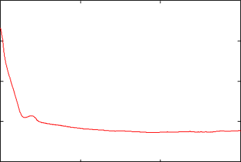
Fig. 3.1 UV-Vis spectroscopy of hibiscus-modified bentonite.
FTIR Analysis
The FTIR spectrum of the Hibiscus-modified bentonite displayed a broad band at~3441 cm⁻¹, assigned to O–H stretching from interlayer water and phenolic groups. Peaks near 2949 cm⁻¹ reflected aliphatic C–H vibrations, whereas the band at 1635 cm⁻¹ indicated H–O–H bending with possible aromatic C=C contributions. The Si–O–Si stretching at~1018 cm⁻¹ confirmed the retention of the bentonite framework. Collectively, these features demonstrate effective intercalation of Hibiscus within the modified bentonite clay.
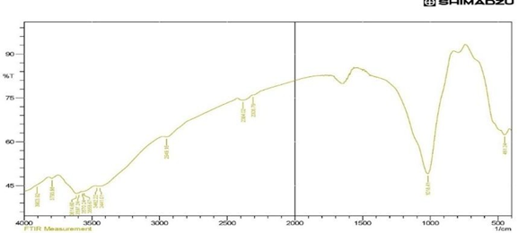
Fig. 3.2 FT-IR spectrum of hibiscus-modified bentonite
XRD Analysis
X-ray diffraction patterns revealed a semi-crystalline structure with reduced intensity and broadening of basal reflections. The characteristic (001) reflection near 7° 2θ was significantly diminished, suggesting expanded interlayer spacing and partial exfoliation due to intercalation. The intense peak near 26.6° indicated structural modification, supporting the semi-crystalline nature of the synthesized hibiscus-modified bentonite.
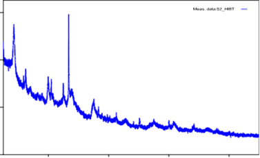 Fig. 3.3 XRD pattern of hibiscus-modified bentonite
SEM Analysis
SEM images confirmed morphological changes. The hibiscus-modified bentonite exhibited sheet-like, fragmented structures with increased surface roughness. These modifications reflect the deposition of Hibiscus phytochemicals, enhanced surface heterogeneity and properties favorable for adsorption and antimicrobial interactions.
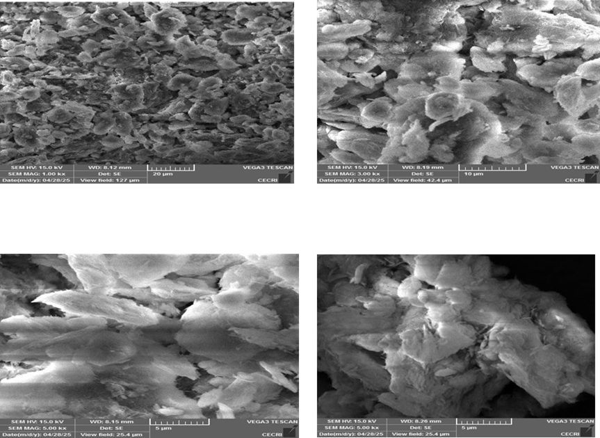
Fig 3.4 SEM images of hibiscus-modified bentonite
Antimicrobial Activity
The organo-bentonite (hibiscus-modified bentonite) displayed notable antibacterial activity against E. coli (16 mm inhibition) and S. aureus (15 mm), comparable to the reference antibiotic. Antifungal tests revealed higher inhibition against Candida albicans (18.5 mm) than the control, whereas Aspergillus niger inhibition (14 mm) was lower than that of the standard. These findings indicate that Hibiscus-modified bentonite has selective antifungal activity, with enhanced efficacy against Candida albicans.
Table 1. Antibacterial activity of hibiscus-modified bentonite
|
Bacteria |
HIBT |
Control (Amikacin) |
|
E. coli |
16 mm |
16 mm |
|
Staphylococcus aureus |
15 mm |
15mm |
Table 2. Antifungal activity of hibiscus-modified bentonite
|
Bacteria |
HIBT |
Control (Amikacin) |
|
Candida albicans |
18.5 mm |
15 mm |
|
Aspergillus niger |
14 mm |
18 mm |
Antioxidant Activity
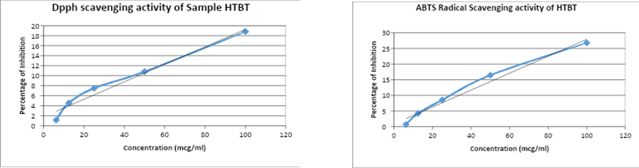
The DPPH assay revealed concentration-dependent radical scavenging, with maximum inhibition of 18.80% at 100 μg/mL and an IC₅₀ value of 277.09 μg/mL, indicating moderate antioxidant potential. The ABTS assay demonstrated stronger activity, with 26.77% inhibition at 100 μg/mL and an IC₅₀ of 182.11 μg/mL. These results suggest that Hibiscus intercalated into the modified bentonite clay retain their redox-active functionality.
Antidiabetic Activity
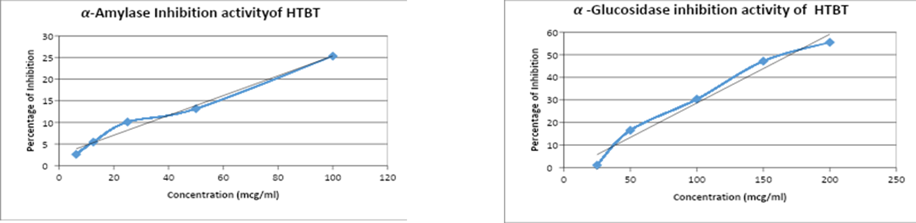
The α-amylase inhibition reached 25.3% at 100 μg/mL, with an IC₅₀ of 208.53 μg/mL, demonstrating moderate activity. α-Glucosidase inhibition was more pronounced, increasing progressively from 1.09% at 25 μg/mL to 55.53% at 200 μg/mL. This indicates stronger inhibitory potential toward α-glucosidase compared with α-amylase, highlighting the material’s relevance as an antidiabetic.
Anticancer Activity
The MTT assay on MCF7 breast cancer cells showed a dose-dependent reduction in viability, with IC₅₀ = 220.58 μg/mL. The SRB assay corroborated these findings, with IC₅₀ = 251.06 μg/mL. Although less potent than doxorubicin, the organo-bentonite demonstrated measurable cytotoxic effects, indicating potential for further exploration as a supplementary anticancer material.
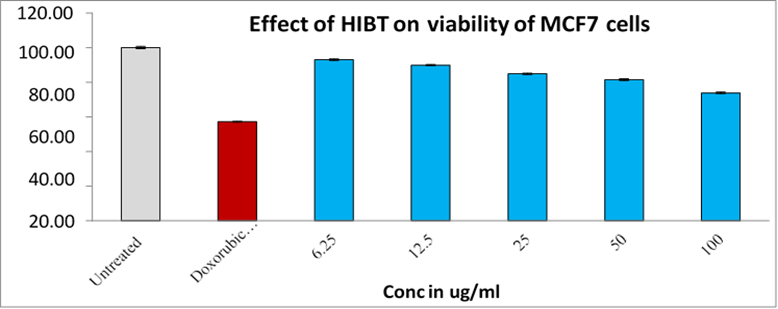 MTT Asaay of hibiscus-modified bentonite
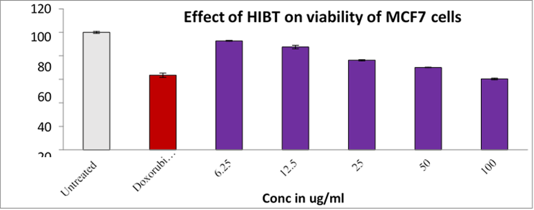
SRB Asaay of hibiscus-modified bentonite
CONCLUSION :
Hibiscus-modified bentonite was successfully synthesized by intercalating Hibiscus rosa-sinensis extract into modified bentonite via a freeze–thaw method. Structural analyses (UV–Vis, FTIR, XRD, SEM) confirmed the incorporation of bioactive phytochemicals and revealed morphological and crystallinity changes. The Hibiscus-modified bentonite exhibited antibacterial and antifungal activity, with Candida albicans showing the highest susceptibility. Antioxidant assays indicated moderate radical scavenging capacity, while enzyme inhibition assays demonstrated significant α-glucosidase inhibitory potential. Furthermore, in vitro anticancer assays revealed dose-dependent cytotoxicity against MCF7 breast cancer cells. These findings suggest that Hibiscus-modified bentonite is a promising multifunctional biomaterial with potential applications in pharmaceuticals, dermo-cosmetics, and regenerative medicine.
BIBLIOGRAPHY:
1. Gurib-Fakim, A., 2006. Medicinal plants: traditions of yesterday and drugs of tomorrow.
2. Molecular Aspects of Medicine, 27, 1–93.
3. Jadhav, V.M., Throat, R.M., Kadam, V.J., Sathe, N.S., 2009. Hibiscus rosa-sinensis Linn – “Rudrapuspa”: a review. Journal of Pharmaceutical Research, 2(7), 1168–1173.
4. Missoum, A., 2018. An updated review on Hibiscus rosa-sinensis phytochemistry and medicinal uses. Journal of Ayurvedic and Herbal Medicine, 4(3), 135–146.
5. Guggenheim, S., Martin, R.T., 1995. Definition of clay and clay minerals: Joint report of the AIPEA and CMS nomenclature committees. Clays and Clay Minerals, 43(2), 225–256.
6. Moosavi, M., 2017. Bentonite clay as a natural remedy: a brief review. Iranian Journal of Public Health, 46(9), 1176–1183.
7. Belghazdis, M., Hachem, E., 2022. Clay and clay minerals: a detailed review. International Journal of Recent Technology and Applied Science, 4(2), 54–75. https://doi.org/10.36079/lamintang.ijortas-0402.367
8. Al-Ani, T., Sarappa, O., 2008. Clay and clay mineralogy: physico-chemical properties and industrial uses. Geologian Tutkuskeskus Report, M19/323/2008/41.
9. Yariv, S., 2002. Organo-clay complexes and interactions. Springer, Berlin.
10. Raj, A., Jeslin, J., 2018. Synthesis of bentonite nanoclay and incorporation of Cassia fistula leaf extract to form organobentonite: Characterisation and biomedical applications. Asian Journal of Pharmaceutical and Clinical Research, 11(9). https://doi.org/10.22159/ajpcr.2018.v11i9.26717
11. Ghadhiri, M., Chrzanowski, W., Rohanizadeh, R., 2015. Biomedical applications of cationic clay minerals. RSC Advances. https://doi.org/10.1039/C5RA03700K
12. Floody, M.C., Bendall, J.S., Jara, A.A., Welland, M.E., Theng, B.K., Rumpel, C., 2011. Nanoclays from an Andisol: extraction, properties and carbon stabilization. Geoderma, 161, 159–167.
13. James, O., Adediran, M., Adekola, F., Odebumni, E., Adekeye, J., 2008. Beneficiation and characterisation of a bentonite from north-eastern Nigeria. Journal of the North Carolina Academy of Science, 124, 154–158.
14. Gowdhami, M., Sarkar, B.L., Ayyasamy, P.M., 2014. Screening of phytochemicals and antibacterial activity of Annona squamosa extracts. International Journal of Pharmaceutical Science Inventions, 3, 30–39.
15. Nooria, S., Kokabia, M., Hassan, Z.M., 2015. Nanoclay-enhanced mechanical properties of poly(vinyl alcohol)/chitosan/montmorillonite nanocomposite hydrogel as wound dressing. Procedia Materials Science, 11, 152–156.
16. Bauer, A.W., Kirby, W.M.M., Sherris, J.C., Turck, M., 1966. Antibiotic susceptibility testing by a standardized single disk method. American Journal of Clinical Pathology, 4(5), 493–496.
17. Blois, M.S., 1958. Antioxidant determinations by the use of a stable free radical. Nature, 181, 1199–1200.
18. Sricharoen, P., Techawongstein, S., Luthria, D., Chanthai, S., 2014. Standardization of DPPH, ABTS and FRAP assays with reference compounds for estimating antioxidant capacity of tomato extracts. Food Chemistry, 178, 271–277.
19. Kwon, Y., Apostolidis, E., Shetty, K., 2006. Inhibitory potential of wine and tea against α- amylase and α-glucosidase for management of hyperglycemia. Journal of Food Biochemistry, 32, 15–31.
20. Dewi, R.T., Iskandar, Y.M., Hanafi, M., Kardono, L.B.S., Angelina, M., Dewijanti, I.D., Banjarnahor, S.D.S., 2007. Inhibitory effect of koji Aspergillus terreus on α-glucosidase activity and postprandial hyperglycemia. Pakistan Journal of Biological Sciences, 10(8), 3131– 3135.
21. Mosmann, T., 1983. Rapid colorimetric assay for cellular growth and survival: application to proliferation and cytotoxicity assays. Journal of Immunological Methods, 65, 55–63.
22. Skehan, P., Storeng, R., Scudiero, D., Monks, A., McMahon, J., Vistica, D., Warren, J.T., Bokesch, H., Kenney, S., Boyd, M.R., 1990. New colorimetric cytotoxicity assay for anticancer-drug screening. Journal of the National Cancer Institute, 82(13), 1107–1112.
23. Vichai, V., Kirtikara, K., 2006. Sulforhodamine B colorimetric assay for cytotoxicity screening. Nature Protocols, 1(3), 1112–1116.